Flank pain in a 60-year-old man

Published on 10/04/2015 by admin

Filed under Surgery

Last modified 22/04/2025

Print this page

rate 1 star rate 2 star rate 3 star rate 4 star rate 5 star
Your rating: none, Average: 0 (0 votes)

This article have been viewed 911 times

Problem 15 Flank pain in a 60-year-old man

The patient has never had pain like this before and is usually in good health apart from type II diabetes for which he takes metformin. His past history is unremarkable. On examination, he looks ill, is pale and diaphoretic, and the pain makes him appear uncomfortable. His temperature is 38.7°C, and he has a heart rate of 95/min, blood pressure of 90/60 mmHg, respiratory rate 23/min and an SaO2 91% breathing room air. His BMI is 30. His abdomen is soft to palpation, with no localizing signs.

His white cell count is 21 (91% neutrophils), creatinine 380 mmol/L, potassium 4.5 mmol/L and blood glucose 12 mmol/L. Urinalysis shows large amounts of red and white blood cells, and nitrites. An ECG is performed and confirms sinus tachycardia with no acute ST changes.

Two large-bore intravenous cannulae are inserted and a broad-spectrum antibiotic (ceftriaxone) is administered after blood and urine cultures have been obtained. Opiate analgesia and an antiemetic are given. A fluid balance chart and insulin sliding scale is commenced. After an hour and 2 litres of isotonic saline the patient is comfortable and his vital signs have stabilized. You can now turn your thoughts to looking for the cause of the sepsis.

Now that the patient’s condition has stabilized some further investigations can be considered.

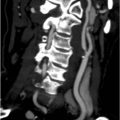
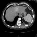
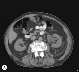
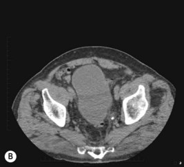
A non-contrast CT scan is performed and two representative slices are shown in Figure 15.1A, B.

The CT scan findings confirm the clinical impression of urosepsis with a stone obstructing the left renal tract.

Once the patient is stabilized, a ureteric stent is inserted under general anaesthetic via cystoscopy and frank pus drains from the left kidney. A specimen is sent for microscopy and culture and grows Escherichia.

The pain resolves rapidly and within 24 hours the patient’s temperature, creatinine and potassium are all within normal limits. Antibiotics are continued intravenously for 5 days total, and the patient is discharged with 10 days of oral antibiotics.

He is readmitted 2 weeks later for a ureteroscopy after his urine is confirmed to be sterile.

The stone fragments are sent to the laboratory for analysis, and are confirmed to be of calcium oxalate composition. The patient is encouraged to maintain a high oral fluid intake to increase his urine volume and reduce the urinary concentration of calcium oxalate. He is given dietetic advice to avoid foods with high oxalate concentrations. The patient was discharged the day following ureteroscopic stone extraction and remained stone-free when reviewed 12 months later.

Answers

A.1

A.2 This man is about to go into septic shock and requires urgent resuscitation with intravenous fluids and supplemental oxygen in a close observation area, such as a high dependency unit. In the absence of any abdominal tenderness or evidence of peritonitis and the finding of microscopic haematuria, the patient is likely to have urosepsis.

A.3 The most common risk factors for sepsis are immunosuppression, malignancy, multiple trauma, diabetes mellitus, malnutrition, elderly age, renal or liver failure.

A.4 The patient almost certainly has an obstructed and infected renal system. A ureteric stone is the likely underlying cause, but the problem could be the result of a sloughed renal papillae, blood clot or acute retroperitoneal pathology. Other non-obstructive urinary tract conditions to consider include pyelonephritis and renal abscess.

Other problems to be considered should include biliary tract sepsis, perforated peptic ulcer disease, pancreatitis and diverticulitis, but these are less likely in the absence of abdominal signs. Aneurysmal disease must always be considered in these circumstances but a leaking or ruptured abdominal aortic aneurysm is not usually accompanied by a fever.

A.5 A non-contrast CT urogram is the investigation of choice for renal colic. This can be performed rapidly and all the required information obtained within a single breath hold. Oral and intravenous contrast materials are not required as virtually all renal tract stones appear densely opaque. In this case two other (and critical) reasons for not using contrast include the renal impairment and the patient’s use of metformin. The use of contrast in such circumstance would risk worsening the renal function. A CT scan will give clear definition of the renal tract and show stones within the system and/or any obstruction. The investigation will also provide information on structures and pathological processes outside the renal tract, such as non-urological causes of flank pain including appendicitis, diverticulitis and dissection of an abdominal aortic aneurysm.

A.6 These are axial views of a non-contrast CT demonstrating (1) a grossly dilated left renal pelvis with surrounding perinephric stranding, and no renal stones are seen; (2) a 6 mm distal left ureteric stone with periureteric oedema and stranding, approximately 1 cm from the vesicoureteric junction.

A.7 Complete ureteric obstruction causes a sudden increase in pressure within the collecting system (ureter, renal pelvis, calyces), which stretches nerve endings resulting in severe, sharp flank pain. Hyperperistalsis of the ureter is responsible for intermittent changes in the intraluminal pressure and subsequent waxing and waning of pain severity. Stones located in the upper ureter tend to radiate pain to the flank and costovertebral angle, and more distal stones radiate pain to the distribution of the ilioinguinal nerve (groin) and genital branch of genitofemoral nerve (inner thigh, scrotum, labia). Stones adjacent to the bladder, such as in this patient, may cause local irritation, which is experienced as urinary frequency and urgency. Haematuria may be present.

A.8 A combination of anti-inflammatories and opioids is effective for renal colic.

NSAIDs are readily administered intramuscularly, intravenously or as a suppository, and are less likely to cause nausea and vomiting than opioids. Recurrent stone forming patients can easily self-administer an indomethacin suppository prior to presenting to an emergency department. Subcutaneous or intravenous morphine is commonly used in conjunction with an antiemetic. Regular assessment of the patient’s pain score is required to ensure adequate analgesia.

A.9

A.10

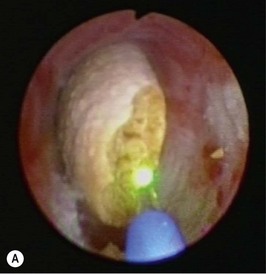
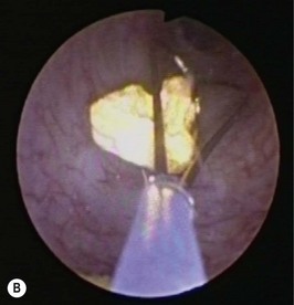
A.11 These are ureteroscopic photographs demonstrating Figure 15.1A after (1) the ureteric stone during LASER lithotripsy, and Figure 15.1B basket extraction of stone fragments.

Revision Points

Ureteric Stones