Originates within esophageal wall but presents as intraluminal polyp or mass
IMAGING
• Fluoroscopic-guided esophagography
Smooth, expansile, sausage-shaped, and intraluminal
Cervical esophageal mass, extending distally to fill esophageal lumen
• CT: Varied density based on content
Fat density: Abundance of adipose tissue
Heterogeneous: Mixture of fat, soft tissue
TOP DIFFERENTIAL DIAGNOSES
• Esophageal carcinoma
May present as large, polypoid intraluminal mass
Margins are irregular and more lobulated
• Esophageal intramural benign tumors
Leiomyoma and lipoma
Rarely as large or long as fibrovascular polyps
PATHOLOGY
• Giant, smooth or lobulated, expansile polyp with discrete pedicle attached to cervical esophagus
• Varying amounts of fibrovascular and adipose tissue covered by normal squamous epithelium
CLINICAL ISSUES
• Uncommonly, regurgitation of mass into pharynx or mouth
May cause laryngeal occlusion, asphyxia, and sudden death
• Fibrovascular polyps may bleed
• Malignant degeneration extremely rare
• Treatment
Small fibrovascular polyps: Endoscopic resection
Gigantic fibrovascular polyps: Surgical resection
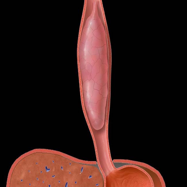
(Left) Graphic shows a long, smooth, sausage-like mass arising from the proximal esophageal wall, filling most of the esophageal lumen.
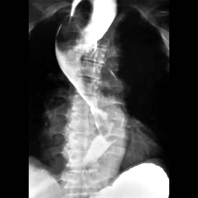
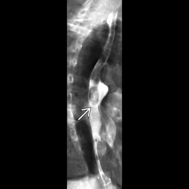
(Right) Barium esophagram demonstrates a large, cylindrical mass in the esophagus. The mass originates from a pedicle near the cricopharyngeal level. The mass is so long and bulky that it might be mistaken for an air bubble or debris within the esophagus.
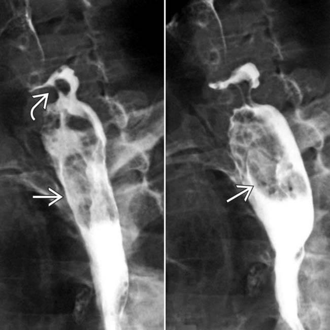
(Left) Barium esophagram demonstrates a huge, lobulated, polypoid filling defect within the esophagus, extending the entire length of the esophagus. The mass is not readily seen within the proximal esophagus, but it distends the lumen.
(Right) Axial CECT in the same patient shows the mass as a mixed soft tissue and fat density lesion within the grossly distended esophagus.
Esophagram shows a large, expansile mass filling the proximal 2/3 of the esophagus. (Courtesy M. Levine, MD.)
Esophagram shows a long, smooth polyp extending from the cervical esophagus into the middle 1/3 in this 67-year-old woman. (Courtesy M. Levine, MD.)
Barium esophagram shows an expansile, sausage-shaped mass extending from the cervical to distal esophagus. (Courtesy M. Levine, MD.)


 in the esophagus. The mass originates from a pedicle
  near the cricopharyngeal level. The mass is so long and bulky that it might be mistaken for an air bubble or debris within the esophagus.
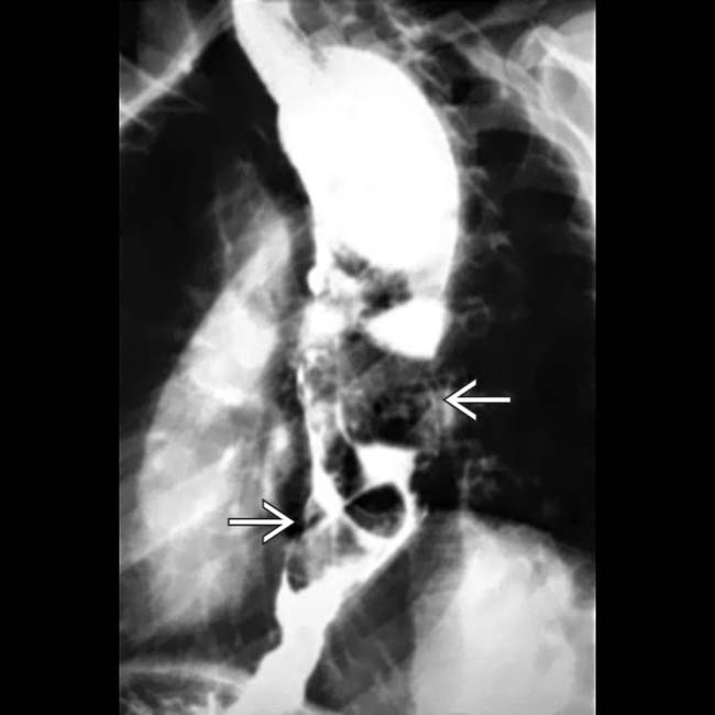
 within the esophagus, extending the entire length of the esophagus. The mass is not readily seen within the proximal esophagus, but it distends the lumen.
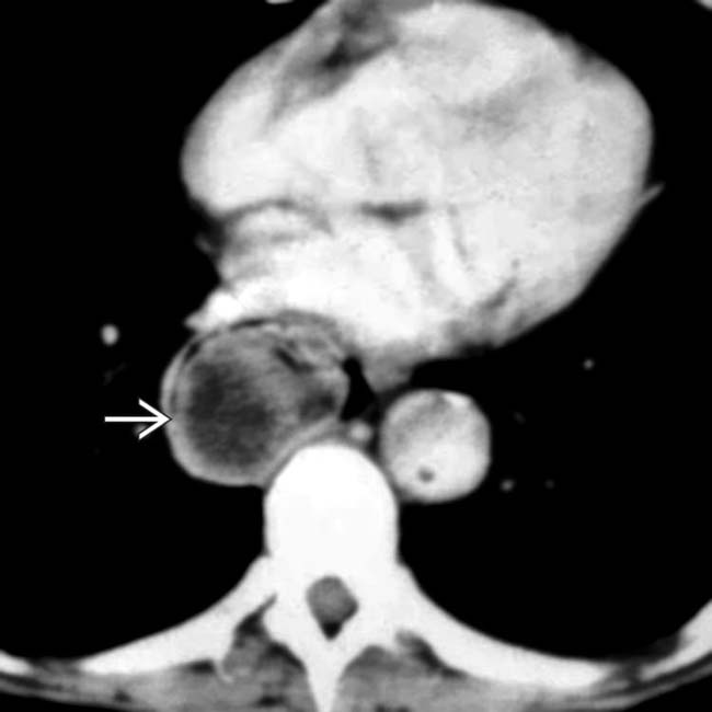
 as a mixed soft tissue and fat density lesion within the grossly distended esophagus.
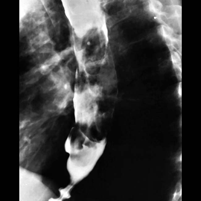
 extending from the cervical esophagus into the middle 1/3 in this 67-year-old woman.