Fiberoptic Bronchoscopy

Published on 22/03/2015 by admin

Filed under Critical Care Medicine

Last modified 22/04/2025

Print this page

rate 1 star rate 2 star rate 3 star rate 4 star rate 5 star
Your rating: none, Average: 0 (0 votes)

This article have been viewed 1616 times

W13 Fiberoptic Bronchoscopy

image Before Procedure

Indications

Equipment

Fiberoptic bronchoscope made up of a few components which are incorporated into a functional unit:

Ancillary technical materials:

image Anatomy

Bronchi

The right main bronchus is wider, shorter, and more vertical than the left main bronchus. The cartilage and mucous membrane of the main bronchi are similar to that in the trachea. At the level where the main bronchi enter the lungs, the membranous region disappears, and the cartilage plates are no longer C-shaped but are smaller, more irregular, and arranged to completely surround the circumference of the airway. At this level, the muscle coat no longer inserts into the cartilage but forms a separate layer of interlacing bundles. Consequently, the airway lumen can be occluded by contraction of the muscle.

The right main bronchus subdivides into three lobar bronchi, while the left main bronchus divides into two, although the lingula of the left lung is analogous to the right middle lobe. The lobar bronchi divide into segmental bronchi, each of which supplies a bronchopulmonary segment. The bronchopulmonary segments are the topographic units of the lung, and they are used to identify regions of the lung either radiologically or surgically. There are 10 bronchopulmonary segments per lung, but some of the segments in the left lung fuse during anatomic development, giving rise to 8 segments. For counting orders or generations of airways, the main bronchi are usually counted as the first generation, the lobar bronchi as the second generation, and so on. Generally in adult subjects, a bronchoscope with an outer diameter of 5 mm cannot be advanced further than the fourth/fifth order bronchi.

Nomenclature of Peripheral Bronchi

The nomenclature commonly used for the bronchial anatomy is that of Jackson and Huber. There are 10 segments in the right lung and 8 in the left. Subdivisions of the bronchial tree correspond to the anatomic segments and are named accordingly.

The right main bronchus gives rise to three lobar bronchi: upper, middle, and lower. The portion of the right main bronchus between the upper lobe bronchus and the origin of the middle and inferior lobe bronchi is known as the lower part of the right main bronchus or bronchus intermedius. The right upper lobe bronchus subdivides into three segmental bronchi: apical, posterior and anterior. The right middle lobe bronchus branches into two segmental bronchi: lateral and medial. The right lower lobe bronchus gives off five segmental bronchi: the superior segmental bronchus, posteriorly directed and just below the orifice of the middle lobe bronchus, and a bit more distally, four basal segmental bronchi: medial, anterior, lateral, and posterior; sometimes the medial basal bronchus is partially separated from the other basal segments by an extra fissure.

The left main bronchus subdivides into two lobar bronchi: upper and lower. The left upper lobe bronchus subdivides into a superior division bronchus and a lingular division bronchus. The superior division has two segmental bronchi: apical-posterior and anterior. The lingular division has two segmental bronchi: superior and inferior. The anatomy of the left lower lobe bronchus is similar to that of the right lower lobe bronchus, except that basal segmental bronchi on the left are usually only three: anteromedial, lateral, and posterior. Compared to the right side, the left lower lobe bronchus has a greater distance between its superior segment and its basal pyramid bronchi.

With the advent of fiberoptic bronchoscopy, Dr Shigeto Ikeda introduced additional nomenclature for the fourth, fifth, and sixth divisions. According to this nomenclature, segmental bronchi are numbered from 1 to 10 on each side and identified with the capital letter B for bronchus, prefixed by either the capital letter R for right or L for left. This way, LB6 identifies the superior segmental bronchus of the left lower lobe. Subsegmental or fourth-order bronchi are designated by the lowercase letters a for posterior and b for anterior; the letter c may be used for additional bronchi. Fifth-order bronchi are identified by the Roman numerals i (posterior) and ii (anterior).

Variations in the bronchial anatomy are not infrequent. Key bronchoscopic anatomic features as viewed by an operator positioned at the head end of a supine patient are:

image Procedure

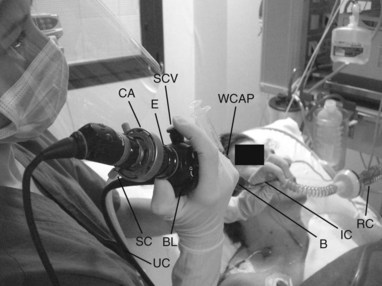
The bronchoscopic procedure requires only three movements: (1) flexion of the tip of the bronchoscope along the plane of the cursor, (2) rotation of the entire endoscope to the left or right, and (3) advancement or withdrawal of the instrument. The goal is to keep the point of interest (vocal cords, bronchial orifice, etc.) in the center of the field. When the bending lever is depressed, the tip rises, whereas when the lever is elevated, the tip points downward. The insertion cord should be kept as straight as possible to either prevent accidental damage to the bronchoscope or improve control over the tip of the instrument. To look right, the tip of the bronchoscope may be turned upward while the control handle is twisted clockwise; alternatively, the insertion cord may be rotated anticlockwise with the tip turned downward. To look left, the insertion cord is rotated clockwise or anticlockwise, with the tip deflected downward or upward, respectively. The operator will decide which maneuvers to perform, depending on the ease of obtaining the desired movements.

In order to avoid distortion of image orientation, the insertion cord should be able to rotate throughout its length when the handle is rotated axially. Orientation distortion resulting from axial rotation usually occurs when the distal end of the bronchoscope is blocked by the rubber airtight seal at the entrance of the endotracheal tube or even by the inside walls of the tube. In these circumstances, the tip of the bronchoscope fails to rotate synchronously with the proximal end of the instrument.

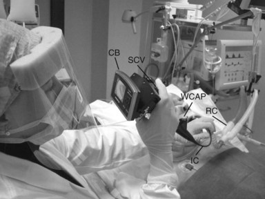
When a camera attachment is used with the bronchoscope, the camera position relative to the bronchoscope must be calibrated by rotating the bronchoscope camera system until a certain movement inside the patient’s airway corresponds with the proper motion on the monitor:

image After Procedure

Complications

Suggested Reading

Jackson C, Huber JF. Correlated applied anatomy of the bronchial tree and lungs with a system of nomenclature. Dis Chest. 1943;9:319-326.

Ikeda S. Atlas of flexible bronchofiberscopy. Tokyo: Igaku Shoin; 1974.

Baughman RP, Golden JA, Keith FM. Bronchoscopy, lung biopsy, and other diagnostic procedures. In: Murray JF, Nadel JA, Mason RJ, et al, editors. Textbook of respiratory medicine. 3rd ed. New York: Saunders; 2000:728-759.

British Thoracic Society Bronchoscopy Guidelines Committee, a Subcommittee of the Standards of Care Committee. British Thoracic Society guidelines on diagnostic flexible bronchoscopy. Thorax. 2001;56:11-21.

Bolliger CT, Mathur PN, Beamis JF, et al. ERS/ATS statement on interventional pulmonology. European Respiratory Society/American Thoracic Society. Eur Respir J. 2002;19:356-373.

Ernst A, Silvestri GA, Johnstone D. Interventional pulmonary procedures: guidelines from the American College of Chest Physicians. Chest. 2003;123:1693-1717.

Prakash UBS, Offord KP, Stubbs SE. Bronchoscopy in North America: the ACCP survey. Chest. 1991;100:1668-1675.

Meduri GU, Chastre J. The standardization of bronchoscopic techniques for ventilator-associated pneumonia. Chest. 1992;102:557S-564S.

Murphy PA. Fibre-optic endoscope used for nasal intubation. Anaesthesia. 1967;22:489-491.

Ovassapian A. Fibreoptic endoscopy and the difficult airway, 2nd ed. Philadelphia: Lippincott-Raven; 1996.

Lindholm CE, Ollman B, Snyder JV, et al. Cardiorespiratory effects of flexible fiberoptic bronchoscopy in critically ill patients. Chest. 1978;74:362-368.

Matsushima Y, Jones RL, King EG, et al. Alteration in pulmonary mechanics gas exchange during routine fiberoptic bronchoscopy. Chest. 1984;86:184-188.

Trouillet J, Guiguet M, Gibert C, et al. Fiberoptic bronchoscopy in ventilated patients: evaluation of cardiopulmonary risk under midazolam sedation. Chest. 1990;97:927-933.

Antonelli M, Conti G, Riccioni L, et al. Noninvasive positive-pressure ventilation via face mask during bronchoscopy with BAL in high-risk hypoxemic patients. Chest. 1996;110:724-728.

Antonelli M, Conti G, Rocco M, et al. Noninvasive positive-pressure ventilation vs conventional oxygen supplementation in hypoxemic patients undergoing diagnostic bronchoscopy. Chest. 2002;121:1149-1154.

Antonelli M, Pennisi MA, Conti G, et al. Fiberoptic bronchoscopy during noninvasive positive pressure ventilation delivered by helmet. Intensive Care Med. 2003;29:126-129.