Fair Game

Published on 03/05/2015 by admin

Filed under Radiology

Last modified 22/04/2025

Print this page

rate 1 star rate 2 star rate 3 star rate 4 star rate 5 star
Your rating: none, Average: 0 (0 votes)

This article have been viewed 1396 times

Chapter 2 Fair Game

Comment

This case of metastatic breast carcinoma illustrates several findings and imaging patterns of metastatic disease. There is a small left subdural effusion that follows the signal characteristics of CSF on all pulse sequences (including FLAIR and diffusion-weighted images, not shown). Note on the axial T2W image that the cortical veins seen as small black flow voids are closely up against the surface of the left cerebrum, consistent with a subdural effusion (in contrast to dilated subarachnoid spaces, as are seen in cerebral atrophy, in which the bridging cortical veins are noted to course through the CSF spaces). There is diffuse thin enhancement of the dura over the cerebral convexities. Axial images also demonstrate two cortical enhancing metastases. Lastly, best appreciated on the sagittal unenhanced T1W image is diffuse decreased signal intensity of the calvarial marrow, with superimposed areas of signal abnormality, consistent with diffuse marrow infiltration or metastases.

Dural metastases usually are secondary to hematogenous dissemination of tumor from extracranial systemic tumors. Dural metastases may also result from direct extension of adjacent calvarial or skull base bone metastases. Breast, lung, and prostate carcinoma as well as melanoma and lymphoma not uncommonly are associated with dural metastases in adults. In children, adrenal neuroblastoma and leukemia are most commonly associated with dural metastases. These malignant neoplasms may also metastasize to the cranial sutures. In infancy, this may present as widened sutures. Inflammatory processes that may mimic dural metastases include granulomatous infections (tuberculosis, syphilis, and fungal), sarcoid, Langerhans’ cell histiocytosis, and Erdheim-Chester disease.

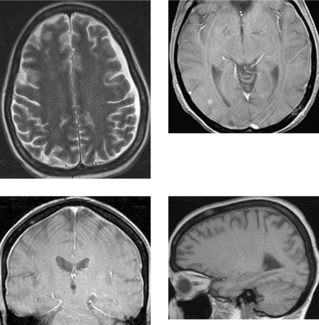
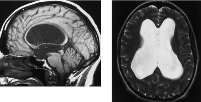
CASE 52 Aqueductal Stenosis

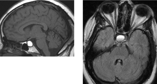
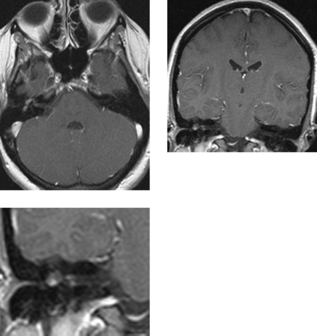
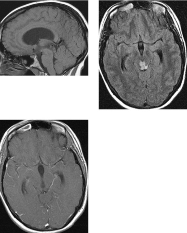
CASE 53 Rathke’s Cleft Cyst

Comment

Rathke’s cleft cysts are embryologic remnants of Rathke’s pouch, the neuroectoderm that ascends from the oral cavity to the sella to form the anterior pituitary lobe and pars intermedia. In the majority of cases, Rathke’s cleft cysts are incidental findings noted at autopsy or identified on imaging studies performed for other reasons. In a recent series of 1000 autopsy specimens, 11% of the pituitary glands had an incidental Rathke’s cleft cyst. These lesions are typically localized within the pituitary sella, although they may also extend into the suprasellar region. In addition, Rathke’s cleft cysts may be centered in the suprasellar cistern anterior to the hypothalamic stalk. A single layer of cuboidal or columnar epithelium, which may contain goblet cells, typically lines these cysts.

The contents within the cysts are mucoid, resulting in their typical MR imaging appearance. Most Rathke’s cleft cysts are circumscribed and hyperintense on both unenhanced T1W and T2W images. Although this is the most common pattern, cysts may also be hypointense or isointense on either T1W or T2W images, depending on their protein concentration and viscosity. In this case, the cyst is hyperintense on unenhanced T1W imaging and hyperintense on T2W imaging, with a fluid–debris level (did you catch that?). These cysts do not enhance (although there may be mild peripheral enhancement). More recently, intracystic nodules have been described that are hyperintense on unenhanced T1W images and hypointense on T2W images. At surgery, these appear as yellow, waxy solid masses, and the pathologic correlate shows a mucin clump.

Although most Rathke’s cleft cysts are incidental findings, symptoms may occur with larger lesions that compress the optic chiasm (resulting in headache and visual disturbances) or pituitary gland (patients may present with diabetes insipidus or hypopituitarism).

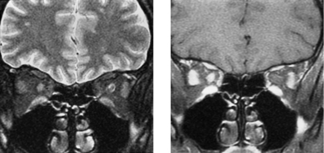
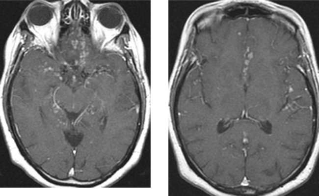
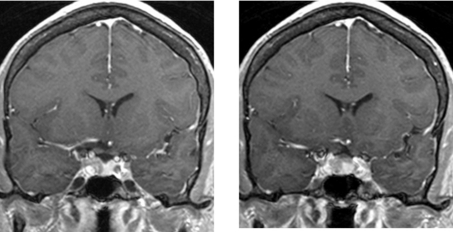
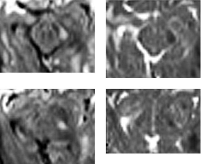
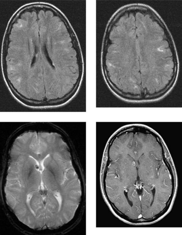
CASE 54 Optic Neuritis (Demyelinating Disease)

Comment

A wide spectrum of disease processes may be associated with optic neuritis, most commonly, demyelinating disease followed by idiopathic disease. Approximately 50% of patients with optic neuritis have multiple sclerosis, and approximately 15% of patients with multiple sclerosis have optic neuritis as their initial clinical presentation. Devic’s disease (neuromyelitis optica) represents a specific syndrome of multiple sclerosis in which patients have isolated transverse myelitis and optic neuritis. These two may present simultaneously or on separate occasions. In older patients, vasculopathies and ischemia are more common causes of optic neuritis.

Patients presenting with optic neuritis can be assessed using high-resolution, fat-suppressed, fast spin-echo T2W and enhanced T1W images, as in this case. Imaging in the coronal plane is particularly useful. Imaging may demonstrate increased T2W signal intensity within the nerve itself, and postcontrast images may demonstrate enhancement of the nerve as well as the nerve sheath due to perivenous inflammation (as in this case). In cases of clinically diagnosed optic neuritis, MR imaging of the brain is especially useful and is recommended because it may help to establish the diagnosis of demyelinating disease. In patients with their first episode of optic neuritis, up to 65% have asymptomatic cerebral white matter lesions on brain MR study. In addition, studies have shown that the presence of brain lesions on MR imaging in patients with optic neuritis may be important in determining prognosis and outcome.

CASE 55 Ocular Trauma—Lens Dislocation and Globe Rupture

Comment

On CT imaging, lens dislocation (Case A) may be differentiated from other intraocular foreign bodies by identifying that the intraocular density has the configuration of a lens and, importantly, observing that the lens in that eye is not located in its normal position. Lens dislocation may be confirmed on physical examination. CT is the imaging modality of choice in the initial assessment of patients with orbital trauma due to its availability as well as the ease and rapidity with which the examination can be performed. In addition, CT readily establishes the presence of orbital foreign bodies, delineates orbital fractures, and assesses for retrobulbar complications of trauma.

Subluxation or dislocation of the lens may be unrelated to trauma. It may be spontaneous or related to infection. There are also hereditary disorders that affect the connective (mesodermal) tissues that may be associated with lens dislocation, such as Marfan syndrome, Ehlers-Danlos syndrome, and homocystinuria. In Marfan syndrome, lens subluxation or dislocation is superior and at the periphery of the globe (and usually bilateral), whereas in homocystinuria, subluxations are inferior (“down and out”).

Globe rupture indicates that the integrity of the outer membranes of the eye is disrupted. Globe rupture may occur when a blunt object impacts the orbit, causing anterior–posterior compression of the globe and raising intraocular pressure such that the sclera tears. Ruptures from blunt trauma usually occur at the sites where the sclera is thinnest, at the insertions of the extraocular muscles, at the limbus, and around the optic nerve. Sharp objects or those traveling at high velocity may perforate the globe directly. Globe rupture represents an ophthalmologic emergency and requires surgical intervention. Damage to the posterior segment of the eye is associated with a very high frequency of permanent visual loss. Early recognition and ophthalmologic intervention are critical to maximizing functional outcome. On CT imaging, the presence of vitreous hemorrhage suggests an associated globe rupture. An enlarged anterior chamber and retraction of the lens are indicative of rupture of the posterior sclera. Other findings of globe rupture are a small, misshapen globe that contains blood or air (as in Cases B and C).

CASE 56 Developmental Venous Anomaly

Comment

Developmental venous anomalies, also referred to as venous malformations or venous angiomas, likely occur late in the first trimester and early in the second trimester of gestation, when the medullary veins are developing. Insults leading to failure in the development of the normal draining venous structures are believed to be the cause of these anatomic variants. Instead of the normal parallel appearance of the medullary veins as they drain into subependymal or superficial cortical veins, a disorganized network of dilated medullary veins converges in a “caput medusa” appearance and drains into an enlarged venous channel that subsequently drains superficially to cortical veins or sinuses, or deeply to subependymal veins of the lateral ventricle and then into the galenic system. Normal cerebral tissue intervenes between the dilated veins. The brain parenchyma surrounding a venous angioma is typically normal, although occasional gliosis, seen as mild T2W hyperintensity, may be present. Developmental venous anomalies are found most commonly in the frontal lobes, followed by the parietal and temporal lobes. Infratentorial developmental venous anomalies occur most commonly in the cerebellum. These can drain to the fourth ventricle and then to the pontomesencephalic vein, or to the precentral cerebellar vein and into the galenic system.

More than 99% of developmental venous malformations are asymptomatic; however, rarely, these lesions may hemorrhage. Hemorrhagic complications are usually secondary to coexisting cavernous malformations. Rare hemorrhage related specifically to the venous angioma is most common with cerebellar lesions. Patients may present with headache, seizure, or focal neurologic symptoms. Asymptomatic (essentially all) developmental venous anomalies are not treated because they represent a compensatory venous drainage route for normal brain. Sacrifice of these lesions could result in venous infarction of the normal brain that they drain.

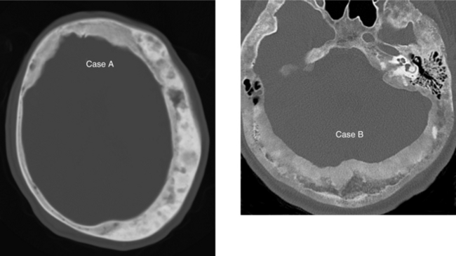
CASE 57 Fibrous Dysplasia of the Calvaria

Comment

Fibrous dysplasia is a developmental bone disorder in which osteoblasts do not undergo normal differentiation and maturation. The cause is unknown. Fibrous dysplasia may be monostotic (a solitary lesion) or polyostotic (multiple bones involved or multiple lesions in one bone). Approximately 75% of cases of fibrous dysplasia are monostotic (25% polyostotic). Polyostotic fibrous dysplasia involves the skull and facial bones more frequently than does monostotic disease. Common areas of calvarial involvement include the ethmoid, maxillary, frontal, and sphenoid bones. Involvement of these bones may result in orbital abnormalities such as exophthalmos, visual disturbances, and displacement of the globe. Involvement of the temporal bone may result in hearing loss or vestibular dysfunction.

Fibrous dysplasia of the skull or facial bones on plain radiography may present as radiolucent or sclerotic lesions. Sclerotic lesions are more common in the calvaria, skull base, and sphenoid bones. Although Paget’s disease may occur in these same locations of the calvaria, unlike fibrous dysplasia, concomitant involvement of the facial bones is less common. In this case, there is a mass in the right frontal bone, with associated focal expansion of the diploic space. There is preservation of the inner and outer cortical tables without destruction or significant thickening of the cortex. These features are typical of fibrous dysplasia. In contrast, although there may be expansion of the diploic space in patients with Paget’s disease, there is normally thickening of the cortex (which may be extensively involved).

CASE 58 Cavernous Sinus Mass—Meningioma

Comment

A variety of lesions can affect the cavernous sinus, including neoplasms primarily arising from bone (chondrosarcoma, chondroma, and chordoma) and bone metastases. Metastases and lymphoma primarily affecting the cavernous sinus and perineural spread of tumor are also common. Benign neoplasms include meningioma, schwannoma, and extension of a pituitary adenoma. A variety of vascular and inflammatory lesions may also involve the cavernous sinus.

Infectious processes include bacterial and fungal (actinomycosis, aspergillus, and mucormycosis) agents. In addition, the cavernous sinus may be affected indirectly by complications of infectious processes (eg, cavernous sinus thrombosis). Tolosa-Hunt syndrome represents an idiopathic granulomatous inflammatory disorder that may affect the orbital apex and cavernous sinus. Histologically, Tolosa-Hunt syndrome is identical to orbital pseudotumor (differing only in location). It may present with painful ophthalmoplegia, deficits of the cranial nerves coursing through the cavernous sinus, and retro-orbital pain. Symptoms may last for days to weeks, and may be recurrent. Like orbital pseudotumor, it responds rapidly to steroid treatment. MR imaging may show abnormal signal intensity or an enhancing mass within the cavernous sinus. There may be extension into the orbital apex. Thrombosis of the cavernous sinus or superior ophthalmic vein may occur.

These images demonstrate a mass within the left cavernous sinus that is hypointense to CSF (isointense to brain parenchyma) on T2W imaging. Neoplastic processes that may be hypointense on T2W imaging include lymphoma, meningioma, and less commonly, plasmacytoma and schwannoma. Sarcoid may also be hypointense on T2W imaging. Enhancement extends along the lateral dural margin, as well as posteriorly along the tentorial margin (an appearance that may be seen with meningiomas, lymphoma, and sarcoid). The marked narrowing of the left cavernous internal carotid artery with preserved antegrade flow makes meningioma the best diagnosis. Biopsy through a left temporal craniotomy revealed a meningioma.

CASE 59 Pyogenic Ventriculitis with Acute Hydrocephalus

Comment

This case demonstrates acute hydrocephalus and fluid–fluid levels (arrows) within the occipital horns of the lateral ventricles, consistent with ventriculitis complicating pyogenic meningitis. Lumbar puncture yielded pus. Ventriculitis is due to the introduction of infectious organisms into the ependyma or ventricles, and may be secondary to bacteremia, extension of an intraparenchymal abscess, trauma, or surgical instrumentation (especially placement of ventricular shunts). In addition to ventriculitis, other complications of ventriculoperitoneal shunts include overdrainage resulting in slit ventricle syndrome, subdural hematomas (which are frequently bilateral), shunt malfunction, surgery-related complications, and metastases in the setting of neoplasm (such as primitive neuroectodermal tumor).

Approximately 20% of patients with pyogenic bacterial meningitis will have complications necessitating neurosurgical intervention (surgery or a ventriculostomy), even after antibiotic therapy. Such complications include subdural empyema; parenchymal brain abscess; ventriculitis with hydrocephalus, as in this case; and encephalitis. The development of such complications may correlate with inadequate treatment or with the duration of meningitis before the initiation of therapy.

Cytomegalovirus (CMV) ventriculitis is unusual, but may occur in patients with HIV infection. Most HIV-infected patients with CMV ventriculitis have already been diagnosed with an AIDS-defining condition. Pathologic findings include inflammation of the ependyma and periventricular structures, as well as ependymal necrosis with CMV intranuclear inclusion bodies. The differential diagnosis in this patient population includes non-Hodgkin’s lymphoma.

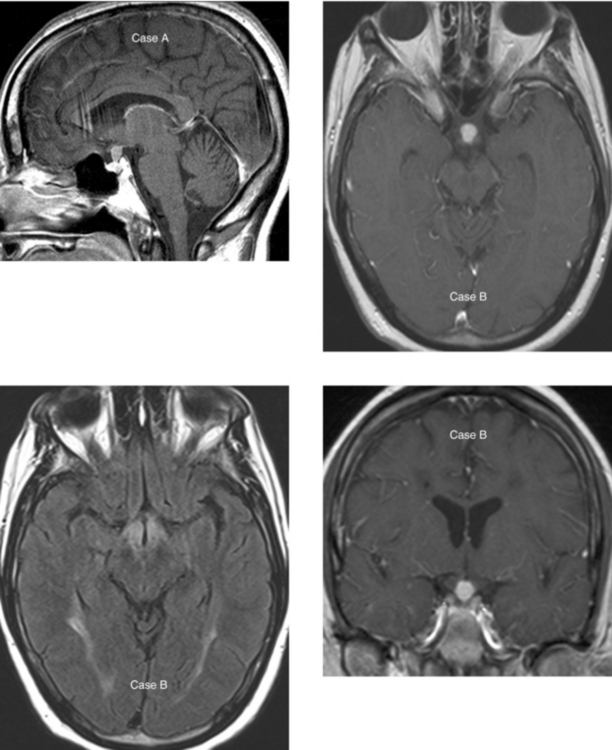
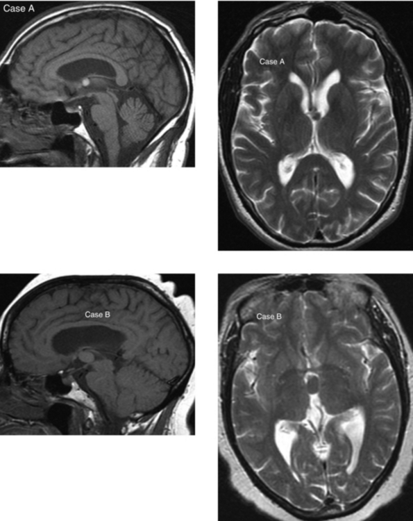
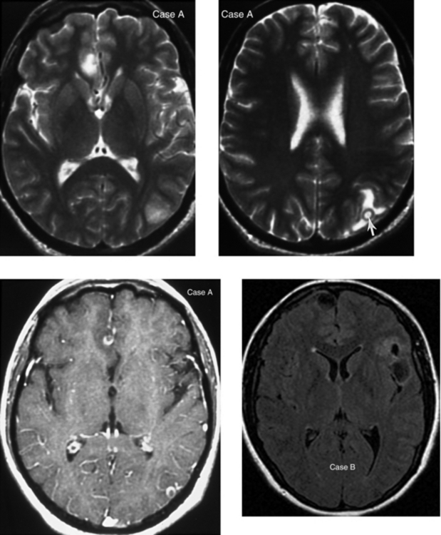
CASE 60 Lesions of the Pituitary Stalk and Hypothalamus: Case A—Sarcoidosis; Case B—Langerhans’ Cell Histiocytosis

Comment

In Case A, the midline sagittal enhanced T1W MR image shows abnormal thickening and enhancement along the pituitary stalk that lacks normal tapering as it extends inferiorly toward the pituitary gland. This represented sarcoid, and responded well to steroid treatment. This patient had a history of sarcoid with pulmonary involvement. Involvement of the CNS by sarcoid occurs in approximately 5% to 15% of patients, with isolated involvement of the pituitary gland or stalk in fewer than 1% to 2% of all patients with sarcoid. When CNS sarcoid presents with leptomeningeal involvement, the pituitary stalk in these cases is commonly involved.

Case B shows an enhancing mass in the hypothalamic region at the level of the upper pituitary stalk, and this represented Langerhans’ cell histiocytosis. The differential diagnosis of an infundibular mass in children includes germinoma, infection (meningitis or tuberculosis) and, less commonly, lymphoma or leukemia and glioma involving the hypothalamic region. In patients with Langerhans’ cell histiocytosis, there may be absence of the normal “bright spot” of the posterior pituitary gland in the sella on unenhanced T1W images. Langerhans’ cell histiocytosis uncommonly affects the CNS. It is a disorder of the reticuloendothelial system. The most common cranial bone abnormality is a lytic, circumscribed, enhancing mass of the calvaria or skull base. In the cranium, it most commonly involves the temporal bone along the mastoid segment. It also may involve the frontal bone along the orbit.

The differential diagnosis of an infundibular lesion in adults includes granulomatous disease (sarcoid or tuberculosis) and metastasis (especially lung and breast). Less commonly, lymphoma or a hypothalamic glioma may present in this way. Lymphocytic adenohypophysitis is a unique condition that almost always affects women. It is most common in the postpartum period or in the late stages (third trimester) of pregnancy. It is characterized by lymphocytic infiltration of the adenohypophysis and is usually self-limited.

CASE 61 Lhermitte-Duclos Disease (Dysplastic Gangliocytoma of the Cerebellum)

Comment

Lhermitte-Duclos disease is also known as dysplastic gangliocytoma of the cerebellum. When symptomatic, patient typically presents with complaints related to mass effect in the second and third decades of life. Lhermitte-Duclos disease has been associated with Cowden disease (multiple hamartoma syndrome), an autosomal dominant disorder associated with an increased incidence of neoplasms in the pelvis, breast, colon, and thyroid. Intracranial meningiomas have also been noted with this syndrome.

Although controversial, Lhermitte-Duclos disease is considered to represent a complex hamartomatous malformation, not a true neoplasm. Dysplastic gangliocytomas often occur in the cerebellar hemispheres. These lesions tend to be poorly demarcated, presenting on CT as a mildly hypodense mass. Calcification has been reported. On MR imaging, these lesions have a more characteristic appearance in which the gray and white matter of the cerebellar hemisphere are both involved and are thickened and hyperintense on T2W imaging, showing a somewhat characteristic laminated or “corduroy” appearance. They may exert mass effect. Hydrocephalus may be present. Dysplastic gangliocytomas do not demonstrate significant enhancement. On pathologic evaluation they usually appear as dysplasia with cellular disorganization of the normal laminar structure of the cerebellum, and hypertrophied granular cell neurons. Histologically there is hypermyelination of axons, and pleomorphic ganglion cells replace the granular and Purkinje cell layers.

CASE 62 Basilar Meningitis—Sarcoidosis

Comment

Sarcoidosis is a systemic disorder characterized pathologically by noncaseating granulomas. Typical presentation is in the third and fourth decades of life, and it is slightly more common in women than in men. CNS involvement has been reported in 5% to 15% of cases, and isolated CNS involvement occurs in fewer than 2% to 4% of cases. Multiple patterns of CNS involvement may occur. The most common is chronic meningitis with a predilection for the leptomeninges of the basal cisterns, as in this case. These patients may present with chronic meningeal symptoms, cranial neuropathies (especially involving the facial and optic nerves), or symptoms related to involvement of the hypothalamus and pituitary stalk. Imaging findings are best demonstrated with MR imaging and include nodular enhancement of the leptomeninges that tends to be most pronounced in the basal cisterns (but may be seen in any of the subarachnoid spaces, as in this case). Another common pattern of CNS sarcoid is brain involvement, with multiple regions of abnormality that may occur as a result of direct extension from leptomeningeal disease; white matter lesions mimicking multiple sclerosis may be present because of disease extension along the perivascular spaces or related to sarcoid-induced small vessel vasculitis. There may also be granulomas in the brain. Less commonly, dural-based disease may be the predominant imaging finding and may be mistaken for a meningioma.

Central nervous system tuberculosis may have a variety of clinical and radiologic presentations, including nodular tuberculous meningitis similar to sarcoid, cerebritis, abscess formation, tuberculoma, or a combination of these. CNS tuberculosis is normally related to hematogenous dissemination from a systemic source, such as the lung, genitourinary system, or gastrointestinal tract. Arteritis may be seen in up to one third of patients with basilar meningitis. This is because the vessels coursing through this inflammatory exudate may become directly involved. Consequences of arteritis include vasospasm and infarction.

CASE 63 Pituitary Microadenoma

Comment

Pituitary adenomas are slow-growing, benign epithelial neoplasms arising from the anterior lobe of the gland. They are typically demarcated with a “pseudocapsule” that separates them from the normal gland. Pituitary adenomas larger than 10 mm are referred to as macroadenomas, and those less than 10 mm in diameter are referred to as microadenomas. Many adenomas are incidental findings (asymptomatic).

The clinical presentation of pituitary adenomas depends on their size, the presence of hormone secretion resulting in endocrine hyperfunction, and the presence of extension beyond the sella (leading to visual symptoms or cranial nerve palsies related to compression). In vivo, approximately 75% of pituitary adenomas are hormonally active (however, in autopsy series, nonsecreting adenomas are much more common).

The most common clinically significant secreting adenoma is the prolactinoma (which arises from the prolactin-secreting cells [lactotrophs]). In women, the most common clinical presentation is irregular menses, galactorrhea, and infertility. In men, impotence may be present. Hormonally active pituitary adenomas arising from somatotrophs, the growth hormone-secreting cells, cause acromegaly in adults and gigantism in children. In acromegaly, endochondral and periosteal bone formation is stimulated, as is proliferation of connective tissue. These changes result in bone overgrowth and increased soft tissue thickness, especially in the “acral” regions (feet, hands, mandible). There is enlargement of the mandible, thickening of the cranial vault, and frontal bossing (the result of enlargement of the frontal sinuses and prominence of the supraorbital ridges).

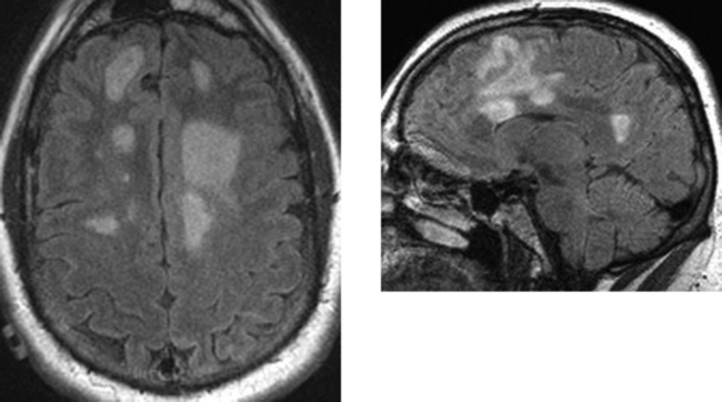
CASE 64 Multiple Sclerosis—Marburg Type

Comment

Although the etiology of multiple sclerosis is unknown, several causative factors have been implicated. These include autoimmune disease, infection (viral agent), and genetic factors. The prevalence of multiple sclerosis varies with geographic location.

Variants of multiple sclerosis may be present on a clinical or imaging basis. A handful of rare borderline types of multiple sclerosis occur, including Marburg type (also known as acute, fulminant, or malignant multiple sclerosis), a form of acute multiple sclerosis usually seen in younger patients that may be preceded by fevers, is typically rapidly progressive, and can result in death. In such cases, there is extensive demyelination and there may be defined rings within or surrounding plaques of acute demyelination. Enhancement is typically seen in the region of these rings. Concentric sclerosis or Balo-type sclerosis is characterized histologically by alternating rings of demyelination and myelination (normal brain or areas of remyelination) and has a characteristic MR imaging appearance. Schilder’s disease is a rare progressive demyelinating disorder that usually begins in childhood. Symptoms may include dementia, aphasia, seizures, personality changes, tremors, balance instability, incontinence, muscle weakness, headache, and visual impairment.

A type of multiple sclerosis that is usually limited to the optic nerves and spinal cord (either simultaneously or separately) is Devic’s disease or neuromyelitis optica. The main symptoms of Devic’s disease are loss of vision and spinal cord dysfunction. The visual impairment can consist of reduced visual fields, diminished light sensitivity, or loss of color vision. Spinal cord dysfunction includes muscle weakness and lack of coordination, reduced sensation, and incontinence. The brain is usually spared.

“Tumefactive” multiple sclerosis on imaging may be mistaken for a neoplasm or occasionally an abscess, particularly in the absence of a clinical history. The age of the patient may be helpful (multiple sclerosis typically occurs in younger patients). In addition, on close questioning, patients often have neurologic symptoms that are spaced in both time and location. MR imaging may show white matter lesions separate from the mass, suggesting multiple sclerosis. Unlike neoplasms, tumefactive multiple sclerosis often has relatively little mass effect for the amount of signal abnormality present.

CASE 65 Transtentorial Herniation

Comment

This case illustrates global cerebral swelling after resuscitation from a cardiac arrest resulting in transtentorial herniation. The brain is confined by the cranial vault and an inelastic dura. CSF surrounding the brain serves as a shock absorber. With increased intracranial pressure, herniation of brain from one compartment to another may occur. There are multiple patterns of herniation. In the posterior fossa, there may be inferior tonsillar and cerebellar herniation, with displacement of these structures into the foramen magnum, or there may be upward herniation (superior vermian herniation) in which cerebellar tissue obliterates the quadrigeminal and superior vermian cisterns. Herniation syndromes in the supratentorial compartment include subfalcine, temporal lobe, and central transtentorial herniation.

Transtentorial herniation is typically caused by supratentorial mass lesions or global cerebral swelling, resulting in vector forces that are directed medially and inferiorly. There is resultant effacement of the basilar cisterns and ventricles, as well as caudal displacement of the upper brainstem. The temporal lobe shifts over the tentorium. Caudal displacement of the brainstem and effacement of the ambient cisterns may result in compression of the oculomotor nerve (resulting in ipsilateral pupillary dilation) and compression of the posterior cerebral arteries, resulting in ischemic changes in the occipital lobes and brainstem or diencephalon. There may be secondary small hemorrhages (Duret), which are typically centrally located within the rostral pons and midbrain. Other findings include pseudosubarachnoid hemorrhage and pseudovenous sinus thrombosis, in which the subarachnoid spaces and venous sinuses appear hyerdense on unenhanced imaging relative to the diffuse hypodensity of the edematous cerebrum, as in this case.

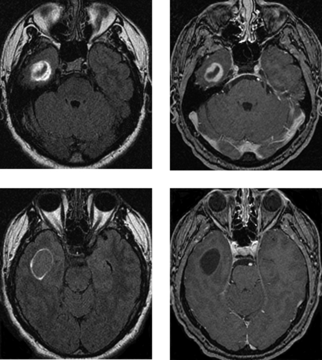
CASE 66 Epidermoid Cyst—Cerebellopontine Cistern

Comment

Epidermoid cysts are congenital lesions that result from incomplete separation of the neural and cutaneous ectoderm at the time of closure of the neural tube. These cysts are lined by a single layer of stratified squamous epithelium, and they contain desquamated epithelium, keratin, and cholesterol crystals. Many of these cysts are incidental findings, but when symptomatic, epidermoid cysts typically present in the third or fourth decade of life. Men and women are equally affected. These are frequently asymptomatic, but in the cerebellopontine cistern, they may present with dizziness, trigeminal neuralgia, and facial nerve weakness.

This case illustrates the typical appearance of an epidermoid cyst. There is a mass in the right cerebellopontine angle cistern that exerts mild mass effect on the adjacent brainstem. On the unenhanced T1W images (not shown), this mass is mildly hyperintense to CSF, with a fine internal architecture. On T2W image, the lesion is isointense to CSF. Unlike arachnoid cysts, which are typically isointense to CSF on proton density and FLAIR images, epidermoid cysts are hyperintense relative to CSF on these pulse sequences. Furthermore, this case illustrates hyperintensity on DW images, consistent with restricted diffusion, characteristic of these lesions. After contrast, there is no enhancement, as in this case. Other characteristic features of epidermoid cysts that distinguish them from arachnoid cysts (the major differential consideration here) are also presented in this case, including the lobulated and scalloped borders of this lesion and its insinuating nature, which fills and conforms to the shape of the space that it occupies.

CASE 67 Cavernous Malformation (Cavernous Hemangioma, Occult Cerebrovascular Malformation)

Comment

Cavernous malformations, also referred to as cavernomas, cavernous hemangiomas, and angiographically occult cerebrovascular malformations, represent a sinusoidal network of blood vessels without intervening normal brain parenchyma. Frequently, gliosis is also present. On unenhanced CT, CMs may be mildly hyperdense as a result of pooling of blood in the sinusoids. They may also be associated with focal calcification. On MR imaging, CMs are recognized by their characteristic appearance, representing blood products of different ages. Typically, cavernomas have a central region of high signal intensity on unenhanced T1W and T2W images, representing methemoglobin, surrounded by a complete rim of hemosiderin that is hypointense on T2W and gradient echo susceptibility images (as in this case). In the absence of recent hemorrhage, there should be no associated T2W signal abnormality (edema) within the surrounding brain parenchyma.

Angiographically, CMs are usually occult. Unlike other occult vascular malformations, such as capillary telangiectasias and venous angiomas, these patients may present clinically with seizures or symptoms related to mass effect in cases in which there has been recent hemorrhage.

Cavernous malformations may be present in as much as 5% of the population. The majority of CMs are located superficially in the cerebrum and are often closely associated with the adjacent subarachnoid space. They may occur deep within the cerebral hemispheres, although this is less common. CMs occur less frequently in the infratentorial compartment. The most common brainstem location is the pons. Symptoms may be related to lesion location or acute hemorrhage. In the cerebrum, the most common presentation is seizures. In the infratentorial compartment, neurologic deficits may occur on the basis of acute hemorrhage, thrombosis, or progressive enlargement of a CM related to recurrent hemorrhage.

CASE 68 Persistent Trigeminal Artery

Comment

The trigeminal, otic, and hypoglossal arteries (named after the cranial nerves with which they course) are the three embryologic anastomoses between the anterior internal carotid artery and the posterior vertebrobasilar circulations. The persistent trigeminal artery is the most common embryonic carotid–vertebrobasilar anastomosis to persist into adulthood, reported in as many as 0.1% to 0.5% of cerebral arteriograms. Persistence of a trigeminal artery may be associated with hypoplasia or absence of the ipsilateral posterior communicating artery, or with hypoplasia of both posterior communicating arteries. In addition, the proximal basilar artery and the distal vertebral arteries are often hypoplastic.

A persistent trigeminal artery usually arises from the precavernous internal carotid artery; however, origin from the intracavernous internal carotid artery has been reported. In some cases, trigeminal arteries may course through the sella turcica before joining the basilar artery (knowledge of this variant is critical in patients before transsphenoidal surgery). Persistent trigeminal arteries are associated with a variety of intracranial vascular abnormalities, including aneurysms and arteriovenous malformations, as well as a spectrum of clinical syndromes, such as tic douloureux (cranial nerve V), other cranial neuropathies, and vertebrobasilar insufficiency. Aneurysms arising from the trigeminal artery itself have been reported. Correct identification and an understanding of this anatomic variation are important because interventional neuroradiologic and neurosurgical procedures (often performed to treat an associated vascular abnormality) may need to be modified appropriately.

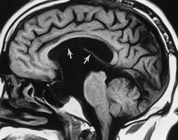
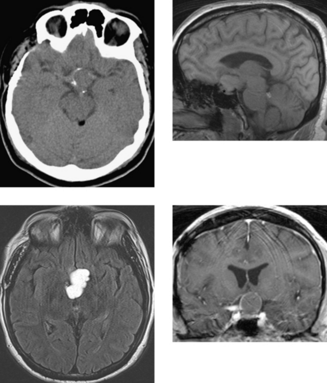
CASE 69 Colloid Cyst

Comment

Colloid cysts are benign masses typically located in the superior aspect of the anterior third ventricle between the columns of the fornices. They are lined by a single layer of epithelium and represent the most common type of neuroepithelial cyst (the origin of these cysts has been debated). Many of these lesions are incidental findings in patients being evaluated for other reasons. Alternatively, signs and symptoms of hydrocephalus (headache, nausea, vomiting) that may be positional in nature may occur intermittently due to obstruction of the foramen of Monro. These benign masses, even if an incidental finding, are usually treated because their mobility with changes in head position put the patient at risk for acute obstructive hydrocephalus. Management options include stereotactic aspiration, surgical resection, and shunting, or a combination of these treatments.

These cystic masses usually are radiologically characteristic, allowing them to be distinguished from other mass lesions in this location that are typically more solid, including those related to the choroid plexus (the occasional choroid plexus papilloma), craniopharyngiomas, gliomas, and occasionally meningiomas. Within colloid cysts is found thick, mucoid material, as well as a variety of other products, including old blood, CSF, other proteins, and paramagnetic ions, such as magnesium. The location of these cysts and their contents give them a characteristic radiologic appearance, including variable density and intensity characteristics on CT and MR imaging, respectively. Depending on the specific contents within an individual cyst, on unenhanced CT, these lesions may range from isodense (low protein concentration) to extremely hyperdense (high protein concentration) to the CSF and brain tissue. Colloid cysts also vary extensively in their signal characteristics on T1W and T2W MR imaging, ranging from very hyperintense (Case A) to isointense or hypointense (Case B) on unenhanced T1W images. The signal characteristics will depend on protein concentration, viscosity, water content, and cross-linking of glycoproteins. A thin wall is usually seen that commonly enhances after contrast administration. With the exception of this mild, thin, peripheral enhancement of the epithelial lining, these lesions should not demonstrate solid or central enhancement.

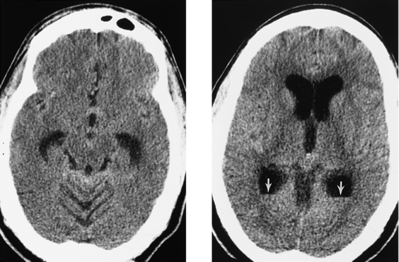
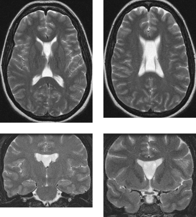
CASE 70 Cavum Septum Pellucidum and Cavum Vergae

Comment

The cavum pellucidum is bordered superiorly by the corpus callosum and posteriorly by the body of the fornix. The cavum extending posterior to the columns of the fornix and the foramen of Monro is called the cavum vergae. The cavum septum pellucidum and cavum vergae are potential cavities that lie between the membranes of the septum pellucidum. They most commonly represent a normal anatomic variant. On obstetric ultrasound, the cavum septum pellucidum is present in essentially all normal fetuses between 18 and 37 weeks’ gestation. During the latter half of gestation, the cavum septum pellucidum decreases in size. At birth, approximately 50% to 80% of term infants have a small residual cavum septum pellucidum that continues to decrease in size with age. Review of the literature shows considerable variation in the reported prevalence of a cavum septum pellucidum and cavum vergae in normal adults. Autopsy studies have also shown significant variability (range, 3%–30%). When a cavum septum pellucidum and cavum vergae are present concomitantly, as in this case, they usually communicate with one another; however, they do not typically communicate with the ventricular system. They are usually asymptomatic. Cysts may arise in this location and exert mass effect. In addition, enlargement of the cavum septum pellucidum, with intermittent obstruction of the foramen of Monro, resulting in hydrocephalus, has rarely been reported. Cysts may be treated with surgical resection, shunting, or fenestration.

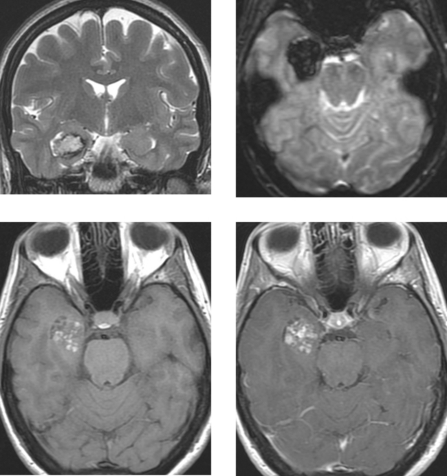
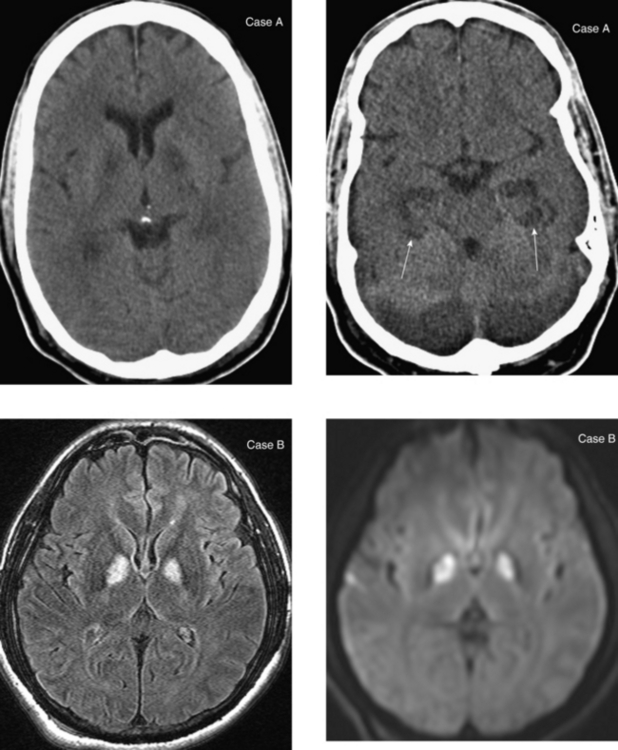
CASE 71 Toxoplasmosis Infection in Acquired Immunodeficiency Syndrome
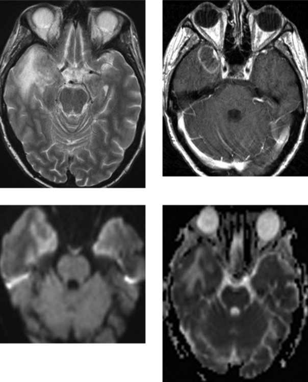
Comment

Central nervous system toxoplasmosis is caused by the intracellular protozoan Toxoplasma gondii. Toxoplasma encephalitis is most commonly seen in immunocompromised patients with impaired cellular immunity, especially in the setting of AIDS. Other immunodeficient conditions associated with increased infection include following organ transplantation, long-term steroid therapy or chemotherapy, and impaired immunity from an underlying malignancy. In the setting of AIDS, radiologic differentiation between toxoplasmosis and lymphoma can be difficult. Both entities may have multiple lesions, and both may have solid or ring enhancement. Toxoplasmosis has a predilection for the basal ganglia and the corticomedullary junction. Lesions are often hyperintense on T2W imaging, but vary widely in their signal characteristics. Lesions may be hemorrhagic. Findings favoring lymphoma are hyperdense masses on unenhanced CT, ependymal spread on enhanced MR imaging (rare in toxoplasmosis), and a periventricular distribution.

Distinguishing these two disease processes is important because they are treated differently. Primary lymphoma responds to radiation therapy; however, the benefit of radiation therapy is diminished when treatment is delayed, as may happen in patients first treated empirically for toxoplasmosis. Thallium-201 SPECT can be effective (sensitive and specific) in distinguishing lymphoma (takes up thallium) from toxoplasmosis (normally does not take up thallium). Positron emission tomography has been shown to be useful in the accurate differentiation of hypometabolic toxoplasmosis lesions versus metabolically active lymphoma.

Diffusion-weighted imaging with apparent diffusion coefficient (ADC) maps has been used to distinguish these two lesions. Toxoplasmosis lesions have demonstrated significantly greater diffusion than lymphoma, with increased diffusion relative to that in normal white matter, in contrast to the restricted diffusion seen within pyogenic abscesses. Increased diffusion in toxoplasmosis lesions has been postulated to reflect relatively decreased viscosity within the central cores of the lesions, perhaps due to an impaired cellular immune response related to the immunocompromised state of these patients. Although considerable overlap of ADC ratios between 1.0 and 1.6 has been reported, ADC ratios greater than 1.6 have been associated solely with toxoplasmosis. The core of the lesion in this case demonstrates increased diffusion relative to white matter: the ADC value was 1.3. Recent investigations with perfusion MR imaging have shown decreased regional blood volumes in toxoplasmosis lesions (attributed to the avascularity of abscesses) compared with increased blood volumes in lymphoma (attributed to increased vascularity in regions of metabolically active tumor).

CASE 72 Encephalocele—Developmental

Comment

This case illustrates a large congenital basal encephalocele associated with bilateral frontal lobe cortical dysplasia. Meningocele (encephalocele) refers to herniation of the meninges, CSF, or brain through an osseous defect in the cranium. Meningoencephaloceles are more common than meningoceles. Congenital encephaloceles are due to an abnormality in the process of invagination of the neural plate. During embryogenesis, the dura around the brain contacts the dermis in the facial or nasion region as the neural plate regresses. When there is failure of dermal regression, an encephalocele, dermoid cyst, sinus tract, or nasal glioma may develop. Dermoid sinus tracts may have an intracranial connection in up to 25% of cases, and may be complicated by infection (osteomyelitis, meningitis, and abscesses). Vietnamese and southeastern Asian women have a higher incidence of congenital nasofrontal and sphenoethmoidal meningoencephaloceles. Nasofrontal and sphenoethmoidal encephaloceles are frequently clinically occult, and the differential diagnosis is broad when this entity is seen through the nasoscope on office examination. Anterior basal encephaloceles have an association with other developmental anomalies, as in this case, including migrational abnormalities, agenesis of the corpus callosum, and cleft lip and palate.

In the setting of trauma or surgery, most meningoencephaloceles involve the nasal cavity and paranasal sinuses or the temporal bone. Patients may present with rhinorrhea.

A combination of imaging modalities, including nuclear scintigraphy, CT, and MR imaging, can be used to assess CSF leaks and meningoencephaloceles. It is important to determine whether the CSF leak is due to a dural laceration or a meningocele (encephalocele). After the placement of pledgets in the nares, intrathecal instillation of indium diethylene triamine pentaacetic acid may be used to confirm and localize the CSF leak. Once a leak is established, coronal CT may be performed for anatomic localization. In the hands of skilled ear, nose, and throat surgeons and radiologists, iodinated contrast CT cisternography is rarely necessary. If an encephalocele is suspected, MR imaging in the sagittal and coronal planes is most useful in establishing this diagnosis by showing direct continuity of the tissue in the sinonasal cavity with the intracranial brain. Although imaging may be useful in detecting CSF leaks, fluorescein injected intrathecally, followed by endoscopic evaluation, may allow direct visualization of an active leak.

CASE 73 Cysticercosis of the Central Nervous System

Comment

Cysticercosis is the most common parasitic infection of the CNS. It usually involves the intracranial compartment, and it may very, very, very rarely involve the spinal contents. Cysticercosis is endemic to Central and South America, parts of Asia, Mexico, Africa, and India. The pork tapeworm (Taenia solium) is the causative agent. Humans may become the definitive host (the parasite sexually reproduces) by eating inadequately cooked pork that harbors the larvae of the pork tapeworm (cysticerci). These larvae develop into tapeworms in the small intestine that release eggs that pass into the stool. If humans ingest food or water contaminated by these ova, they may serve as an intermediate host. In the stomach, the ova release oncospheres (primary larvae), which enter the bloodstream through the gastrointestinal mucosa. These primary larvae may deposit within muscle and subcutaneous tissue, although they have a propensity to infect the CNS. There are multiple patterns of neurocysticercosis, including the parenchymal pattern (the larvae penetrate directly into the brain), the intraventricular pattern (involves the ependyma or choroid plexus), and the subarachnoid pattern (involves the meninges). In mixed neurocysticercosis, there is involvement of the parenchyma, ventricles, or subarachnoid spaces. Patients with parenchymal involvement may present with seizures and neurologic signs (confusion, dementia, paresis, paraesthesias, visual disturbances). Intraventricular involvement may be symptomatic if there is obstructive hydrocephalus, and meningeal involvement may result in communicating hydrocephalus.

There is a spectrum of radiologic appearances, depending on the stage of disease; however, imaging findings are frequently characteristic. In the initial stage of cerebral infection, the larvae result in small, edematous lesions that are hypodense on CT and hyperintense on T2W images. The cysticerci then develop into cysts that range in size from millimeters to centimeters and contain a scolex. There may be mild surrounding edema in the brain, as in Case B. On the more cephalad T2W image in Case A, the left parietal lobe lesion has a characteristic appearance, with a defined capsule that has a hypointense rim and a small (1 mm), hypointense focus (arrow), representing the scolex. As the cysts die, there is an intense inflammatory reaction in the adjacent brain parenchyma that may result in prominent edema and mass effect. It is at this time that patients may be most symptomatic, presenting with seizures or focal neurologic signs. After years of infestation, the cysts finally collapse and often calcify. Rim enhancement has been described in as many as 38% of calcified lesions.

CASE 74 Facial Nerve—Inflammation (Viral)

Comment

This case shows enlargement and thickening of the right facial nerve (cranial nerve VII) along the tympanic portion, as well as enhancement of the facial nerve in the fundus of the internal auditory canal. Enhancement of the right cochlear nerve below the facial nerve in the anteroinferior internal auditory canal is also seen. The patient presented with Bell’s palsy. Statistically speaking, inflammatory processes (ie, viral) are most likely. Other infectious etiologies associated with cranial nerve VII involvement, in addition to viral causes, include Lyme disease. In immunocompromised patients, especially those with HIV infection, cytomegalovirus can affect the nerves in the internal auditory canal. Other inflammatory processes that affect the cranial nerves here include sarcoidosis.

Neoplasms that may involve cranial nerve VII include schwannoma (unlikely in this case because there are multiple cranial nerves involved and this patient’s clinical presentation was acute in onset). Subarachnoid seeding of tumor may involve the internal auditory canals and can be seen in lymphoma and carcinomatosis (lung, breast, or seeding of primary brain tumors). Perineural spread of malignancies along the facial nerve is often associated with destructive changes in the temporal bone. Perineural spread of temporal bone squamous cell carcinoma, primary parotid malignancies, and skin cancers of the ear and cheek are probably most common.

Frequently, imaging is not necessary in the workup of Bell’s palsy. However, when Bell’s palsy is bilateral, is recurrent, or does not show significant improvement in 6 to 8 weeks, imaging is indicated to assess for causes other than viral disease.

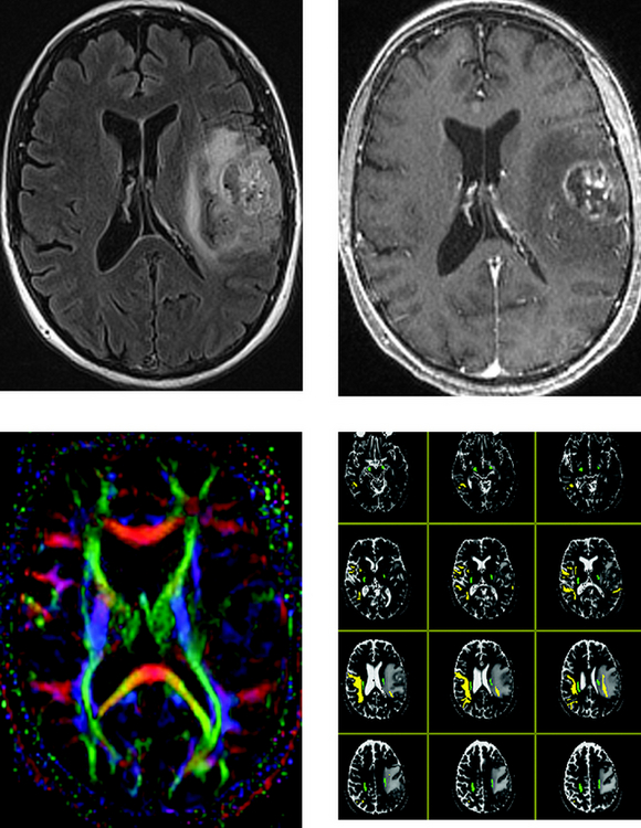
CASE 75 Magnetic Resonance Tractography— Preoperative Mapping of the Corticospinal Tracts for Glioblastoma Multiforme Resection

Comments

White matter tractography based on diffusion tensor imaging is an MR technique that can depict white matter tracts in the brain in vivo. Diffusion tensor sequences may be analyzed with combined volume analysis and tractography extraction software, giving indirect visualization of white matter connections. Rapid data processing allows imaging of the normal and diseased fiber pathways to be part of a routine MR imaging examination.

Diffusion tensor imaging enables the measurement of the restricted diffusion of water in tissue. The principal application currently is in the imaging of white matter, where the location, orientation, and anistrophy of the tracts can be measured. The architecture of the axons and their myelin sheaths facilitates the diffusion of water molecules preferentially along their main direction, referred to as anisotropic diffusion.

The corticospinal tract (CST) is the main white matter connection between the motor cortex and the spinal cord, serving as the main conduit of information between the higher cortical structures and voluntary muscular motion. The location and internal organization of the CST, as it passes through the corona radiata and the posterior limb of the internal capsule, have important clinical applications. Precise localization of the CST is useful for planning functional neurosurgery in patients with Parkinson disease and in the management of stroke, and importantly, preoperative localization of the CST may avoid inadvertent iatrogenic injury of these structures in patients with brain tumors, as in this case. Accurate depiction of the relationship of the CST and other main white matter tracts relative to a tumor preoperatively may improve neurosurgical planning. The superior longitudinal fasciculus (SLF) is a pair of long, bidirectional bundles of neurons connecting the front and the back of the cerebrum. Each association fiber bundle is lateral to the centrum semiovale and connects the frontal, occipital, parietal, and temporal lobes. Note the left SLF (yellow) passing through the tumorigenic edema in this case.

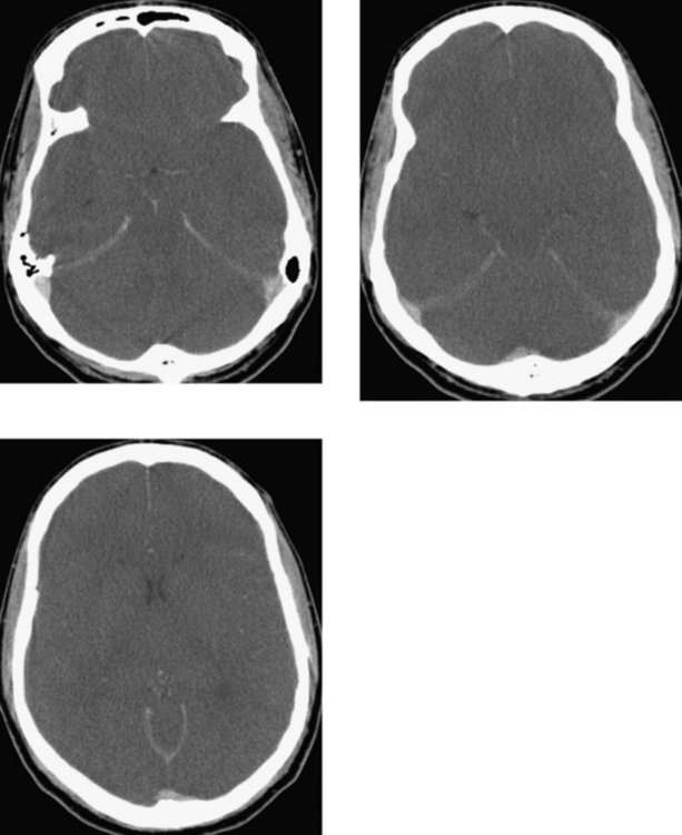
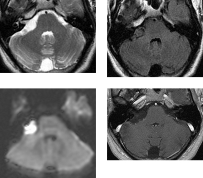
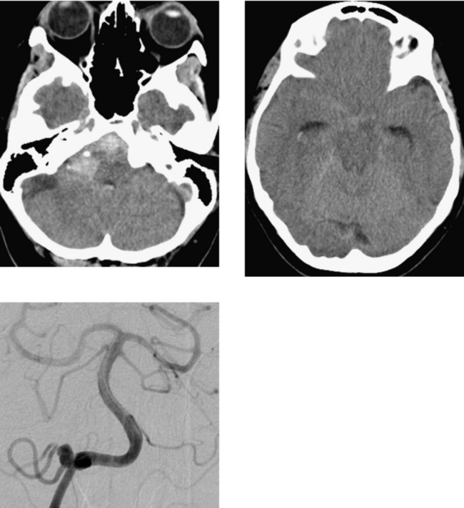
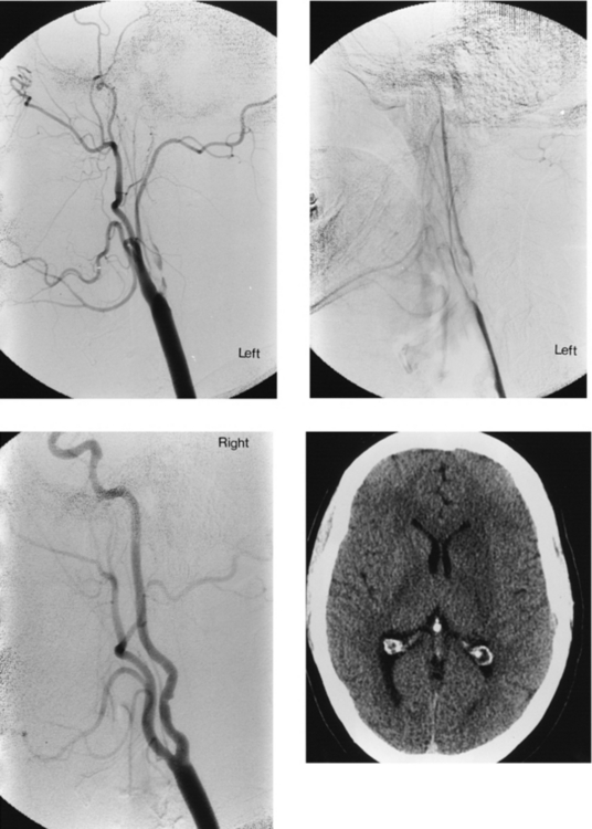
CASE 76 Right Posterior Inferior Cerebellar Artery Aneurysm Rupture

Comment

The majority of intracranial aneurysms arise from the supraclinoid segment of the internal carotid artery and its branches. More than 80% of saccular aneurysms arise from the anterior communicating artery, the distal internal carotid artery at the origin of the posterior communicating artery, the bifurcation of the supraclinoid internal carotid artery, and the middle cerebral artery bifurcation. Intracranial aneurysms are multiple in approximately 20% of cases. Aneurysms arising from the distal internal carotid artery at the origin of the ophthalmic artery account for 5% of intracranial aneurysms and have interesting features, including a preponderance in women, multiplicity in 10% to 20% of cases, and bilaterality in up to 20% of cases.

Aneurysms arising from the posterior circulation are not uncommon; however, they occur much less frequently than do their anterior counterparts. Most originate from the tip of the basilar artery. Basilar tip aneurysms may become quite large, and not uncommonly, the origins of one or both posterior cerebral arteries may be incorporated into the aneurysm. The next most common site for aneurysms in the posterior circulation is the origin of the posterior inferior cerebellar artery. Rarely, aneurysms may arise from the superior cerebellar artery or the anteroinferior cerebellar artery.

Aneurysms arising from distal arterial branches are usually acquired rather than congenital. They are frequently secondary to infection of the arterial wall (mycotic) or to trauma (aneurysms arising from the posterior circulation may be related to compression along the tentorium), and occasionally, they may be related to tumor.

CASE 77 Ganglioglioma

Comment

Gangliogliomas and ganglioneuromas are slow-growing, benign tumors that most commonly affect children and young adults. Gangliogliomas have a predominance of glial tissue and typically occur in the cerebrum, most commonly arising in the temporal lobe, followed by the frontal lobe, parietal lobe, occipital lobe, and region of the hypothalamus and third ventricle. They may also be infratentorial, arising within the cerebellum or brainstem. Gangliogliomas are typically circumscribed tumors that occur superficially in the brain parenchyma, with little or no surrounding edema. They are usually cystic (purely cystic or cystic with solid components), although solid tumors without cyst formation may occur. Calcification is frequently present, and these neoplasms may demonstrate variable contrast enhancement, ranging from mild to marked. However, contrast enhancement need not be present.

Because gangliogliomas are composed of both glial (usually astrocytes) and neural elements, they may undergo malignant degeneration. When neuronal elements make up the majority of the mass, the neoplasm is referred to as a ganglioneuroma. Gangliocytomas are composed of mature ganglion cells. They rarely have glial elements and therefore have no potential for malignant change.

In children and young adults, the main differential considerations for ganglioglioma on imaging studies include low-grade astrocytoma, juvenile pilocytic astrocytoma, dysembryoplastic neuroepithelial tumor, and pleomorphic xanthoastrocytoma.

CASE 78 Paget’s Disease—Osteitis Deformans

Comment

In Paget’s disease, there is malfunction in the normal process of bone remodeling. When an area of bone is destroyed, the new bone replacing it is soft and porous. Bone affected with Paget’s disease also has increased vascularity. The cause of Paget’s disease is unknown; however, a viral etiology is favored. Paget’s disease is more common in men than in women, and it usually presents after the age of 40 years. It is often an incidental finding detected on radiographs obtained for other reasons. Patients may be symptomatic, depending on the distribution of disease. Involvement of the calvarium (Case A) may present with enlarging head size. Involvement of the skull base, resulting in platybasia, may lead to neurologic symptoms (weakness and paralysis). Conductive hearing loss may result if there is involvement of the ossicles. Compression of the eight cranial nerves due to overgrowth of bone may cause sensorineural hearing loss. There is usually relative sparing of the otic capsule (Case B). Paget’s disease has multiple stages, including an initial osteolytic phase characterized by osteoclastic activity with resorption of normal bone. This phase is followed by excessive and sporadic new bone formation as a result of osteoblastic activity. Eventually, Paget’s disease enters its inactive stage.

Neoplastic involvement within pagetoid bone is not uncommon and includes sarcomatous degeneration, giant cell tumors, superimposed hematologic neoplasms (myeloma, lymphoma), and metastatic disease. Giant cell tumors are typically confined to the skull and less often to the facial bones. It is speculated that the increased blood flow within pagetoid bone may make it more susceptible to deposition of metastases. Clinically, the development of neoplastic disease in pagetoid bone should be suspected if there is increased pain or an associated soft tissue mass.

The differential diagnosis of Paget’s disease of the skull includes other sclerotic bone lesions, hyperostosis frontalis, fibrous dysplasia, and metastatic disease. In the elderly, metastatic disease may have the cotton-wool appearance of Paget’s disease (prostate cancer in men and breast cancer in women).

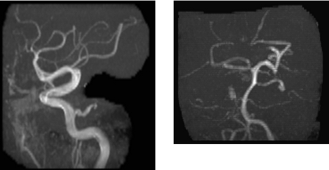
CASE 79 Fibromuscular Dysplasia—Complicated by Acute Stroke

Comment

Conventional catheter angiography demonstrates findings consistent with an extracranial dissection of the proximal left internal carotid artery, distal to the carotid bifurcation. There is marked narrowing and irregularity of the proximal internal carotid artery, with poor distal filling and very slow antegrade flow seen on the delayed image. An extracranial dissection may result from major or minor trauma, including chiropractic manipulation. Alternatively, extracranial dissection may result from an underlying vascular abnormality or dysplasia. In this case, injection of the right internal carotid artery demonstrates a normal appearance of the proximal internal carotid artery distal to the carotid bifurcation; however, there is irregularity, with regions of narrowing alternating with dilation, producing a “string of beads” appearance that is commonly described in fibromuscular dysplasia. There are many subtypes (medial, intimal, and adventitial) of fibromuscular dysplasia in which one or all of the layers of the arterial wall may be involved; however, involvement of the media with hyperplasia or dysplasia is most common, seen in up to 90% of cases. Thinning of the media is associated with abnormalities in the internal elastic lamina, resulting in the dilations seen in this condition. The cervical internal carotid artery is most commonly affected, and the proximal 2 cm of this vessel is usually spared due to architectural differences in this part of the vessel’s wall. The extracranial vertebral artery and external carotid arteries may be involved; however, intracranial fibromuscular dysplasia is relatively uncommon. The incidence of intracranial aneurysms is increased in patients with this condition.

CASE 80 Carbon Monoxide Poisoning

Comment

Injury to the brain with a particular predilection for the basal ganglia may be seen in a variety of toxic exposures, neurodegenerative processes, and metabolic disorders. Of the toxic insults, carbon monoxide, cyanide, and trichloroethane (typewriter correction fluid) poisoning have a particular predilection to involve the globus pallidus bilaterally. The pathologic and microscopic correlate of the abnormal hypodensity on CT or T2W hyperintensity on MR imaging within the bilateral globus pallidus is that of necrosis caused by anoxic injury. Although carbon monoxide toxicity has a characteristic imaging appearance, the diagnosis is usually established by the clinical circumstances in which the patient is found. The diagnosis of carbon monoxide toxicity is confirmed by identification of carboxyhemoglobin in the blood.

In addition to CT density and MR intensity alterations in the globus pallidus bilaterally, other imaging manifestations of diffuse anoxic brain injury may be identified, including injury to the hippocampus (Ammon’s horn) bilaterally (as in Case A, arrows) and global cerebral swelling manifested as sulcal effacement and accentuation of the gray–white matter differentiation. Abnormalities in the cerebellum may also be noted (as in Case A). Patients with carbon monoxide poisoning may experience sudden neurologic deterioration and coma approximately 2 to 3 weeks after the initial injury. Imaging often shows accompanying white matter disease, which may be extensive and which pathologically represents acute demyelination. On delayed imaging performed months to years after carbon monoxide injury, T2W hypointensity in the deep gray matter, especially the putamen, may be present and is likely due to iron deposition.

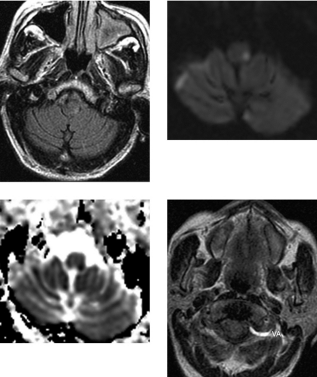
CASE 81 Lateral Medullary Syndrome—Wallenberg’s Syndrome

Comment

Infarction in the posterior inferior cerebellar artery territory can produce the lateral medullary syndrome (Wallenberg’s) illustrated in this case, which causes loss of pain and temperature sensation on the contralateral side of the body and the ipsilateral side of the face. Other symptoms may include ataxia, cranial nerve IX and X neuropathies, nystagmus, and vertigo.

In older patients, the most common cause of posterior circulation ischemia is thromboembolic disease resulting from accelerated atheromatous disease or embolic disease from a cardiac source. However, because the vasculature (including the vertebral arteries) becomes more tortuous as a patient ages, in the elderly population, in the setting of embolic disease from a systemic source, it is less common to have embolic disease isolated to the posterior fossa (usually patients also have embolic disease to the anterior circulation). In my experience, isolated posterior fossa embolic disease is more common in young patients in whom the vertebral arteries course vertically with little tortuosity. In young patients with posterior fossa ischemia, in addition to embolic disease, the diagnosis of a dissection should also be considered. This can be evaluated with MR imaging and MR angiography, or with CT angiography. MR imaging should include unenhanced axial T1W images through the distal vertebral arteries with fat suppression, when possible, to look for clot in the vessel wall, which is hyperintense due to methemoglobin. In addition, the dissected vessel may be enlarged due to mural hematoma, and occlusion (absence of signal void) or luminal narrowing (reduced size of the signal void) may be seen. In this case, the patient’s ischemic disease was related to thrombosis of the distal left vertebral artery due to dissection.

CASE 82 Wallerian Degeneration

Comment

Wallerian degeneration is a secondary manifestation of brain injury from a spectrum of causes. Secondary antegrade degenerative changes of axons and their myelin sheaths occur along the distal axonal segment as a result of injury to the proximal axon or neuronal cell body. Among the causes of degeneration of the corticospinal tract pathways, the most common is cortical infarction. Other injuries and neurodegenerative processes (eg, amyotrophic lateral sclerosis) may result in wallerian degeneration. It may occur as a result of trauma (as in this patient); hemorrhage; white matter disease, including demyelination; or neoplasia and its treatment (radiation injury). Histologically, wallerian degeneration represents several stages of progressive axonal degradation, ultimately resulting in gliosis and volume loss.

Magnetic resonance imaging is superior to CT in detecting wallerian degeneration, especially in the acute stages. CT may show the later changes of atrophy of the involved corticospinal pathways within the brainstem, but it does not show the earlier changes. On MR imaging, signal alteration on T2W or T1W images may be seen as early as 4 weeks after injury (some studies have shown these changes even earlier), and in the late stages (weeks to months), signal alteration and atrophy are invariably present. In the late stages, T2W hyperintensity is accompanied by hypointensity on corresponding T1W images. More recent work has shown that diffusion-weighted imaging and corresponding apparent diffusion coefficient (ADC) maps may reflect the changes of acute wallerian degeneration within a week, manifest as restricted diffusion (hyperintensity on diffusion-weighted images) and hypointensity on corresponding ADC images (low ADC values), as in this case.

CASE 83 Dandy-Walker Malformation

Comment

The Dandy-Walker complex (which includes Dandy-Walker malformation and its variants) is a congenital anomaly believed to be related to an in utero insult to the fourth ventricle leading to complete or partial outflow obstruction of CSF. As a result, there is cyst-like dilation of the fourth ventricle, which protrudes up between the cerebellar hemispheres to prevent their fusion, and there is incomplete formation of all or part of the inferior vermis. The spectrum of Dandy-Walker variant depends on the time in utero at which the insult occurs, as well as the severity of the insult (the degree of fourth ventricular outflow obstruction). Dandy-Walker malformations are associated with hydrocephalus in approximately 75% of cases that usually develops in the postnatal period. Dandy-Walker malformations may be associated with atresia of the foramen of Magendie and, possibly, the foramen of Luschka; however, atresia of the cerebellar outlet foramina is not an essential feature of the condition. In addition, 70% of patients have associated supratentorial anomalies, including dysgenesis of the corpus callosum, migrational anomalies, and encephaloceles.

This case demonstrates characteristic MR findings of a Dandy-Walker malformation including a large retrocerebellar cyst, enlargement of the posterior fossa with osseous remodeling, abnormally high position of the straight sinus, torcular herophili, and tentorium, and torcular lambdoid inversion. The radiologic hallmark of Dandy-Walker malformation is communication of the retrocerebellar cyst with the fourth ventricle, which is readily appreciated on the sagittal MR image.

A mega cisterna magna consists of an enlarged posterior fossa secondary to an enlarged cisterna magna, but with a normal cerebellar vermis and fourth ventricle. Retrocerebellar arachnoid cysts of developmental origin are clinically important. They displace the fourth ventricle and cerebellum anteriorly and show significant mass effect. Differentiation of posterior fossa arachnoid cyst from Dandy-Walker malformation is essential as surgical therapy differs between the two entities.

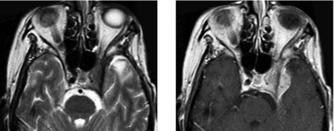
CASE 84 Thyroid Ophthalmopathy—with Optic Nerve Compression in the Orbital Apex

Comment

Thyroid ophthalmopathy is more common in women by a ratio of 4:1 and is frequently asymptomatic; however, when present, it may be seen in euthyroid or hyperthyroid states. Clinical signs and symptoms may include proptosis, lid retraction, decreased ocular range of motion, visual loss resulting from compression of the optic nerve in the orbital apex, and corneal exposure caused by eyelid retraction. Pain is uncommon. The most common cause of unilateral or bilateral exophthalmos in adults is thyroid ophthalmopathy. The incidence of bilateral disease may be as high as 90% of cases. Most patients evaluated with CT or MR imaging carry a known diagnosis of thyroid ophthalmopathy, and the role of imaging is to assess for the presence of optic nerve compression in the orbital apex by enlarged muscles, as in this case. When there is compromise of vision in patients who do not respond to medical therapy, orbital decompression by removal of the osseous walls around the orbital apex may be necessary.

Magnetic resonance imaging may also be useful in evaluating patients with ophthalmopathy without laboratory or clinical evidence of thyroid disease. The most common patterns of extraocular muscle involvement are enlargement of all of the extraocular muscles or of the inferior and medial rectus muscles only. Isolated involvement of the lateral rectus muscle is unusual, and when present, it should raise suspicion for a different disease process, such as myositis or pseudotumor. Characteristically, in thyroid ophthalmopathy, there is enlargement of the muscle bellies, with sparing of the tendinous insertions. In late stages of disease, fibrosis resulting in contraction of the muscle bellies may be evident, and there may be fatty replacement of the muscles.

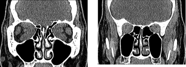
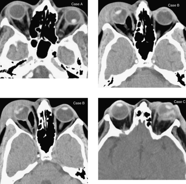
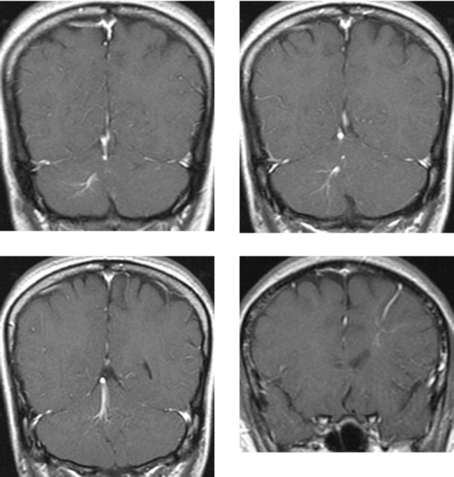
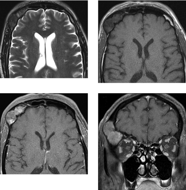
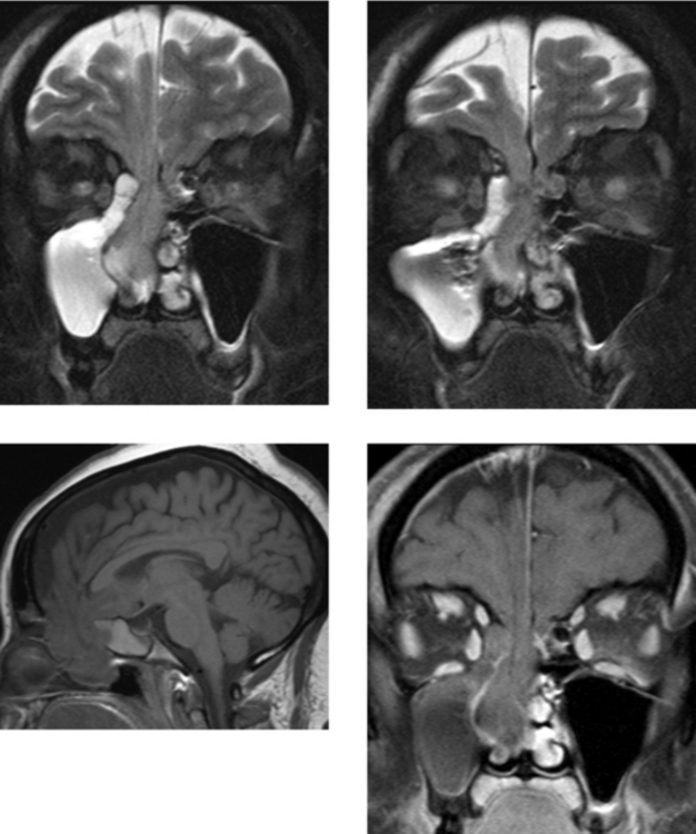
CASE 85 Bilateral Frontal Sinus Mucoceles
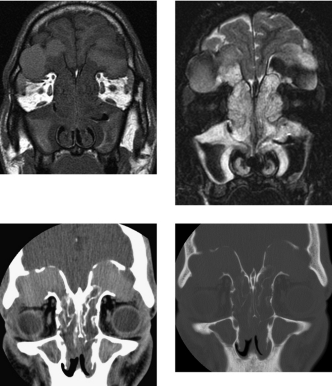
Comment

This case shows multiple, expansile, slow-growing lesions in the bilateral frontal sinuses (the remainder of the paranasal sinuses are opacified in this patient with chronic, recurrent sinus infections). The material within the expansile lesions is hyperdense on unenhanced CT, consistent with mucoid material, and corresponding MR images show the material to be hyperintense on T1W (very common) and heterogeneous on T2W images.

Mucoceles develop from obstruction of the sinus ostia or septated compartments of a sinus and represent mucoid secretions encased by mucus-secreting epithelium (sinus mucosa). In more than 90% of cases, mucoceles occur in the frontal sinuses or the ethmoid air cells (the anterior is more common than the posterior). Although the reported literature suggests that they are least common in the sphenoid sinus, in my experience, mucoceles are least common in the maxillary sinus. Patients frequently have a history of chronic sinusitis, trauma, or sinus surgery. When symptomatic, mucoceles present with signs and symptoms related to mass effect, including frontal bossing, headache, and orbital pain. Orbital extension, as in this case, may result in proptosis and diplopia. Secondary infection (mucopyocele) and direct extension into the anterior cranial fossa are not infrequent complications. Extension into the anterior cranial fossa or orbit is more likely if there is an associated fracture of an involved sinus wall. Alternatively, as a mucocele expands, it may directly erode a sinus wall, allowing for extrasinus extension. Advances in endoscopic sinus surgery have led to an acceptance of simple drainage procedures, even for some seemingly very complicated mucoceles.

In the radiologic evaluation of mucoceles, CT best demonstrates the osseous changes of the sinus walls, which may be remodeled and expanded, thinned, and with large mucoceles, partially dehiscent. However, MR imaging best detects the interface of the mucocele with the intraorbital and intracranial structures. When necessary, enhanced MR imaging is useful in distinguishing a mucocele (which shows peripheral enhancement) from a neoplasm (which typically shows solid enhancement).

CASE 86 Suprasellar Cistern Arachnoid Cyst

Comment

Arachnoid cysts are common and frequently asymptomatic, especially when located in the middle cranial fossa or over the cerebral convexities. Arachnoid cysts positioned in strategic locations are the lesions most likely to be symptomatic. Large cysts in the posterior fossa may present with ataxia or other symptoms of mass effect related to hydrocephalus. Similarly, when large enough, cysts in the quadrigeminal cistern may cause hydrocephalus by compressing the aqueduct of Sylvius. As in this case, arachnoid cysts in the region of the suprasellar cistern or third ventricle may cause headache, visual symptoms (related to compression of the optic chiasm), or symptoms related to mass effect and hydrocephalus. Arachnoid cysts of the suprasellar cistern may be mistaken for third ventricular cystic masses, such as seen with parasitic infection (cysticercosis), ependymal cysts, or epidermoid cysts.

Usually, arachnoid cysts follow the signal characteristics of CSF on all MR pulse sequences and are isodense to CSF on CT. Occasionally, CSF stasis or protein within the cyst may result in different intensities on MR imaging. These are nonenhancing lesions. When looking at a lesion in this location, localization to the suprasellar cistern rather than the third ventricle may be determined by looking for elevation of the floor of the third ventricle (arrows). Arachnoid cysts should have smooth margins, and calcification is rarely present. Most asymptomatic arachnoid cysts are left alone. In cases of strategically positioned cysts that result in symptoms, shunting or surgical resection may be necessary.

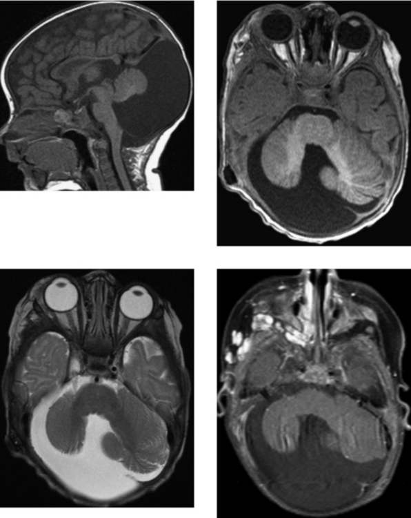
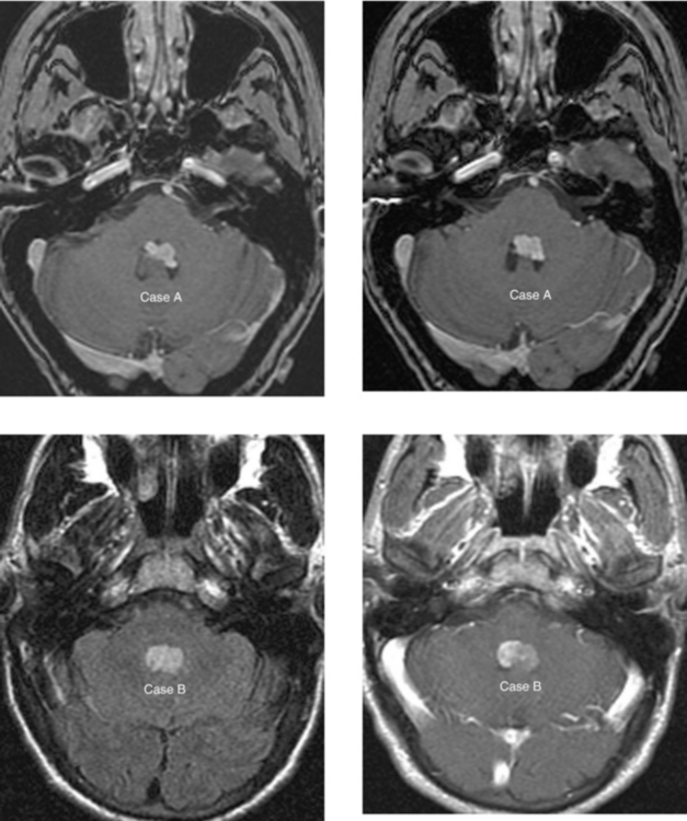
CASE 87 Fourth Ventricular Neoplasms—Choroid Plexus Papilloma and Ependymoma

Comment

Choroid plexus papillomas (Case A) are epithelial tumors arising from the surface of the choroid plexus. They occur most commonly in the lateral ventricles (45%). They may also arise within the fourth ventricle (40%) and the third ventricle (10%). In adults, the majority of choroid plexus papillomas occur in the fourth ventricle, whereas in children, 80% arise in the atria or trigone of the lateral ventricles. Choroid plexus papillomas may cause hydrocephalus as a result of overproduction of CSF or obstructive hydrocephalus related to adhesions from proteinaceous or hemorrhagic material blocking the subarachnoid cisterns or ventricular outlets. In addition, large tumors will cause focal expansion of the ventricle they fill; they may also cause trapping. These tumors are composed of vascularized connective tissue and frond-like papillae lined by a single layer of epithelial cells.

Intracranial ependymomas occur above or below the tentorium. The tumor arises in rests of ependymal cells lining the ventricles that extend into adjacent white matter. Infratentorial ependymomas occur in approximately two thirds of cases, and 75% of these posterior fossa tumors are located in the fourth ventricle (Case B). Approximately 15% arise in the cerebellopontine angle, and the remaining small percentage within the cerebellar hemisphere. Approximately 50% of infratentorial ependymomas extend into the cerebellopontine angle cisterns and foramen magnum via the lateral recesses of the fourth ventricle (foramina of Magendie and Luschka). Of infratentorial ependymomas, 12% present with subarachnoid seeding, especially those demonstrating anaplastic histology.

Calcification, hemorrhage, and cysts are frequently present in both choroid plexus papillomas and ependymomas. Calcification is readily identified on CT, as are large regions of cyst formation. On MR imaging, hypointensity may correspond to calcium, vessels, or blood products. Tumors that are very cystic will be hyperintense on T2W images, whereas those with large areas of old blood products may be hypointense. Both neoplasms may enhance avidly or heterogeneously, depending on the degree of cyst formation, calcification, and hemorrhage. When contained in the fourth ventricle, choroid plexus papillomas and ependymomas may be identical in appearance, as in these cases. Age is the best clinical distinguishing factor.

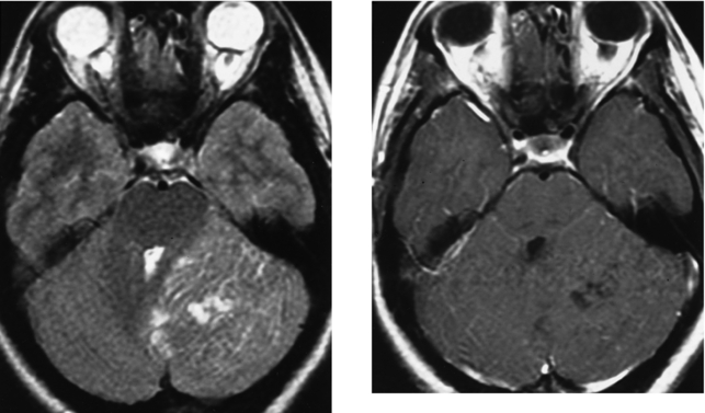
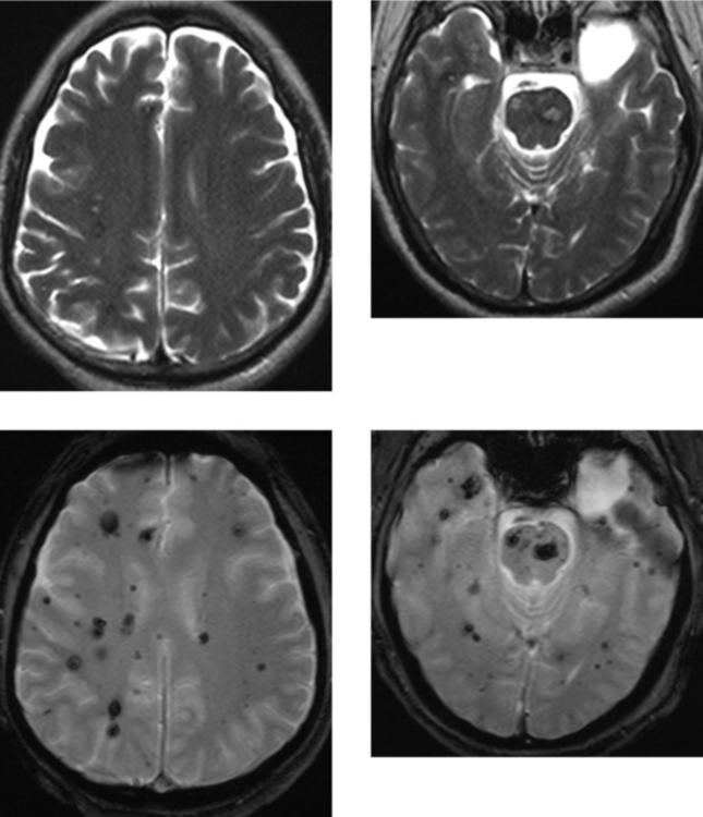
CASE 88 Multiple Cavernous Malformations—Familial Pattern

Comment

Cavernous malformations have a characteristic appearance on MR imaging. Specifically, they have central high signal intensity on unenhanced T1W and T2W imaging and are surrounded by a rim of hemosiderin that is hypointense on T2W and blooms on gradient echo imaging. In approximately 25% of cases, these lesions are multiple, and in a small percentage, there is a familial pattern. Many patients with multiple cavernomas have lesions too numerous to count. As in this case, many of the lesions may be quite small and only identifiable on gradient echo susceptibility images. Although cavernomas are believed to be congenital lesions, de novo development is not uncommon in patients with a familial pattern, as well as after radiation therapy and in association with developmental venous anomalies (venous angiomas).

Gradient echo imaging has increased sensitivity to magnetic susceptibility due to the lack of a 180-degree rephasing pulse. Therefore, blood products (hemosiderin), calcium, iron, and other ions are more readily seen on this sequence as areas of marked hypointensity. The T1 or T2 weighting of a gradient echo scan may be determined by selection of the flip angle and the time to echo. A small flip angle and a long time to echo will result in more T2 weighting. Similarly, a smaller flip angle and a longer time to echo will result in greater sensitivity to susceptibility effects. In addition to increased sensitivity to magnetic field inhomogeneities, gradient echo scanning has several useful features. It is generally faster than conventional spin-echo imaging. In addition, flow-related enhancement (bright blood) may be attained and is the basis of MR angiography. Another advantage of gradient echo scanning is that three-dimensional imaging allows for very thin sections. Such thin sections may also be acquired with three-dimensional fast spin-echo imaging.

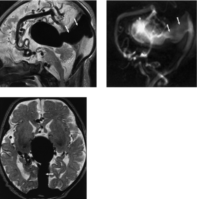
CASE 89 Vein of Galen Aneurysm

Comment

The CT appearance of a vein of Galen aneurysm in an infant is characteristic. On unenhanced images, the vein of Galen appears as a hyperdense, demarcated mass at the level of the posterior third ventricle and diencephalon. After intravenous contrast administration, there is marked homogeneous enhancement of the malformation. MR imaging not only confirms the presence of flow within this abnormality, but also better delineates both the arterial and venous anatomy. In combination with MR angiography, MR imaging may show large choroidal arteriovenous fistulas or the presence of a parenchymal AVM. In this case, a vein of Galen aneurysm is associated with a large AVM confirmed by MR angiography. Because many women undergo obstetric ultrasound as part of their prenatal care, many of these malformations are detected in utero with color flow Doppler sonography.

Early in embryologic development, the deep brain structures and diencephalon are drained by the median prosencephalic vein. As the internal cerebral veins begin to develop, this vein slowly regresses. A caudal remnant of the median prosencephalic vein will become the normal vein of Galen. In patients with vein of Galen aneurysms related to either a parenchymal AVM or a direct arteriovenous fistula between the choroidal vessels, apersistent median prosencephalic vein may occur. Because this provides diencephalic venous drainage, the straight sinus may not form. Instead, a falcine sinus is frequently noted, as in this case (arrows). Vein of Galen malformations resulting from direct arteriovenous fistulas are frequently associated with venous obstruction and venous hypertension. High-output heart failure in newborns and hydrocephalus due to obstruction of the aqueduct of Sylvius by the enlarged vein of Galen with macrocephaly are common clinical scenarios in infants.

Treatment of these malformations is catheter angiography and glue embolization of the shunt. Several embolization procedures may be necessary to completely obliterate the vascular shunts and communications. This is often done over a several-month period before the child is a toddler.

CASE 90 Tuberous Sclerosis (Bourneville’s Disease)

Comment

Tuberous sclerosis most commonly occurs as a sporadic mutation (chromosome 11); it may also be an autosomal dominant disorder transmitted by a mutation on chromosome 9. There are a spectrum of clinical signs and symptoms in these patients; however, the imaging manifestations should be sufficient to make the diagnosis in the majority of cases. Seizures, mental retardation, and adenoma sebaceum are the classic clinical triad described in tuberous sclerosis; however, the three together are seen in fewer than 50% of patients with this diagnosis.

The CNS manifestations of tuberous sclerosis are numerous and include subependymal nodules, cortical tubers, white matter lesions (believed to represent dysplastic white matter or foci of hypomyelination), subependymal giant cell astrocytomas, and ventriculomegaly. Subependymal nodules are seen in essentially all patients with tuberous sclerosis, and more than 75% are calcified. Because of the high incidence of calcification, subependymal nodules are easily detected on CT, but they can be difficult to detect on MR imaging. Gradient echo and unenhanced T1W imaging are best for detection of these lesions, which are hypointense and often mildly hyperintense to brain, respectively.

Cortical tubers are present in approximately 50% of these patients, and approximately half are calcified. Cortical tubers are hyperintense on long TR images and are frequently bilateral and symmetric (as in this case). They affect the frontal, parietal, occipital, and temporal lobes, in descending order of frequency. Up to 33% of subependymal nodules and 5% of cortical tubers may enhance.

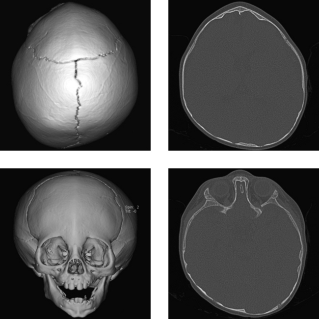
CASE 91 Craniosynostosis—Metopic Suture

Comment

Craniostenosis, or craniosynostosis, refers to premature closure of one or more of the cranial sutures. Isolated premature closure of the sagittal suture is most common, occurring in more than 50% of cases of craniosynostosis. Unilateral or bilateral premature closure of coronal sutures is the next most common, followed by premature closure of the metopic suture. The lambdoid suture undergoes premature closure in fewer than 1% of cases of craniosynostosis. Depending on the sutures involved, there are characteristic deformities of the skull and orbit. Premature closure of the sagittal suture results in a head that has limited growth in the transverse dimension. This results in dolichocephaly (scaphocephaly), in which there is an increase in head size in the anteroposterior dimension.

Plagiocephaly refers to premature closure of a single coronal or lambdoid suture (or, occasionally, a temporosquamous suture). In the majority of cases, plagiocephaly is seen with closure of a single coronal suture, resulting in elevation of the lesser wing of the sphenoid bone and leading to a “harlequin” appearance of the orbit. Premature closure of both coronal sutures results in brachycephaly. Early fusion of the metopic suture in the frontal region results in trigonocephaly, or simply a “triangular” configuration of the head, as in this case. Craniosynostosis is usually an isolated abnormality, although it may be associated with a variety of congenital syndromes. Such conditions include Apert’s syndrome, which is associated with a “cloverleaf” deformity of the skull resulting from closure of the coronal, lambdoid, and sagittal sutures. Other syndromes associated with craniostenosis are hypophosphatasia, Crouzon’s disease (craniofacial dysostosis), and Treacher Collins syndrome (mandibulofacial dysostosis).

CASE 92 Glioma of the Tectum (Quadrigeminal Plate)

Comment

These images show a mildly expansile mass lesion of the tectal plate that is predominantly cystic. There is no significant pathologic enhancement after contrast administration. There is resultant compression of the aqueduct of Sylvius, as is typically seen in patients with tectal gliomas. In many patients, the clinical presentation is that of obstructive hydrocephalus. Gliomas arising from the tectum are usually low-grade astrocytomas. They may be solid or cystic masses and have a wide spectrum of enhancement characteristics, ranging from none to prominent. Because most of these are low-grade neoplasms, the absence of enhancement is not surprising.

The midbrain is separated into the tegmentum and the tectum, which are portions of the midbrain anterior and posterior to the aqueduct of Sylvius, respectively. The tectum (roof) consists of the quadrigeminal plate that contains the paired superior and inferior colliculi. The tectum is affected more frequently by extrinsic rather than intrinsic lesions. It is often compressed (particularly the superior colliculi), along with the aqueduct of Sylvius, by pineal region masses, such as meningiomas in adults, germ cell tumors (germinoma, embryonal carcinoma, choriocarcinoma, and teratoma), tumors of pineal origin (pineoblastoma and pineocytoma), and aneurysms of the vein of Galen, which may result in Parinaud’s syndrome. In this case, * denotes the normal pineal gland. Occasionally, the tectum may be affected by demyelinating disease, vascular abnormalities, or trauma. In addition, the tectum may be abnormal in congenital malformations, most notably, Chiari II malformation, in which there may be a spectrum of abnormalities, ranging from collicular fusion to tectal beaking.

CASE 93 Craniopharyngioma—Recurrent Adamantinomatous Type

Comment

Craniopharyngiomas arise from metaplastic squamous epithelial rests (Rathke’s pouch) along the hypophysis or from ectopic embryonic cell rests, and are seen in children and adults. Histologically, they are characterized by palisading adamantinous epithelium, keratin, and calcification. They account for 1% to 3% of all intracranial neoplasms and 10% to 15% of all supratentorial tumors. They are more common in boys and men. The majority of craniopharyngiomas arise within the suprasellar cistern (80%–90%), as in this case; however, they also may arise within the sella turcica and, occasionally, the third ventricle. Clinical presentation includes visual disturbances related to compression of the optic chiasm, pituitary hypofunction related to compression of the gland or hypothalamus, and symptoms of increased intracranial pressure.

Imaging findings typically include a cystic or a solid and cystic mass lesion. Approximately 80% to 90% of all craniopharyngiomas have a cystic component. Smaller lesions may be purely solid. The majority (90%) of craniopharyngiomas have calcification or regions of avid homogeneous enhancement (in solid portions of tumor) or peripheral enhancement (around cystic portions). Because MR imaging may not be sensitive in detecting the presence of calcification, CT may be quite useful in establishing the diagnosis of craniopharyngioma. On MR imaging, the signal characteristics may be quite variable, depending on the contents and viscosity within the cysts. Whereas the cystic portion is frequently hyperintense on T2W and FLAIR imaging, it may be hypointense, isointense, or hyperintense on T1W imaging. High signal intensity on T1W images may be due to high concentrations of protein or methemoglobin (rather than cholesterol or lipid products).

Incidentally noted, there is a small lipoma in the perimesencephalic cistern on the left adjacent to the tectum. It is hypodense on CT, and hyperintense on the unenhanced sagittal T1 W images, typical of lipomas.

CASE 94 Intradiploic Epidermoid Cyst

Comment

Epidermoid and dermoid cysts of the skull are rare. Both are proposed to occur as a result of inclusion of epithelial cells during closure of the neural tube between the third and the fifth week of gestation. However, development of epidermoid cysts secondary to implantation of epithelial cells after trauma has been suggested as the cause in approximately 25% of cases. Epidermoid cysts account for fewer than 2% of intracranial and cranial tumors. Approximately 25% occur in the skull, whereas the remaining 75% are intradural. Dermoid cysts are even less common. Epidermoid cysts tend to present in young adults, whereas dermoid cysts present in children and young adolescents.

Approximately 10% of calvarial epidermoid cysts are incidental lesions; the remaining 90% are symptomatic. The most common presentation is an enlarging scalp mass; however, pain or headache is present in approximately 20% of cases. The most common location of these cysts is the parietal bone, followed by the frontal and temporal bones. Approximately 70% of these lesions involve both the inner and outer tables of the skull. Involvement of only the outer cortical table or, less commonly, the inner table may occur. On plain films, epidermoid cysts typically appear as lytic lesions with sclerotic borders. The differential diagnosis includes hemangioma, eosinophilic granuloma, and leptomeningeal cysts, especially in childhood. Other lytic lesions, particularly in adults, cannot be definitively differentiated on plain films alone. These lesions are typically hyperintense on T2W imaging; however, on unenhanced T1W imaging, their signal characteristics are variable. High T1W signal intensity may be due to the presence of blood products, protein, debris, crystals, or fat.