F
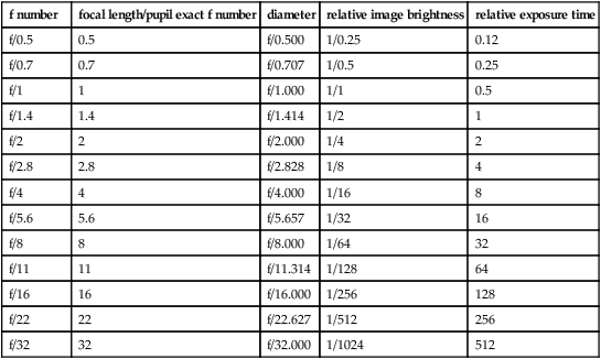
f number Designation for a photographic lens which gives the ratio of the focal length to the diameter of the effective aperture or entrance pupil. Example: f/8 means that the lens has a focal length eight times the diameter of the entrance pupil. Syn. f/stop; f-value; focal ratio; lens speed.
facet, corneal See corneal facet.
factor, daylight The ratio of daylight illuminance received at a given point inside a room to the simultaneous illuminance on a horizontal plane outside exposed to an unobstructed sky. It is usually expressed as a percentage, 5% or more being considered a well-lit room.
factor, reflection See reflectance.
factor, spectral transmission See spectrophotometer.
facultative hyperopia See hyperopia, facultative.
Faden procedure See procedure, Faden.
fallen eye syndrome See syndrome, fallen eye.
false macula See macula, false.
false negative See sensitivity.
false positive See specificity.
familial Pertaining to a condition or trait, either hereditary or acquired, which is found in more members of a family than would be expected by chance.
See acquired; congenital; hereditary.
familial autonomic dysfunction See syndrome, Riley–Day.
Table F1
Sequence of f numbers used in photography with the corresponding relative image brightness and exposure time to maintain constant film exposure. The relative image brightness is equal to the square of the f number fraction
| f number | focal length/pupil exact f number | diameter | relative image brightness | relative exposure time |
| f/0.5 | 0.5 | f/0.500 | 1/0.25 | 0.12 |
| f/0.7 | 0.7 | f/0.707 | 1/0.5 | 0.25 |
| f/1 | 1 | f/1.000 | 1/1 | 0.5 |
| f/1.4 | 1.4 | f/1.414 | 1/2 | 1 |
| f/2 | 2 | f/2.000 | 1/4 | 2 |
| f/2.8 | 2.8 | f/2.828 | 1/8 | 4 |
| f/4 | 4 | f/4.000 | 1/16 | 8 |
| f/5.6 | 5.6 | f/5.657 | 1/32 | 16 |
| f/8 | 8 | f/8.000 | 1/64 | 32 |
| f/11 | 11 | f/11.314 | 1/128 | 64 |
| f/16 | 16 | f/16.000 | 1/256 | 128 |
| f/22 | 22 | f/22.627 | 1/512 | 256 |
| f/32 | 32 | f/32.000 | 1/1024 | 512 |
fan and block test See test, fan and block.
fan chart See chart, astigmatic fan.
far point of accommodation See accommodation, far point of.
far point of convergence See convergence, far point of.
far point of the eye See accommodation, far point of.
far point sphere See sphere, far point.
far sight See hyperopia.
Farnsworth–Munsell 100 Hue test See test, Farnsworth.
Farnsworth test See test, Farnsworth; test, lantern.
fascia A sheet of connective tissue covering, partitioning or binding together muscles and certain other organs, such as the lacrimal sac, the orbital septum and other organs within the orbit, the sclera (e.g. Tenon’s capsule), etc.
fascia bulbi See Tenon’s capsule.
fascia, palpebral See orbital septum.
fasciculus, medial longitudinal One of a pair of nerve fibres, one on each side of the midline and extending from the upper midbrain to the cervical spinal cord. It is composed largely of ascending fibres from the vestibular nuclei ascending to the motor nuclei (third, fourth and sixth) and innervating the extraocular muscles; and to a lesser extent of descending fibres from the medial vestibular nuclei, the reticular formation, the superior colliculi and nucleus of Cajal innervating the musculature of the neck. Syn. medial longitudinal bundle; posterior longitudinal bundle.
fast eye movements See movements, saccadic eye.
fatigue, visual A feeling of weariness resulting from a visual task. It can be of ocular, muscular or psychic origin. However, there does not seem to be objective proof of a reduction in visual aptitude (e.g. visual acuity) accompanying visual fatigue.
See asthenopia.
feathers A cluster of fine bubbles or particles commonly arising from foreign material or from a fold in the glass in a molten or plastic state (British Standard).
Fechner’s colours See Benham top.
Fechner’s law See law, Fechner’s.
Fechner’s paradox Subjective impression of a decrease in the brightness of a field when viewing it binocularly, after one eye that was closed looks through a dark filter (about 5% transmission). This is paradoxical since more light is received by the eyes when the field is viewed binocularly, as compared to monocularly.
felderstruktur muscle fibres See fibres, felderstruktur muscle.
fenestration See lens, fenestrated; lens, scleral contact.
Fermat’s law See law, Fermat’s.
Ferry–Porter law See law, Ferry–Porter.
fibre A long thread or filament constituting human and animal tissues (e.g. nerve axon, muscle fibre, the filament of connective tissue).
arcuate f’s. Axons of the ganglion cells of the retina which are temporal to the optic disc and pass above and below the papillomacular bundle in an arcuate course. Syn. arcuate nerve fibres bundle.
See retinal raphe; scotoma, arcuate.
cilio-equatorial; cilio-posterior capsular f’s. See Zinn, zonule of.
circular f’s. See muscle, ciliary.
felderstruktur muscle f’s. A type of extraocular muscle fibres whose effect is to produce slow, and tonic contraction. They are mainly responsible for maintaining smooth pursuit movements. The fibres are located in the superficial portions of the extraocular muscles and are unique to this type of muscle.
fibrillenstruktur muscle f’s. A type of extraocular muscle fibres whose effect is to produce fast and twitch type of contractions. They are mainly responsible for saccadic eye movements. The fibres are located deep within the extraocular muscles and are the type usually found in skeletal muscles.
Henle’s f. See layer of Henle, fibre.
lens f’s. Long, six-sided bands containing few organelles and mostly lacking a nucleus, derived from epithelial cells just within the capsule of the crystalline lens and attached to an anterior and to a posterior suture. New lens fibres are continuously produced throughout life thus contributing to lens growth with age.
longitudinal f’s. See muscle, ciliary.
macular f’s. See fibres, papillomacular.
medullated nerve f’s. See fibres, myelinated nerve.
meridional f’s. See muscle, ciliary.
f’s. of Mueller See cell, Mueller’s.
myelinated nerve f’s. Anomalous congenital extension onto the retina of the myelin sheaths covering the optic nerve fibres. This myelination beyond the lamina cribrosa normally disappears soon after birth. Ophthalmoscopically, it appears as whitish, striated feather-shaped patches which may or may not obscure retinal vessels. Vision in these areas may be reduced, although visual acuity is not affected as the patches are most frequently located adjacent to the optic disc and sometimes in the periphery. The most characteristic sign may be an enlargement of the blind spot. Syn. medullated nerve fibres; opaque nerve fibres.
See cribriform plate.
orbiculo-anterior capsular; orbiculo-posterior capsular f’s. See Zinn, zonule of.
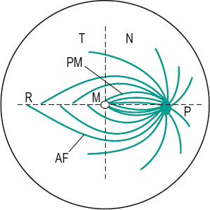
papillomacular f’s. Axons of the ganglion cells of the macular region of the retina which enter the temporal portion of the optic disc and travel in the central region of the optic nerve. In the optic chiasma, the temporal macular fibres remain on the same side, while the nasal ones cross to the other side. These fibres make up the papillomacular bundle (Fig. F1).
pupillary f’s. Axons of the optic nerve which branch off from the visual portion of the optic tract, before the lateral geniculate body, to run in the superior brachium towards the pretectal region anterior to the superior colliculus. They mediate the pupillary reflexes.
See pretectum; reflex, pupil light.
radial f’s. See muscle, ciliary.
visual f’s. Axons from the ganglion cells of the retina, making up the optic nerves and optic tracts. They synapse in the lateral geniculate body and then project to the region of the calcarine fissure of the cortex conveying the nervous impulses associated with vision.
zonular f’s. See Zinn, zonule of.
fibrillenstruktur muscle f’s. See fibres, fibrillenstruktur muscle.
fibrinoplatelet See plaques, Hollenhorst’s.
fibrocyte, corneal See corneal corpuscle.
fibroplasia, retrolental See retinopathy of prematurity.
fibrosis, preretinal macular Proliferation of glial cells over the surface of the internal limiting membrane of the macular region of the retina. Ophthalmoscopically the retina presents a glinting reflex. The condition may occur after trauma, eye surgery, retinal vascular disease (e.g. branch retinal vein occlusion) and inflammation and with any of the causes of retinitis proliferans and most commonly in elderly patients. Initially the patient is asymptomatic or reports some distortion of vision (metamorphopsia). This stage is often called cellophane maculopathy. As the condition develops, visual acuity diminishes, there is retinal wrinkling and the preretinal membrane becomes denser obscuring some retinal vessels in ophthalmoscopy. Some patients may also develop a macular hole and posterior vitreous detachment. If vision is significantly reduced, the main treatment is by vitreous surgery with removal of the layer of preretinal proliferative tissue. Syn. epiretinal membrane; macular epiretinal membrane; macular pucker; premacular fibrosis; preretinal membrane; preretinal vitreous membrane; surface wrinkling retinopathy.
See retinopathy, proliferative.
Fick, axes of See axes of Fick.
Fick’s phenomenon See Sattler’s veil.
field A limited area.
binocular visual f. An approximately circular zone of radius about 60° centred on the point of fixation (slightly larger in the lower part of the field) in which an object stimulates both retinas simultaneously. Beyond that area on each side, the visual field is monocular.
f. of excursion See field of fixation.
f. of fixation The area in space over which an eye can fixate when the head remains stationary. The field of fixation is smaller than the field of vision. It extends to approximately 47° temporally, 45° nasally, 43° upward and 50° downward. Syn. field of excursion; motor field.
See field of view, apparent; field of view, real; field, visual.
f. glasses See binoculars.
keyhole visual f. A term used to describe a visual field defect in which there is a bilateral homonymous hemianopia with macular sparing. An occipital lobe lesion sparing the posterior tips of the occipital lobe usually causes this lesion.
f. lens See eyepiece.
motor f. See field of fixation.
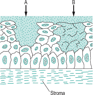
receptive f. The retinal area within which a light stimulus can produce a potential difference in a single cell. Retinal ganglion
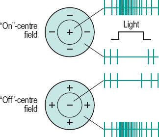
receptive fields are circular, often with a response different in the centre than in the periphery (also referred to as on-centre/off-centre or centre/surround organization). Ganglion cell receptive fields are very small in the macular region and large in the periphery of the retina. Receptive fields also exist in the lateral geniculate bodies where they are similar to those of the retina. In the visual cortex they have various shapes and sizes and may only respond to either a vertical bar or a black dot moving in a given direction and at a given speed, etc. Receptive fields reflect the interaction between excitation and inhibition between neighbouring neurons. The term can also describe the region of space that induces these neural responses (Fig. F2).
See cell, complex; cell, hypercomplex; cell, simple; inhibition, lateral; summation.
f. stop See diaphragm.
surrounding f. That area of the field of view surrounding any object.
f. of view The extent of an object plane seen through an optical instrument.
f. of view, apparent Angle subtended by the exit port of a sighting instrument or an empty frame aperture at the centre of the entrance pupil of the eye. Syn. apparent peripheral field of view. Note: when referring to the apparent field of fixation, the reference point is the centre of rotation of the eye. Syn. apparent macular field of view (Fig. F3).
See field of fixation.
Table F2
Average extent of the normal visual field (in degrees) of one eye of a young adult looking in the straight-ahead position, and measured with a white target subtending 1.0° under normal room illumination
| temporally | 94° |
| down and temporally | 88° |
| down | 70° |
| down and nasally | 54° |
| nasally | 60° |
| up and nasally | 56° |
| up | 54° |
| up and temporally | 64° |
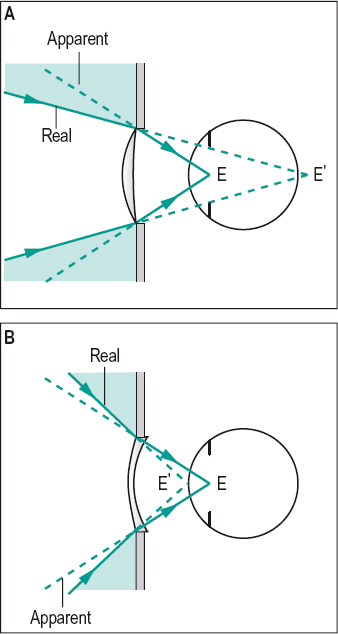
f. of view, real Angle subtended by the effective diameter of a lens at the point conjugate with the centre of the entrance pupil of the eye. Syn. real peripheral field of view; true field of view. Note: when referring to the real field of fixation, the reference point is the centre of rotation of the eye. Syn. real macular field of view (Fig. F3).
See phenomenon, jack-in-the-box.
f. of vision See field, visual.
visual f. (VF) The extent of space in which objects are visible to an eye in a given position. The extent of the visual field tends to diminish with age. The visual field can be measured either monocularly or binocularly. In the horizontal plane meridian the visual field extends to nearly 190° with both eyes open, the area seen binocularly, that is the region where both eyes can see the simulus is about 120°, and the area seen by one eye only is about 154°. Syn. field of vision.
See field, binocular visual; perimetry, kinetic; perimetry, static; test, confrontation; vision, island of.
visual f. expander An optical system designed to enlarge the field of vision. The most common types are reverse telescopes (e.g. looking through the objective of a galilean telescope), which minify objects being viewed but present more information by means of the enlarged visual field. They are usually of low power because of the reduction in visual acuity induced by the minification of the image. Prisms can also be used to expand the visual field. These systems are used mainly to improve mobility in patients with glaucoma and retinitis pigmentosa who have constricted visual fields or tunnel vision.
fifth cranial nerve See nerve, trigeminal.
figure A part or pattern in the visual field which has the perceptual attribute of completeness and is perceived as distinct from the rest of the field which forms the ground. Example: a printed word against a background page.
ambiguous f. An image or drawing arranged in such a way that its perception oscillates or flips involuntarily between, usually, two interpretations even though the retinal image remains constant, thus indicating that higher cortical processing are involved. Syn. reversible figure.
See figure, Blivet; figure, Kanizsa; illusion; Necker cube; Rubin’s vase; Schroeder’s staircase.
Blivet f. An ‘impossible’ figure in which three apparently solid tubes are attached at one end of a rectangular base which projects only two bars (Fig. F4).

See Necker cube; Schroeder’s staircase; Rubin’s vase.
fortification f. See scotoma, scintillating.
Kanizsa f. An ambiguous figure in which the illusory contour of a square (or triangle) appears in the middle of four (or three) truncated solid squares (or circles). It is an illustration of the perceptual ability to make sense of an incomplete figure by creating a ‘whole’ image from the separate elements (Gestalt organization). Some people cannot perceive the contour. Syn. Kanizsa square (Fig. F5).
reversible f. See figure, ambiguous.
filamentary keratitis See keratitis, filamentary.
film, anti-reflection See anti-reflection coating.
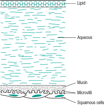
film, precorneal The field covering the anterior surface of the cornea which consists of lacrimal fluid and of the secretion of the meibomian and conjunctival glands. Its total thickness was thought to be about 9 μm but recent investigations have questioned that value and point to a much larger figure. It is composed of three layers: (1) The deepest and densest is the mucin layer (or mucous layer) which derives from the conjunctival goblet cells, as well as some secretion from the lacrimal gland. (2) The watery lacrimal fluid is the middle layer, called the lacrimal (or aqueous layer). It is secreted by the lacrimal gland and the accessory glands of Krause and Wolfring. It forms the bulk of the film and contains most of the bactericidal lysosyme and other proteins, inorganic salts, sugars, amino acids, urea, etc. (3) The oily layer (or lipid layer) is the most superficial and is derived principally from the meibomian glands in the lids as well as some secretion from the glands of Zeis. It greatly slows the evaporation of the watery layer and may provide a lubrication effect between lid and cornea (Fig. F6). Note: Some authors have suggested that the precorneal film is made up of only two layers; an innermost aqueous and mucin gel layer and an outer lipid layer. Syn. lacrimal layer; preocular tear film; tear film; tear layer.
See hyperlacrimation; mucin; tear secretion; Tearscope; test, break-up time.
filter Material or device used to absorb or transmit light of all wavelengths equally (neutral density filter which is abbreviated ND filter) or selectively, such as the coloured filters (blue filter transmits only blue light, green filter transmits only green light, etc.).
See density, optical; lens, absorptive; test, neutral density filter; wedge, optical.
bandpass f. A filter that allows the passage of radiations only within a narrow band of wavelengths around a central wavelength. This is done by multilayer coating, which produces destructive interference.
green f. A filter which transmits only green light. It may be used in ophthalmoscopy to increase the contrast of the blood vessels to the background facilitating the visibility of retinal circulation defects, haemorrhages and microaneurysms and the distinction between retinal and choroidal lesions. However, ophthalmoscopes actually use a filter that transmits a certain amount of red light, as otherwise the observation would be so dark as to make it extremely difficult. Syn. red-free filter.
interference f. A coloured filter consisting of five layers, two outside glass, two intermediate evaporated metal films and one central evaporated layer of transparent material. These filters act not by absorption of light but by destructive interference for all except a very narrow band of wavelengths, which are transmitted. Syn. coloured filter.
neutral density f. See filter.
red f. A filter that transmits only red light. It may be used in ophthalmoscopy to facilitate viewing the yellow macular pigment, but other structures are seen with less contrast. It also produces a larger pupil allowing observation of a larger fundus area.
red-free f. See filter, green.
filtration surgery A surgical procedure aimed at lowering the intraocular pressure by producing an outflow of aqueous humour through a drainage passage created in the sclera between the anterior chamber and the subconjunctival space.
See glaucoma surgery ; sclerectomy, deep; trabeculectomy; viscocanalostomy.
Fincham, coincidence optometer of See optometer of Fincham, coincidence.
Fincham’s theory See theory, Fincham’s.
finished lens See lens, finished.
first-degree fusion See vision, Worth’s classification of binocular.
first-order optics See optics, paraxial.
fisheye lens See lens, fisheye.
fissure A cleft or a groove found in an organ. In the brain, it usually applies to the deepest cleft.
See sulcus.
calcarine f. Fissure on the medial aspect of the occipital lobe separating the upper and lower halves. Its anterior portion is in front of the parieto-occipital fissure and the posterior portion extends round the occipital pole and even appears for a short distance on the lateral surface where it ends at the lunate sulcus. Syn. calcarine sulcus.
See area, visual; line of Gennari.
embryonic f. See fissure, optic.
inferior orbital f. An elongated opening lying between the lateral wall and the floor of the orbit. It is bounded anteriorly by the maxilla and the orbital process of the palate bone and posteriorly by the greater wing of the sphenoid bone. Syn. sphenomaxillary fissure.
See artery, infraorbital; nerve, zygomatic; Table O4.
interpalpebral f. See aperture, palpebral.
optic f. An invagination of the inferior portion of the optic stalk of the embryo. The hyaloid vessels pass through that fissure to supply the developing crystalline lens. In cases in which the invagination (or fissure) fails to fully close, colobomas will be formed. Syn. embryonic fissure; choroidal fissure.
See artery, hyaloid; cup, optic.
palpebral f. See aperture, palpebral.
sphenoidal f. See fissure, superior orbital.
sphenomaxillary f. See fissure, inferior orbital.
superior orbital f . An elongated opening lying between the roof and the lateral walls of the orbit, that is, between the two wings of the sphenoid bone. Syn. sphenoidal fissure.
See nerve, abducens; nerve, oculomotor; nerve, ophthalmic; nerve, trochlear; vein, superior ophthalmic; Table O4.
fistula An unnatural passage from an organ to the body surface or from one organ to another.
carotid-cavernous f. An abnormal interconnection between the internal carotid artery and the cavernous sinus. It may be caused by trauma to the skull or orbit, vascular disease or systemic hypertension. Common signs are pulsating proptosis, eye redness, diplopia, dilated epibulbar vessels and bruit. The pressure in the orbital veins is elevated as a result of the flow of arterial blood into the cavernous sinus.
lacrimal f. An abnormal opening from the skin onto any part of the lacrimal passage, although most often the lacrimal sac. It may follow a severe acute dacryocystitis.
fit-over See clipover.
fitted on K Refers to a contact lens in which the back optic zone radius is the same as that of the flattest meridian (or the mean of the two principal meridians) of the cornea. K is a symbol referring to the keratometer reading of the principal meridians of the cornea. Syn. alignment fit.
fitting Technique and art of selecting and adjusting spectacles or contact lenses following a visual examination.
See dispensing, optical.
fixation The act of directing the eye to a given object so that its image is formed on the foveola.
anomalous f. See fixation, eccentric.
bifoveal f. See bifixation.
binocular f. Fixation on an object with both eyes simultaneously.
f. disparity See disparity, retinal; fusional movements.
f. disparity unit, Mallett See Mallett fixation disparity unit.
eccentric f. Monocular condition in which the image of the point of fixation is not formed on the foveola. In this condition, the patients feel that they are looking straight at the object stimulating the non-foveolar retinal area and the visual acuity of that eye is reduced. The condition occurs most commonly in strabismic amblyopia but can also occur when the fovea has been destroyed by some pathological process. Syn. anomalous fixation.
See Haidinger’s brushes; occlusion treatment; penalization; pleoptics; spot, Maxwell’s; test, after-image transfer; viewing, eccentric; Visuscope.
foveal f. Normal fixation in which the image of an object falls on the foveola.
line of f. See axis, fixation.
f. movements See movements, fixation.
parafoveal f. Fixation by a retinal area located outside the fovea but within the macula (or fovea centralis), i.e. within 5 degrees of the central visual field. It may occur in amblyopia.
plane of f. See plane of regard.
point of f. Point in space upon which the eye is directed, either monocularly or binocularly. If there is no eccentric fixation, the image of that point is formed on the foveola. Syn. object of regard.
See point of regard.
f. reflex See reflex, fixation.
f. response Eye movement aimed at placing the image of a point of fixation on the foveola.
voluntary f. Conscious fixation of an object as distinguished from the fixation reflex.
flame haemorrhage See haemorrhage, preretinal.
flare, aqueous Scattering of light seen when a slit-lamp beam is directed, obliquely to the plane of the iris, into the anterior chamber. It occurs as a result of increased protein content, and usually inflammatory cells, in the aqueous humour. Visual impairment depends on the intensity of the flare. It is a sign of intraocular inflammation.
See effect, Tyndall; iritis; uveitis.
flash An intense light of short duration.
flash blindness See keratoconjunctivitis, actinic.
flashes Perception of sudden and transient bright spots of light in the absence of light stimuli. Flashes may occur as a result of rhegmatogenous retinal detachment, posterior vitreous detachment, proliferative diabetic retinopathy, papilloedema, retinal break or migraine.
See photopsia.
flavimaculatus fundus See fundus, flavimaculatus.
Fleischer’s ring See ring, Fleischer’s.
flexure, lens See lens flexure.
flicker Perception produced when the retina is stimulated by an intermittent light stimulus which fluctuates between a frequency of a few hertz and the critical fusion frequency.
flicker photometer See photometer, flicker.
flight of colours The temporal sequence of after-images of different colours that follow a brief exposure to a bright source of light. It is most easily seen in dark-adapted eyes. The duration of the sequence is much shorter or absent in some patients with retrobulbar optic neuritis or multiple sclerosis.
floaters Heterogeneities in the vitreous humour which may be of embryonic origin or pathological (e.g. in posterior vitreous detachment, retinal detachment, vitritis, asteroid hyalosis). The patient sees spots which float as the eye moves. Floaters are common in normal old eyes. Syn. vitreous floaters.
See iritis; muscae volitantes; myiodesopsia; photopsia; retina, lattice degeneration of the; retinitis, cytomegalovirus; uveitis; vitritis.
floccules of Busacca See Koeppe’s nodules.
fluconazole See antifungal agent.
flucytosine See antifungal agent.
fluid, lacrimal See film, precorneal; tears.
fluorescein A fluorescent, weak dibasic acid with a molecular weight of 376 whose sodium salt is used in dilute solution as a dye in the fitting of contact lenses, in the detection of corneal abrasions, etc. It is a yellowish-red compound, which fluoresces a brilliant yellow-green under ultraviolet or blue illumination (Fig. F7). Syn. sodium fluorescein.
See fluorexon; lamp, Burton; light, Wood’s; rose bengal; staining; test, break-up time; test, fluorescein.
fluorescein angiography See angiography, fluorescein.
fluorescence Property of a substance that, when illuminated absorbs light of a given wavelength and re-emits it as radiations of a longer wavelength. Example: fluorescein.
See law, Draper’s; light, Wood’s; luminescence.
fluorescent lamp See lamp, fluorescent.
fluorexon A staining agent similar to fluorescein but with a much higher molecular weight (710) and which is less readily absorbed by soft contact lens material. It is used in the fitting of soft or hybrid lenses (e.g. a piggyback lens). It stains a pale yellow-brown. However, it is not recommended for use with high water contact lenses (above 65%).
fluorometholone See antiinflammatory drug.
fluoroquinolone See antibiotic.
flurbiprofen sodium See antiinflammatory drug.
flush bridge See bridge, flush.
flush, choroidal See choroidal flush.
flutter, ocular An involuntary, rapid, horizontal saccadic oscillation of both eyes while attempting to fixate an object. It is a sign of cerebellar disease.
See myoclonus, ocular; opsoclonus.
flux, luminous Flow of light which produces a visual sensation. It is measured in lumens.
flux, radiant Power emitted in the form of radiation. It is measured in watts or ergs per second.
FM 100 Hue test See test, Farnsworth.
focal interval See Sturm, interval of.
focal length See length, focal.
focal length, equivalent See length, equivalent focal.
focal length, vertex See vertex focal length.
focal line; plane See under the nouns.
focal point See focus, principal .
focal power See paraxial equation, fundamental; power, refractive.
focal ratio See f number.
foci, conjugate See distances, conjugate.
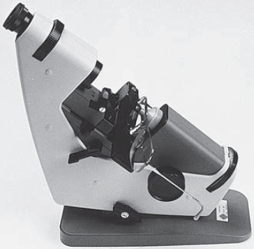
focimeter An optical instrument for determining the vertex power, axis direction and optical centre of an ophthalmic lens (Fig. F8). The instrument can be either manual or automated. Syn. Lensometer (a tradename); vertexometer; Vertometer (a tradename).
See lens measure; neutralization; power, back vertex.
focus 1. The point at which rays of light converge after passing through a convex lens to form a real image (real focus), or diverge from (virtual focus) after passing through a concave lens. 2. The centre or starting point of a disease process. 3. To adjust an optical system (e.g. camera or projector) in order to obtain a sharp image. Plural: foci. Syn. focusing.
See confocal; focus, principal; line, focal.
aplanatic foci A pair of conjugate object and image points for which an optical system is free of spherical aberration. Syn. aplanatic points.
dark f. See accommodation, resting state of.
depth of f. See depth of focus.
principal f. The axial image point produced by an optical system of an infinitely distant object (the second principal focus or posterior principal focus), or that axial object point for which the image will be formed at infinity (the first principal focus or anterior principal focus). A converging optical system or lens has two principal foci that are real. A diverging optical system or lens has a second principal focus that is virtual. In curved mirrors the two principal foci coincide. Depending upon whether the object is at infinity or at the principal focus, this same focal point becomes either the second principal focus or the first principal focus, respectively. Syn. focal point.
See length, focal; power, equivalent; sign convention.
real f. See focus.
sagittal f.; tangential f. See astigmatism, oblique.
virtual f. See focus.
focusing See focus.
fogging method See method, fogging.
fold, conjunctival See conjunctiva.
fold, corneal See oedema.
fold, semilunar See plica semilunaris.
folds, choroidal See choroidal folds.
follicle 1. A small gland. 2. A small cavity or deep narrow depression with excretory or secretory function. 3. A small nodule of lymphocytes and other cells occurring as a result of chronic inflammation.
See papilla.
conjunctival f. Small localized aggregation of lymphocytes, plasma and other cells appearing as white or grey elevations on the palpebral conjunctiva (tarsal area) as a result of chronic irritation (allergic, viral or mechanical such as contact lenses).
See conjunctivitis, adult inclusion; conjunctivitis, follicular.
palpebral f’s. See glands, meibomian.
follicular conjunctivitis See conjunctivitis, follicular.
footcandle Non-metric unit of illuminance. It is equal to a flux of 1 lumen per ft2. Symbol: fc. 1 fc = 10.764 lux.
footlambert Non-metric unit of luminance. It is equal to the average luminance of a surface emitting or reflecting 1 lumen per ft2. Symbol: fL. 1 fL = 3.426 cd/m2.
foramen, optic See canal, optic.
forced duction test See test, forced duction.
former A pattern, usually made of plastic, used to guide the automatic machines which cut and edge lenses. Syn. lens pattern.
See edging.
fornix See conjunctiva.
Forster–Fuchs spot See spot, Fuchs’.
fortification spectrum See scotoma, scintillating.
fossa A depression or cavity below the surface level of a part.
hyaloid f. A cup-shaped depression in the anterior vitreous body that accommodates the posterior part of the crystalline lens. It is actually separated from the lens itself by the postlenticular space of Berger. Syn. lenticular fossa; patellar fossa.
See ligament of Wieger.
f. for the lacrimal gland A depression in the frontal bone in which rests the orbital portion of the lacrimal gland, as well as some orbital fat which itself lies in the posterior part of the fossa called the accessory fossa of Rochon–Duvigneaud. The fossa is located behind the zygomatic process of the frontal bone in the anterior and lateral part of the orbital roof.
f. for the lacrimal sac A vertical groove, some 5 mm deep and about 14 mm high, formed by the frontal process of the maxilla and lacrimal bones and which contains the lacrimal sac. The fossa is bounded by the anterior and posterior lacrimal crests coming from the maxilla (frontal process) and lacrimal bone respectively, with no definite boundary above. It leads downward to the nasolacrimal canal, which contains the nasolacrimal duct.
patellar f. See fossa, hyaloid.
trochlear f. A small depression in the frontal bone which contains the pulley (or trochlea), a cartilaginous structure surrounded by a thick fibrous sheath 1 mm thick and through which passes the superior oblique muscle. The fossa is located about 4 mm behind the medial upper margin of the orbit.
Foster Kennedy syndrome See syndrome, Foster Kennedy.
Foucault grating See grating.
four prism dioptre base out test See test, four prism base out.
Fourier analysis The mathematical breakdown of waveforms into simple sine wave constituents. Any complex waveform consists of sine waves of different frequencies: the slowest (fundamental) frequency and harmonics thereof (these are frequencies which are odd multiples of the fundamental frequency). It is used in analysis and reconstruction of waveforms as, for example, analysing the spatial frequency components of a visual image. Syn. Fourier transform.
See sensitivity, contrast.
fourth cranial nerve See nerve, trochlear.
fourth nerve paralysis See paralysis of the fourth nerve.
fovea See foveola.
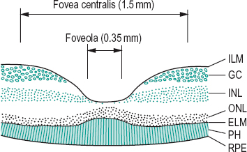
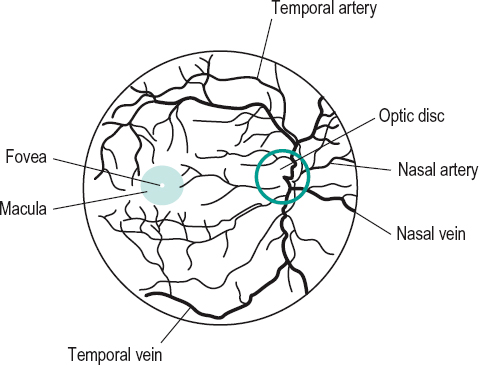
fovea centralis A small area of the retina of approximately 1.5 mm in diameter situated within the macula lutea. At the fovea centralis, the retina is the thinnest as there are no supporting fibres of Mueller, no ganglion cells and no bipolar cells. These cells are shifted to the edge of the depression. The fovea centralis contains mainly cone cells, each one being connected to only one ganglion cell and thus contributing to the highest visual acuity of the retina. The visual field represented by the fovea centralis is equal to about 5° (Fig. F9). Syn. foveal pit; macula (term often used by clinicians).
See acuity, central visual; image, retinal; macula lutea.
foveal avascular zone See foveola.
foveal fixation See fixation, foveal.
foveal pit See fovea centralis.
foveola The base of the fovea centralis with a diameter of about 0.35 mm (or about 1° of the visual field). The image of the point of fixation is formed on the foveola in the normal eye. The foveola contains cone cells only (rod-free area). The foveal avascular zone is slightly larger (about 0.5 mm in diameter) (Fig. F9). Syn. fovea (term often used by clinicians).
See eccentricity; fixation; umbo.
foveation period In congenital nystagmus it is the amount of time when the image of an object of regard is on or near the fovea and moving so slowly as to enable the patient to acquire useful visual information. The longer the foveation period and the lower the retinal image velocity, the higher the visual acuity.
Foville’s syndrome See syndrome, Foville’s.
fracture, orbital A term relating to any break in the bony integrity of the orbital walls, most commonly occurring in the orbital floor or less frequently in the medial wall (called ‘blow-out’ fracture). Although most frequently resulting from blunt trauma, it may also result as a necessary step in surgical treatment (e.g. orbital decompression). There is periorbital bruising, oedema, haemorrhage, or enophthalmos. Diplopia and limited upward movement are present usually as a result of a muscle or its fascia being entrapped in the break. There may be concomitant intraocular injury if caused by trauma (e.g. tennis ball). Fractures in the orbital apex or roof are less common. The lateral wall of the orbit is very tough as it protects the globe and is only involved in very severe maxillofacial trauma.
fragile X syndrome See syndrome, fragile X.
frame A structure in metal, plastic, tortoiseshell, wood, leather, etc. for enclosing or supporting ophthalmic lenses but usually considered without the lenses.
See spectacles.
frame, eyeglass 1. Synonym for spectacles. 2. Synonym for rimless spectacles.
frame heater A device used to warm plastic spectacle frames in order to soften its material sufficiently to allow insertion of lenses and/or adjustment. Some frame heaters warm air, which is directed to the area of the frame that is to be altered. Others heat salt or glass beads to a given temperature and the part of the frame that is to be altered is placed into the heated material. Syn. frame warmer.
See spectacle frame, plastic.
frame markings, spectacle See spectacle frame markings.
framycetin sulfate See antibiotic.
Fraunhofer’s lines See lines, Fraunhofer’s.
free-space Refers to natural viewing conditions, that is viewing an object that is neither housed inside an instrument, (e.g. in a synoptophore), nor viewed through an optical system. Syn. open environment; true-space.
frequency, critical fusion (CFF) Frequency of a light stimulation at which it becomes perceived as a stable and continuous sensation. That frequency depends upon various factors: luminance, colour, contrast, retinal eccentricity, etc. Syn. critical flicker frequency.
See effect, Brücke–Bartley; flicker; law, Ferry-Porter; law, Granit–Harper; law, Talbot–Plateau; photometer, flicker.
frequency, cut-off See sensitivity, contrast.
frequency of light See hertz; spectrum, electromagnetic; wavelength.
frequency, spatial The rate of alternation of the luminance in a visual stimulus as a function of length, usually expressed in cycles per degree.
See cycle per degree; Fourier analysis; function, contrast sensitivity.
Fresnel’s bi-prism; lens; Press-On prism See under the nouns.
Fresnel’s formula Formula used to determine the amount of light reflected at the interface between two transparent media. The reflection is
< ?xml:namespace prefix = "mml" />
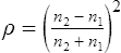
where n1 is the index of refraction of the first medium and n2 that of the second medium. For a lens surface in air, the percentage of light reflected is given by
Example: the reflection from the two surfaces of a glass lens made of crown (n = 1.523) is equal to 8.6%.
See image, ghost; reflection, surface.
Table F3
Percentage of light reflected in normal incidence ρ at the surface of several transparent substances of varying refractive indices, in air
| refractive index | ρ (%) |
| 1.333 | 2.04 |
| 1.4 | 2.78 |
| 1.45 | 3.35 |
| 1.5 | 4.0 |
| 1.523 | 4.3 |
| 1.55 | 4.65 |
| 1.6 | 5.32 |
| 1.65 | 6.02 |
| 1.7 | 6.72 |
| 1.75 | 7.44 |
| 1.8 | 8.16 |
Friedmann visual field analyser See analyser, Friedmann visual field.
Friedreich’s ataxia See ataxia, hereditary spinal.
fringes, diffraction A pattern of alternate dark and light bands produced by diffracted light passing the edge of an opening.
See diffraction.
fringes, interference See interference fringes.
Frisby stereotest See stereotest, Frisby.
front The part of a spectacle frame without the sides.
front silvered mirror See mirror, front surface.
front vertex focal length See vertex focal length.
front vertex power See power, front vertex.
frontal plane See plane, frontal.
frontoparallel plane See plane, frontoparallel.
frosted lens See lens, frosted.
Fuchs’ adenoma See adenoma, Fuchs’.
Fuchs’ coloboma See crescent, congenital scleral.
Fuchs, crypts of Pit-like depressions or openings found near the collarette and near the periphery of the iris. They allow the passage of aqueous humour from the anterior chamber into the stroma of the iris as the volume of the iris changes with dilatation and contraction.
Fuchs’ endothelial dystrophy See dystrophy, Fuchs’ endothelial.
Fuchs’ heterochromic iridocyclitis See iridocyclitis, Fuchs’ heterochromic.
Fuchs’ spur A few fibres located about midway along the length of the sphincter muscle which join with a few fibres of the dilator muscle of the iris.
Fuchs’ syndrome See syndrome, Fuchs’.
function 1. The particular action of an organ or tissue. 2. Any two variables in which the value of one depends upon the value of the other.
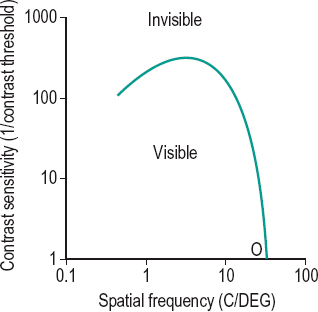
contrast sensitivity f. (CSF) The graphical representation of contrast sensitivity for the detection of a sine wave grating from a uniform field as a function of its spatial frequency. It is done by reducing the contrast of a grating until it can no longer be resolved (this point represents the contrast threshold) and repeating the procedure for a number of different spatial frequencies. The contrast sensitivity (1/contrast threshold) is plotted against spatial frequency (Fig. F10). The CSF is greatest at a spatial frequency around 3 cycles/degree and the point where the curve intercepts the spatial frequency axis (called the cut-off frequency) represents the standard visual acuity of the subject at 100% contrast.
See chart, contrast sensitivity; resolution, spurious; sensitivity, contrast.
line-spread f. A mathematical description of the distribution of light across the image of a very thin bright line object. On the retina the image of a thin bright slit spreads over a distance subtending about six minutes of arc at which point the intensity is less than two log units below the maximum.
modulation transfer f. (MTF) A relationship between the spatial frequency of an image (e.g. in number of cycles per degree or lines per inch) and the modulation amplitude (i.e. the difference between the luminance at the peaks and troughs of a grating). This gives an indication of the ability of a lens to resolve a grating. The greater the quality of a lens, the higher the spatial frequency at which the modulation amplitude falls to zero. At this point the lens can no longer transfer spatial modulation of intensity from the object to the image and the image appears as a uniform intensity distribution. This technique has been applied to assess the quality of the retinal image by measuring the contrast sensitivity function.
point-spread f. (PSF) The mathematical description of the light distribution across the image of a point source. The shape and width of the function depends upon the amount of diffraction, aberrations and scatter and in the eye, the shape of the pupil. Its shape, which resembles a normal distribution, is conventionally defined by its ‘half-width’, being the width of the curve at half the peak luminance. If only diffraction is considered the point-spread function is known as Airy’s disc.
See Fig. D5
functional Pertaining to a disorder with no detectable lesion to account for the symptoms.
functional visual loss Reduced vision experienced by an individual, but which is not based on objective measurements that could explain the symptom.
See amblyopia, hysterical; malingering.
fundoscopy The act of examining the fundus of the eye, as with an ophthalmoscope or with a biomicroscope and slit-lamp.
fundus The bottom or back surface of an organ. The term is used here to refer to the posterior portion of the interior of the eye visible with an instrument (e.g. ophthalmoscope).
f. albipunctatus A recessively inherited, nonprogressive tapetoretinal degeneration. It is characterized by a multitude of small, grey or whitish dots scattered throughout the fundus at the level of the pigment epithelium and accompanied by night blindness. The macula is spared and the retinal blood vessels, optic disc, visual field, colour vision and visual acuity are normal.
f. examination See biomicroscope; slit-lamp; ophthalmoscope, direct; ophthalmoscope, indirect.
f. flavimaculatus A retinal degeneration characterized by prominent, irregularshaped whitish or yellow flecks scattered throughout the posterior fundi of both eyes. There is usually no loss of vision unless one of the flecks involves the fovea. It is a variant of Stargardt’s disease. The electrooculogram is useful in diagnosing this condition.
leopard f. An ocular fundus marked with dark blotches on its surface as a result of a tapetoretinal degeneration, such as retinitis pigmentosa. Syn. leopard retina.
ocular f. The interior of the eye (as may be seen with the aid of an ophthalmoscope) consisting of the retina, the retinal blood vessels and even sometimes the choroidal vessels when there is little pigment in the pigment epithelium (e.g. albinos), the foveal depression, and the optic disc. The fundus appears red, owing mainly to the choroidal blood supply. The colour is lighter in fair people than in darker races and is dependent upon the amount of pigment in the pigment epithelium and in the choroid. In dark races the fundus is almost dark grey (Fig. F11). Plural: fundi.
See biomicroscopy, fundus; camera, fundus; plaques, Hollenhorst’s; tapetum lucidum.
salt and pepper f. The appearance of the ocular fundus characterized by a stippling of dark pigmented spots and yellowish-red spots of atrophy, as is found in congenital syphilis, choroideremia, Leber’s congenital amaurosis, rubeola, poliomyelitis, etc.
tessellated f. A normal ocular fundus in which the choroidal pattern appears as roughly polygonal dark areas in between choroidal vessels because the retinal pigment epithelium layer is thin and the choroid heavily pigmented. Syn. tessellated retina; tigroid fundus; tigroid retina.
tigroid f. See fundus, tessellated.
fungal keratitis See keratitis, fungal.
furrow, marginal A degenerative process occurring in the peripheral cornea characterized by corneal thinning. It is frequently associated with rheumatoid arthritis in which case there is epithelial defects and progressive ulceration. Furrows may also occur in corneal arcus without epithelial defect or vascularization. Syn. limbal furrow.
fused bifocal See lens, bifocal.
fusidic acid A product used as an antibiotic agent in solution 1%. It is synthesized as sodium fusidate from the microorganism Fusidium coccineum. It is effective against gram-positive bacteria. It is used in the treatment of bacterial blepharoconjunctivitis, especially staphylococcal infection.
fusion The act or process of mixing or uniting.
binocular f. See fusion, sensory.
central f. See fusion, sensory.
chiastopic f. Fusion obtained by voluntary convergence on two targets separated in space and such that the right eye fixates the left target and the left eye the right target. This is often facilitated by fixating a small mark above a single aperture placed in front of the two targets and then slowly shifting one’s gaze to the targets. The procedure is aimed at improving positive fusional convergence.
See convergence, fusional; fusion, orthopic.
critical f. frequency See frequency, critical fusion.
first-degree f.; flat f. See vision, Worth’s classification of binocular.
f. field An area around the fovea of each eye within which the fusion reflex is initiated. If the disparate images fall within this area motor fusion will occur, but if the disparity is too great there will be no fusional movement. This field is much larger horizontally than vertically.
flat f. Binocular fusion in which the single percept is two-dimensional and without stereoscopic effect. Syn. second-degree fusion.
See vision, Worth’s classification of binocular.
f. lock See lock, binocular; heterophoria, associated.
motor f. One of the components of convergence in which the eyes move until the object of regard falls on corresponding retinal areas (e.g. the foveas) in response to disparate retinal stimuli. Syn. disparity vergence; fusion reflex.
See convergence, fusional; fusion, sensory; retinal corresponding points; vergence facility.
orthopic f. Fusion obtained by voluntary divergence on two targets separated in space and such that the right eye fixates the right target and the left eye the left target. This is often facilitated by looking beyond the targets and then slowly shifting one’s gaze to the targets through double apertures placed in front of them. This procedure is aimed at improving negative fusional convergence.
See convergence, fusional; fusion, chiastopic.
peripheral f. See fusion, sensory.
second-degree f. See fusion, flat.
sensory f. The neural process by which the images in each retina are synthesized or integrated into a single percept. In normal binocular vision, this process occurs when corresponding (or nearly corresponding) regions of the retina are stimulated. This process can occur when the images are either in the central part of the retinae (central fusion) or in the peripheral part of the retinae (peripheral fusion). Syn. binocular fusion.
See anaglyph; convergence, fusional; haploscope; response, SILO; retinal corresponding points; stereogram, random-dot; test, bar reading; test, diplopia; test, Worth’s four dot.
third-degree f . See vision, Worth’s classification of binocular.
fusional convergence See convergence, fusional; convergence, relative.
fusional divergence See divergence, fusional.
fusional movements Reflex movements of the eyes occurring in response to retinal disparity (even though it may be below the threshold for diplopia to be seen) in order to produce a single image. If the fusional movements are such that although diplopia is eliminated there is still some disparity, this is fixation disparity.
fusional reserve, convergence; vergence See convergence, relative.
f-value See f number.


