[level-membership-for-emergency-medicine-category]
Chapter 55 Eye Injuries
3 List the important aspects of the history in all patients with eye injuries


4 What is included in the routine physical examination for all pediatric patients with eye injuries (except when temporarily deferred in absolute emergencies)?


5 What are the common pitfalls in evaluating children with eye injuries?


9 For patients without their glasses or contact lenses, how can you differentiate between decreased acuity and baseline refractive error?


14 Which eye injuries are possible when a child is a restrained passenger in the front seat and is involved in a car crash with air bag deployment?
20 How are hyphemas diagnosed and treated?
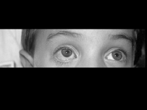
A hyphema (Fig. 55-1) should be suspected in any child with a tearing, painful eye and injected bulbar conjunctiva immediately after blunt eye trauma. It is often diagnosed by careful physical examination—many are visible without microscopic slit-lamp examination. Smaller hyphemas may require microscopic slit-lamp examination. All hyphemas require urgent ophthalmologic consultation for management, although in 66–97% of isolated hyphemas the hemorrhage resorbs without complication.
Hertle RW, Bacal D: Traumatic hyphema: Evaluation and management. Contemp Pediatr 14:51–68, 1997.
22 When and how does traumatic iritis present?
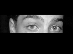
Traumatic iritis (Fig. 55-2), which may accompany other ocular injury or be the sole manifestation of blunt eye trauma, usually presents later than microscopic hyphema (24–72 hours after injury). Physical examination reveals a painful, red eye (typically perilimbal conjunctival injection), tearing, and pain with pupillary constriction (on accommodation or concentric constriction to light). The affected eye also may have slight miosis and a decreased or sluggish pupillary response. The pain is secondary to inflammation in the anterior chamber. Slit-lamp evaluation is diagnostic when it reveals white cells and a protein “flare” in the aqueous humor.
26 What physical examination findings are consistent with a ruptured or perforated globe?
 Significantly decreased visual acuity and severe pain
 Characteristic tear-drop pupil, pointing toward perforation

 Large, overlying subconjunctival hemorrhage (often fully around iris) or hyphema

KEY POINTS: REASONS TO SUSPECT GLOBE PERFORATION OR RUPTURE
1 History of hammering/grinding metal
2 Significant eye trauma causing decreased vision
3 Physical examination findings of a subconjunctival hemorrhage encircling the iris or cornea, large hyphema, posttraumatic corneal conjunctival edema, enophthalmos, extruded vitreous, iris, or choroid
4 The more difficult it is to examine the patient after trauma (because of pain, edema, hyphema, or vitreous hemorrhage), the greater the concern for globe rupture
27 What aspects of the history or physical examination place patients at high risk for globe perforation or rupture?


29 Describe the emergency initial management of chemical burns to the eye


31 An emergency physician attempts to repair a forehead laceration of a young child using cyanoacrylate glue. Some of the glue drips down the child’s face and the child’s eyelashes are noted to be stuck together. The child’s parent is quite concerned. Which eye injuries are likely?
[/level-membership-for-emergency-medicine-category][not-level-membership-for-emergency-medicine-category]
Chapter 55 Eye Injuries
