[level-membership-for-basic-science-category]
Eye and Adnexa


Anatomy and Physiology
 CM Guideline Alert
Ocular Adnexa
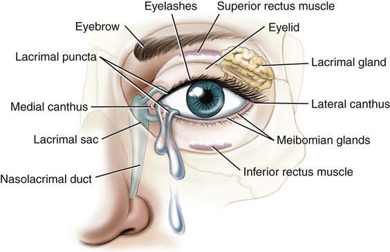
Each of our paired eyes is encased in a protective, bony socket called the orbit or orbital cavity.
Within the orbit, the eyeball is protected by a cushion of fatty tissue. The eyebrows mark the supraorbital area and provide a modest amount of protection from perspiration and sun glare. Further protection is provided by the upper and lower eyelids and the eyelashes that line their edges (Fig. 13-1).
 Note
Note
This chapter includes all the anatomy necessary to assign ICD-10 eye and adnexa codes, including detail on the zonule of Zinn, the pars plana, and the scleral venous sinus. See Appendix F for a complete list of body parts and how they should be coded.
 Be Careful!
 Be Careful!
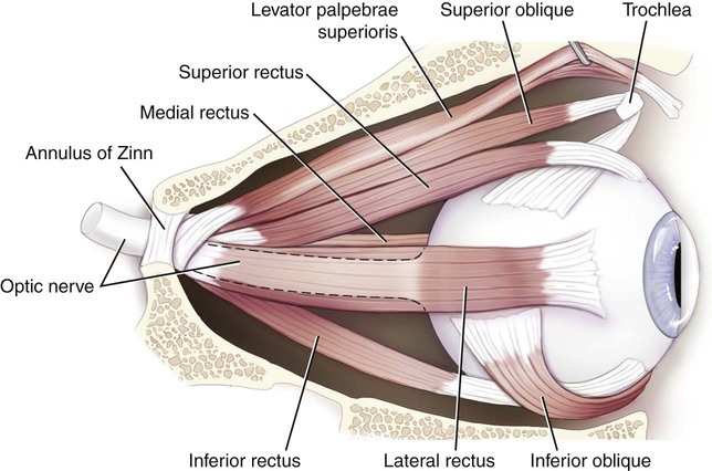
The extraocular muscles attach the eyeball to the orbit and, on impulse from the cranial nerves, move the eyes (Fig. 13-2). These six voluntary (skeletal) muscles are made up of four rectus (straight) and two oblique (diagonal) muscles. The origin of five of these muscles is in a ringlike structure surrounding the optic nerve behind the eyeball called the annulus of Zinn (also referred to as the annular tendon). This is mentioned only because later, when the lens of the eye is described, another structure in the lens called a zonule of Zinn will be named. Note that the muscle to raise the eyelids, the levator palpebrae superior muscle, is also labeled. “Levator” is used for any muscles whose function it is to elevate a structure. When this muscle is dysfunctional, it can result in an eyelid that droops (ptosis). The orbicularis oculi are the sphincter (ringlike) muscles that close the eye.
 Exercise 1:
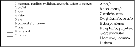
 To practice labeling the structures of the eye, click on Label It.
The Eyeball
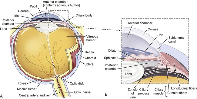
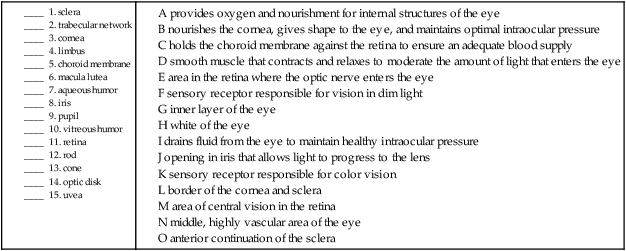
The anatomy of the eyeball itself is traditionally explained in three layers or tunics. The outer layer, or fibrous tunic, consists of the sclera and cornea. The middle layer, or vascular tunic, is composed of the choroid, ciliary body, and iris. The inner layer, or nervous tunic, consists of the retina (Fig. 13-3).
 Be Careful!
 PCS Guideline Alert
 Be Careful!
The Inner/Nervous Layer (Retina)
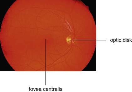
The inner layer of the eye, called the retina, is composed of several parts. The pars optica retinae contain the sensory receptors (rods and cones), the optic disk, the ora serrata, the macula lutea, and the fovea centralis. This layer is nourished by the retinal vessels that radiate from the optic nerve (Fig. 13-4).
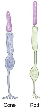
The sensory receptors for the images carried by the light rays are named for their appearance. They are the rods, which appear throughout the retina and are responsible for vision in dim light and the cones, which are concentrated in the central area of the retina and are responsible for color vision (Fig. 13-5). Three types of cones, termed L, M, and S (for long, medium, and short) cones, are endowed with photopigments that react to different wavelengths of light that produce the perception of red, green, and blue vision. Those individuals who have difficulty with their color vision (through inheritance or trauma) have deficiencies in one or more of these cones.
Vision
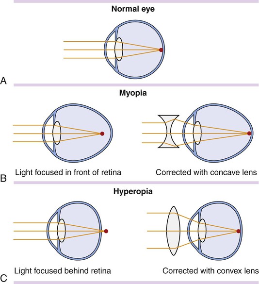
Two important mechanisms contribute to the ability to see. As light hits the eye, it passes first through the cornea, which bends the rays of light (refraction) so that they are projected properly onto the receptor cells in the eye. The muscles in the ciliary body adjust the shape of the lens to aid in this refraction. The lens flattens to adjust to something seen at a distance, or thickens for close vision (a process called accommodation). Errors of refraction are the most common reason for lens prescriptions. See Figure 13-6 for an example of refraction in normal vision, as well as in nearsightedness (myopia) and farsightedness (hypermetropia), and how they are corrected through the use of corrective lenses.
 Exercise 2:
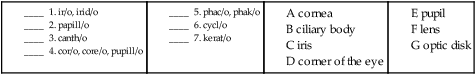
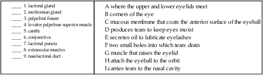
B. Match the structure with its definition or function.
Combining Forms for the Anatomy and Physiology of the Eye
| Meaning | Combining Form |
| choroid | choroid/o |
| ciliary body | cycl/o |
| conjunctiva | conjunctiv/o |
| cornea | corne/o, kerat/o |
| corner (of eye) | canth/o |
| eye | ophthalm/o, ocul/o |
| eyelid | blephar/o, palpebr/o |
| iris | irid/o, ir/o |
| lacrimal (tear) gland | dacryoaden/o |
| lacrimal (tear) sac | dacryocyst/o |
| lens | phak/o, phac/o |
| limbus | limb/o |
| macula lutea | macul/o |
| notched | serrat/o |
| optic disk | papill/o |
| orbit | orbit/o |
| pupil | pupill/o, core/o, cor/o |
| retina | retin/o |
| sclera | scler/o |
| tarsal plate (of the eyelid) | tars/o |
| tears | lacrim/o, dacry/o |
| uvea | uve/o |
| vision | opt/o, optic/o |
| vitreous humor | vitre/o, vitr/o |
 You can review the anatomy of the eye by clicking on Body Spectrum Electronic Anatomy Coloring Book, then Senses.
Pathology
Terms Related to Disorders of the Eyelid, Lacrimal System, and Orbit (HØØ-HØ5)
| Term | Word Origin | Definition |
| blepharitis | blephar/o eyelid -itis inflammation |
Inflammation of an eyelid. |
| blepharochalasis | blephar/o eyelid -chalasis relaxation, slackening |
Hypertrophy of the skin of the eyelid. |
| blepharoptosis | blephar/o eyelid -ptosis drooping, prolapse, falling |
Drooping of the upper eyelid. |
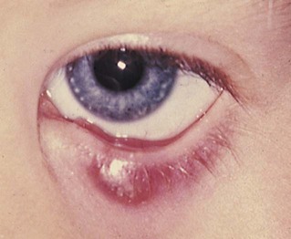
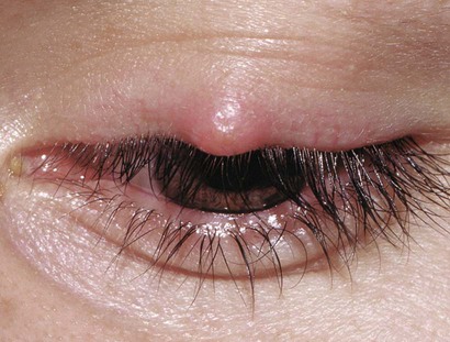
| chalazion | Hardened swelling of a meibomian gland resulting from a blockage. Also called meibomian cyst (Fig. 13-7). | |
| dacryoadenitis | dacryoaden/o lacrimal gland -itis inflammation |
Inflammation of a lacrimal gland. |
| dacryocystitis | dacryocyst/o lacrimal sac -itis inflammation |
Inflammation of a lacrimal sac. |
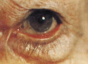
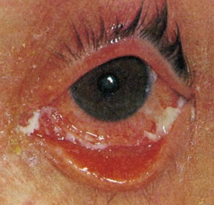
| ectropion | ec- out trop/o turning -ion process of |
Turning outward (eversion) of the eyelid, exposing the conjunctiva (Fig. 13-8). |
| entropion | en- in trop/o turning -ion process of |
Turning inward of the eyelid toward the eye (Fig. 13-9). |
| epiphora | Overflow of tears; excessive lacrimation. | |
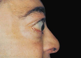
| exophthalmos | ex- out ophthalm/o eye -os condition |
Protrusion of the eyeball from its orbit; may be congenital or the result of an endocrine disorder (Fig. 13-10). |
| hordeolum | Infection of one of the sebaceous glands of an eyelash (Fig. 13-11). Also called a stye. | |
| lacrimal canaliculitis | lacrim/o tear -al pertaining to canalicul/o little canal -itis inflammation |
Inflammation of the tear ducts, especially the lacrimal canaliculi. |
Terms Related to Conjunctiva Disorders (H1Ø-H11)
| Term | Word Origin | Definition |
| conjunctivitis | conjunctiv/o conjunctiva -itis inflammation |
Inflammation of the conjunctiva, commonly known as pinkeye, a highly contagious disorder (Fig. 13-12). |
| pinguecula | A yellowish, noncancerous growth on the conjunctiva covering the eyeball in the area of the palpebral fissure. Usually asymptomatic; if irritated is termed pingueculitis. | |
| pterygium | pteryg/o wing -ium structure |
A winglike growth of the conjunctiva at the medial canthus of the eye, usually as a result of excessive exposure to wind/weather. |
Terms Related to Disorders of Sclera, Cornea, Iris, and Ciliary Body (H15-H22)
| Term | Word Origin | Definition |
| hyphema | hypo- under hem/o blood -a noun ending |
Blood in the anterior chamber of the eye as a result of hemorrhage due to trauma. |
| iridocyclitis | irid/o iris cycl/o ciliary body -itis inflammation |
Inflammation of the anterior uvea, specifically the iris and ciliary body. Symptoms include photophobia (sensitivity to light), miosis (constriction of the pupil), and synechia (adhesion of the cornea to the lens). |
| keratitis | kerat/o cornea -itis inflammation |
Inflammation of the cornea. |
| keratomalacia | kerat/o cornea -malacia softening |
Literally a softening of the cornea, this condition is the result of a vitamin A deficiency and malnutrition. Often leads to xerophthalmia (dry eye) and nyctalopia (night blindness). |
| scleritis | scler/o sclera -itis inflammation |
Inflammation of the sclera (white of the eye); usually associated with autoimmune disorders. |
| synechia | syn- together | Adhesion of the lens to the cornea. |
| uveitis | uve/o uvea -itis inflammation |
Inflammation of the uvea (iris, ciliary body, and choroid). |
 Exercise 3:
Disorders of the Eyelid, Lacrimal System, Orbit, Conjunctiva, Sclera, Cornea, Iris, and Ciliary Body
A infection of a sebaceous gland of an eyelash
B inflammation of the conjunctiva
C blood in the anterior chamber of the eye due to trauma
D hardened swelling of a meibomian gland due to blockage
F yellowish, noncancerous growth on the conjunctiva in the palpebral fissure area
G inflammation of the lacrimal sac
H inflammation of the anterior uvea, specifically the iris and ciliary body
K turning outward of the eyelid
M inflammation of the tear ducts
N adhesion of the lens to the cornea
17. drooping of the upper eyelid _____________________________________________________________________
18. inflammation of a tear gland _____________________________________________________________________
19. softening of the cornea __________________________________________________________________________
20. slackening of the eyelid __________________________________________________________________________
Terms Related to Disorders of the Lens (H25-H28)
| Term | Word Origin | Definition |
| aphakia | a- no, not, without phak/o lens -ia condition |
Condition of no lens, either congenital or acquired. |
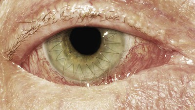
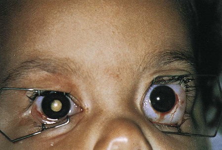
| cataract | Progressive loss of transparency of the lens of the eye (Fig. 13-13). Age-related (senile) cataracts can be classified as opacities of the lens in the center (nuclear) or on the periphery (cortical). |
Terms Related to Disorders of Choroid and Retina (H3Ø-H36)
| Term | Word Origin | Definition |
| age-related macular degeneration (ARMD or AMD) | Progressive destruction of the macula, resulting in a loss of central vision. This is the most common visual disorder after the age of 75 (Fig. 13-14). Appears as “wet” (exudative) or “dry” (nonexudative) form depending on whether or not there is bleeding and leaking under the macula. | |
| posterior cyclitis | cycl/o ciliary body -itis inflammation |
Inflammation of the ciliary body. Note that iridocyclitis is categorized with disorders of the ciliary body, whereas posterior cyclitis is with the choroid and retina. Also referred to as pars planitis because the pars plana is a structure within the ciliary body. |
| retinal ischemia | retin/o retina -al pertaining to isch/o hold back, suppress -emia blood condition |
Lack of blood flow to the retina. |
| retinal tear, retinal detachment | retin/o retina -al pertaining to |
Separation of the retina from the choroid layer. May be due to trauma, inflammation of the interior of the eye, or aging. A hole in the retina allows fluid from the vitreous humor to leak between the two layers. |
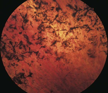
| retinitis pigmentosa | retin/o retina -itis inflammation |
Hereditary, degenerative disease marked by nyctalopia and a progressive loss of the visual field (Fig. 13-15). |
Terms Related to Glaucoma (H4Ø-H42)
| Term | Word Origin | Definition |
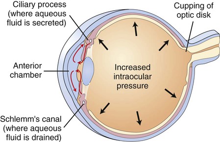
| glaucoma | glauc/o gray, bluish green -oma mass, tumor |
Group of disorders characterized by abnormal intraocular pressure due to obstruction of the outflow of the aqueous humor. Chronic or primary open-angle glaucoma (Fig. 13-16) is characterized by an open anterior chamber angle. Angle-closure or narrow-angle glaucoma is characterized by an abnormally narrowed anterior chamber angle. |
Terms Related to Disorders of Vitreous Body and Globe (H43-H44)
| Term | Word Origin | Definition |
| panophthalmitis | pan- all ophthalm/o eye -itis inflammation |
Inflammation of the entire eye. |
| purulent endophthalmitis | endo- within ophthalm/o eye -itis inflammation |
Infection within the eyeball usually caused by a bacterial infection. Purulent means “pertaining to pus.” |
Terms Related to Disorders of Optic Nerve and Visual Pathways (H46-H47)
| Term | Word Origin | Definition |
| optic neuritis | opt/o vision -ic pertaining to neur/o nerve -itis inflammation |
Inflammation of the optic nerve; often mentioned as a predecessor to the development of multiple sclerosis. |
| optic papillitis | opt/o vision -ic pertaining to papill/o optic disk -itis inflammation |
Inflammation of the optic disk usually accompanied by varying degrees of visual deficiencies. |
| papilledema | papill/o optic disk -edema swelling |
A swelling of the optic disk, usually secondary to intracranial pressure. |
| retrobulbar neuritis | retro- behind bulb/o globe -ar pertaining to |
Inflammation of the optic nerve behind the eyeball. A type of optic neuritis, the etiology is unknown. |
 Exercise 4:
Disorders of Choroid, Retina, Lens, Glaucoma, Vitreous Body, Globe, Optic Nerve, and Visual Pathways
A progressive destruction of the macula, resulting in a loss of central vision
B hereditary, degenerative disease resulting in nyctalopia and progressive visual field loss
C group of disorders characterized by abnormal intraocular pressure due to obstruction of outflow of aqueous humor
D inflammation of the optic disk
E separation of the retina from the choroid layer
F bacterial infection within the eyeball
G inflammation of the optic nerve behind the eyeball
H inflammation of the optic nerve
I progressive loss of transparency of the lens of the eye
12. condition of no lens _____________________________________________________________________________
13. swelling of the optic disk ________________________________________________________________________
14. inflammation of all the eye ______________________________________________________________________
Terms Related to Disorders of Ocular Muscles, Binocular Movement, Accommodation, and Refraction (H49-H52)
| Term | Word Origin | Definition |
| astigmatism (Astig, As, Ast) | Malcurvature of the cornea leading to blurred vision. If uncorrected, asthenopia (muscle weakness or fatigue) may result. | |
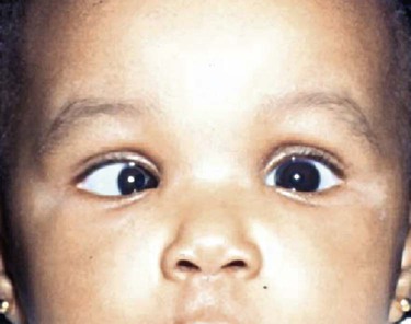
| esotropia | eso- inward trop/o turning -ia condition |
Turning inward of one or both eyes (Fig. 13-17). |
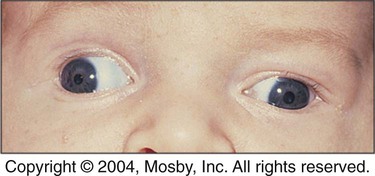
| exotropia | exo- outward trop/o turning -ia condition |
Turning outward of one or both eyes (Fig. 13-18). |
| hypermetropia | hyper- excessive metr/o measure -opia vision condition |
Farsightedness; refractive error that does not allow the eye to focus on nearby objects (also termed hyperopia) (see Fig. 13-6). |
| myopia (MY) | my/o to shut -opia vision condition |
Nearsightedness; refractive error that does not allow the eye to focus on distant objects (see Fig. 13-6). |
| presbyopia | presby- old age -opia vision condition |
Progressive loss of elasticity of the lens (usually accompanies aging), resulting in hyperopia. |
| strabismus | General term for a lack of coordination between the eyes, usually due to a muscle weakness or paralysis. Sometimes called a “squint,” which refers to the patient’s effort to correct the disorder. |
Terms Related to Visual Disturbances and Blindness (H53-H54)
| Term | Word Origin | Definition |
| achromatopsia | a- no, not, without chromat/o color -opsia vision condition |
Impairment of color vision. Inability to distinguish between certain colors because of abnormalities of the photopigments produced in the retina. Also called color blindness. Protanopia, deuteranopia, and tritanopia are types of achromatopsia due to respective defective L, M, and S cones. |
| amblyopia ex anopsia | ambly/o dull, dim -opia vision condition ex without an- no, not, without -opsia vision condition |
Dull or dim vision due to disuse. Also called lazy eye. |
| diplopia | dipl/o double -opia vision condition |
Double vision. Emmetropia (EM, Em) means normal vision. |
| hemianopsia | hemi- half an- no, not, without -opsia vision condition |
Loss of half the visual field, often as the result of a cerebrovascular accident. |
| nyctalopia | nyctal/o night blindness -opia vision condition |
Inability to see well in dim light. May be due to a vitamin A deficiency, retinitis pigmentosa, or choroidoretinitis. |
| photophobia | phot/o light -phobia condition of fear, sensitivity |
Extreme sensitivity to light. The suffix -phobia here means “aversion,” not fear. |
| scotoma | scot/o darkness -oma mass, tumor |
Area of decreased vision in the visual field. Commonly called a “blind spot.” |
Terms Related to Other Disorders of Eye and Adnexa (H55-H57)
| Term | Word Origin | Definition |
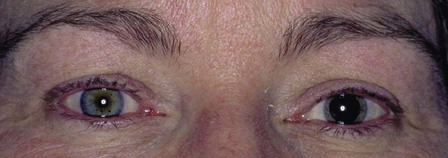
| anisocoria | an- no, not, without is/o equal cor/o pupil -ia condition |
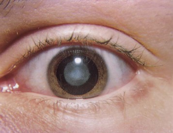
Condition of unequally sized pupils, sometimes due to pressure on the optic nerve as a result of trauma or lesion (Fig. 13-19). |
| miosis | mi/o to close, constrict -sis state |
Excessive and/or prolonged constriction of the pupil. |
| mydriasis | mydr/o dilation -iasis state |
Excessive and/or prolonged dilation of the pupil. |
| nystagmus | Involuntary, back-and-forth eye movements due to a disorder of the labyrinth of the ear and/or parts of the nervous system associated with rhythmic eye movements. |
 Be Careful!
 Be Careful!
Terms Related to Benign Neoplasms
| Term | Word Origin | Definition |
| choroidal hemangioma | choroid/o choroid -al pertaining to hemangi/o blood vessel -oma tumor, mass |
Tumor of the blood vessel layer under the retina (the choroid layer). May cause visual loss or retinal detachment. |
Terms Related to Malignant Neoplasms
| Term | Word Origin | Definition |
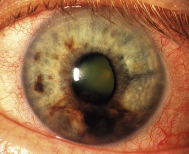
| intraocular melanoma | intra- within ocul/o eye -ar pertaining to melan/o dark, black -oma tumor, mass |
Malignant tumor of the choroid, ciliary body, or iris that usually occurs in individuals in their 50s or 60s (Fig. 13-20). |
| retinoblastoma | retin/o retina blast/o embryonic, immature -oma tumor, mass |
An inherited condition present at birth that arises from embryonic retinal cells (Fig. 13-21). |
 Exercise 5:
Disorders of Ocular Muscles, Accommodation, Refraction, Visual Disturbances, Blindness, Neoplasms, and Other Disorders of the Eye and Adnexa
A turning inward of one or both eyes
B term for lack of coordination between the eyes
D turning outward of one or both eyes
E malignant tumor of the choroid, ciliary body or iris
F extreme sensitivity to light
H excessive or prolonged constriction of the pupil
I malcurvature of the cornea leading to blurred vision
L loss of half the visual field
15. visual condition of double _______________________________________________________________________
16. mass of darkness ________________________________________________________________________________
17. condition of not equal pupil _____________________________________________________________________
18. state of dilation _________________________________________________________________________________
19. tumor of embryonic retina _______________________________________________________________________
20. vision condition of old age _______________________________________________________________________
Procedures
A, A microkeratome is used to create a hinged cap of tissue, which is lifted off the cornea. B, An excimer laser is used to vaporize and reshape underlying tissue. C, Tissue cap is replaced.
Terms Related to Diagnostic Procedures
| Term | Word Origin | Definition |
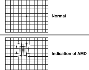
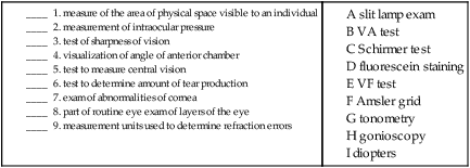
| Amsler grid | Test to assess central vision and to help diagnose age-related macular degeneration (Fig. 13-22). | |
| diopters | Level of measurement that quantifies refraction errors, including the amount of nearsightedness (negative numbers), farsightedness (positive numbers), and astigmatism. | |
| fluorescein staining | Use of a dye dropped into the eyes that allows differential staining of abnormalities of the cornea. | |
| gonioscopy | goni/o angle -scopy viewing |
Visualization of the angle of the anterior chamber of the eye; used to diagnose glaucoma and to inspect ocular movement. |
| ophthalmoscopy | ophthalm/o eye -scopy viewing |
Any visual examination of the interior of the eye with an ophthalmoscope. |
| Schirmer tear test | Test to determine the amount of tear production; useful in diagnosing dry eye (xerophthalmia). | |
| slit lamp examination | Part of a routine eye examination; used to examine the various layers of the eye. Medications may be used to dilate the pupils (mydriatics), numb the eye (anesthetics), or dye the eye (fluorescein staining). | |
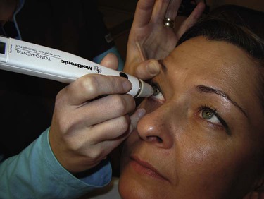
| tonometry | ton/o tone, tension -metry measuring |
Measurement of intraocular pressure (IOP); used in the diagnosis of glaucoma. In Goldmann applanation tonometry, the eye is numbed and measurements are taken directly on the eye. In air-puff tonometry, a puff of air is blown onto the cornea (Fig. 13-23). |
| visual acuity (VA) assessment | Test of the clearness or sharpness of vision; also called the Snellen test. Normal vision is described as being 20/20. The top figure is the number of feet the examinee is standing from the Snellen chart; the bottom figure is the number of feet a normal person would be from the chart and still be able to read the smallest letters. Thus if the result is 20/40, the highest line that the individual can read is what a person with normal vision can read at 40 feet. | |
| visual field (VF) test | Test to determine the area of physical space visible to an individual. A normal visual field is 65 degrees upward, 75 degrees downward, 60 degrees inward, and 90 degrees outward. |
 Exercise 6:
Diagnostic Procedures
| Term | Word Origin | Definition |
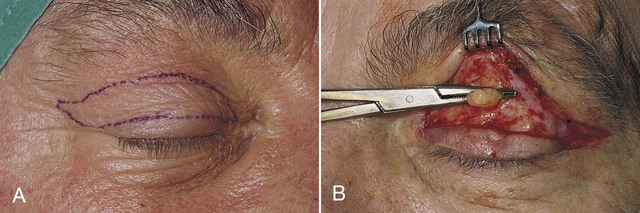
| blepharoplasty | blephar/o eyelid -plasty surgically forming |
Forming a new eyelid or restoring an eyelid. May be done to correct blepharoptosis or blepharochalasis (Fig. 13-24). |
| canthorrhaphy | canth/o corner (of eye) -rrhaphy suturing |
Suturing the upper and lower eyelids to prevent them from opening. Also called tarsorrhaphy or blepharorrhaphy. |
| conjunctivoplasty | conjunctiv/o conjunctiva -plasty surgically forming |
Forming a new or restored conjunctiva that may require the use of grafting procedures from the tissue of the cheek or other eye. |
| cyclodiathermy | cycl/o ciliary body dia- through therm/o temperature, heat -y process of, condition |
Use of heat to destroy part of the ciliary body for the treatment of glaucoma. Destruction of the ciliary body reduces the amount of aqueous humor, reducing intraocular pressure. If light is used (instead of heat) the procedure is called cyclophotocoagulation. |
| enucleation of eyeball | e- out nucle/o nucleus -ation process of |
Removal of the entire eyeball. |
| evisceration of eyeball | e- out viscer/o organ -ation process of |
Removal of the contents of the eyeball, leaving the outer coat (the sclera) intact. |
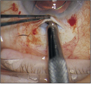
| iridectomy | irid/o iris –ectomy cutting out |
Cutting out all or part of the iris to allow aqueous humor to flow out of the anterior chamber. Used to treat closed angle glaucoma. |
| iridoplasty | irid/o iris -plasty surgically forming |
Forming a new or restored iris with laser treatment that allows the drainage of aqueous humor through an enhanced opening. Used to treat closed angle glaucoma. |
| keratectomy | kerat/o cornea –ectomy cutting out |
Cutting out part or all of the cornea to remove a lesion (Fig. 13-25). |
| keratoplasty | kerat/o cornea –plasty surgically forming |
Forming a new or restored cornea. A transplantation of corneal tissue from a donor or the patient’s own (autograft) cornea. May be either a full- or partial-thickness graft. |
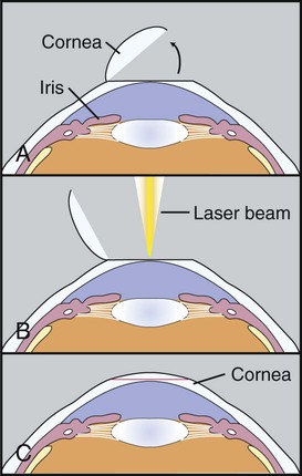
| laser-assisted in-situ keratomileusis (LASIK) | kerat/o cornea –mileusis Greek word meaning “carving” |
Flap procedure in which an excimer laser is used to remove material under the corneal flap. Corrects astigmatism, myopia, and hyperopia (Fig. 13-26). |
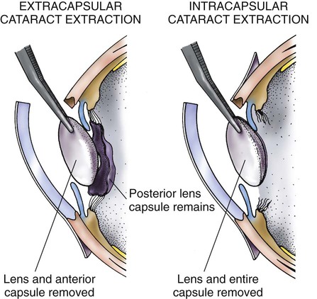
| phacoemulsification with/without intraocular lens (IOL) | phac/o lens -emulsification breaking down |
Breaking down and removing the lens (with/without lens implant) to treat cataract. May be intracapsular (ICCE), in which the entire lens and capsule are removed, or extracapsular (ECCE), in which the lens capsule is left in place (Fig. 13-27). |
| radial keratotomy | kerat/o cornea -tomy cutting |
Cutting the cornea in a spokelike fashion in order to flatten it and correct myopia. |
| vitrectomy | vitr/o vitreous body, glassy -ectomy cutting out |
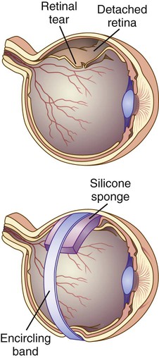
Removal of part or all of the vitreous humor. Usually done as part of the procedure (scleral buckling) to treat a retinal detachment. In scleral buckling the vitreous is removed and a buckle is attached to the sclera to hold it away from the detached retinal layer. The sclera then is able to return to its normal proximity to the choroid layer and heal (Fig. 13-28). |
 Exercise 7:
Procedures
A breaking down and removing the lens to treat cataract
B removal of the entire eyeball
C cutting out part or all of the cornea to remove a lesion
D removal of the contents of the eyeball
E transplantation of corneal tissue from a donor or an autograft
F suturing the upper and lower eyelids to prevent them from opening
G forming a new or restored iris with laser treatment
H use of heat to destroy part of the ciliary body to treat glaucoma
I cutting the cornea in spokelike fashion in order to correct myopia
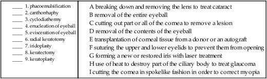
10. blepharoplasty __________________________________________________________________________________
11. iridectomy ______________________________________________________________________________________
12. conjunctivoplasty _______________________________________________________________________________
13. vitrectomy ______________________________________________________________________________________
Pharmacology
antibiotics: Medications used to treat bacterial infections. Examples include gentamicin (Garamycin) and ciprofloxacin (Ciloxan).
antiglaucoma drugs: Decrease the intraocular pressure by decreasing the amount of fluid in the eye or increasing the drainage. Examples include carbonic anhydrase (dorzolamide), cholinergics (pilocarpine), prostaglandin agonists (latanoprost), beta blockers (levobunolol), and alpha-2 agonists (brimonidine).
antihistamines: Drugs used to treat allergic conditions such as itchy or watery eyes. Diphenhydramine (Benadryl) is a common oral OTC product used to treat allergies. Ketotifen (Zaditor) is an example of OTC eye drops.
cycloplegics: Induce paralysis of the ciliary body to allow examination of the eye. One example is atropine eye drops.
lubricants: Keep the eyes moist, mimicking natural tears.
miotics: Cause the pupils to constrict; often used to treat glaucoma. An example is echothiophate iodide (Phospholine Iodide).
mydriatics: Cause the pupils to dilate; used in diagnostic and refractive examination of the eye. An example is cyclopentolate (Cyclogyl).
ophthalmics: Drugs applied directly to the eye. These may be in the form of solutions or ointments.
topical anesthetics: Temporarily anesthetize the eye for the purpose of examination.
 Exercise 8:
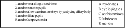
Recognizing Suffixes for PCS
Suffixes and Root Operations for the Eye and Adnexa
| Suffix | Root Operation |
| -ectomy | excision, resection |
| -plasty | reposition, repair, replacement, supplement |
| -rrhaphy | repair |
| -tomy | drainage, repair |
| Abbreviation | Meaning |
| ARMD, AMD | age-related macular degeneration |
| Astigm, As, Ast | astigmatism |
| ECCE | extracapsular cataract extraction |
| EM, Em | emmetropia |
| ICCE | intracapsular cataract extraction |
| IOL | intraocular lens |
| IOP | intraocular pressure |
| LASIK | laser-assisted in-situ keratomileusis |
| MY | myopia |
| OD | right eye |
| OS | left eye |
| OU | each eye |
| VA | visual acuity test |
| VF | visual field test |
 Exercise 9:
Operative Report
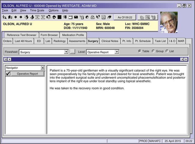
Using the operative report above, answer the following questions.
1. What is the condition that the procedure is intended to correct? ____________________________________
2. What does “preoperatively” mean? ________________________________________________________________
3. Phacoemulsification means that the lens was ______________________________________________________.
4. How do you know that he received a new lens? ____________________________________________________
5. What type of anesthetic was used during the surgery? ______________________________________________
 Exercise 10:
Healthcare Report
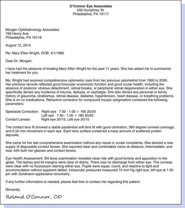
Using the healthcare report on the previous page, answer the following questions.
1. Mary Ellen denies diplopia and cephalgia. Explain these terms. ________________________________________________________________________________________________________________________________________
2. Mary Ellen has been diagnosed with myopic astigmatism. In your own words, explain this visual disorder. _________________________________________________________________________________________
3. What is the name of the test for glaucoma? ________________________________________________________
4. Explain the term paralimbal. _______________________________________________________________________
 Go to Evolve to interactively:
 [/level-membership-for-basic-science-category][not-level-membership-for-basic-science-category]
Eye and Adnexa
Anatomy and Physiology
Ocular Adnexa
Each of our paired eyes is encased in a protective, bony socket called the orbit or orbital cavity.
Within the orbit, the eyeball is protected by a cushion of fatty tissue. The eyebrows mark the supraorbital area and provide a modest amount of protection from perspiration and sun glare. Further protection is provided by the upper and lower eyelids and the eyelashes that line their edges (Fig. 13-1).
Note
This chapter includes all the anatomy necessary to assign ICD-10 eye and adnexa codes, including detail on the zonule of Zinn, the pars plana, and the scleral venous sinus. See Appendix F for a complete list of body parts and how they should be coded.
The extraocular muscles attach the eyeball to the orbit and, on impulse from the cranial nerves, move the eyes (Fig. 13-2). These six voluntary (skeletal) muscles are made up of four rectus (straight) and two oblique (diagonal) muscles. The origin of five of these muscles is in a ringlike structure surrounding the optic nerve behind the eyeball called the annulus of Zinn (also referred to as the annular tendon). This is mentioned only because later, when the lens of the eye is described, another structure in the lens called a zonule of Zinn will be named. Note that the muscle to raise the eyelids, the levator palpebrae superior muscle, is also labeled. “Levator” is used for any muscles whose function it is to elevate a structure. When this muscle is dysfunctional, it can result in an eyelid that droops (ptosis). The orbicularis oculi are the sphincter (ringlike) muscles that close the eye.
The Eyeball
The anatomy of the eyeball itself is traditionally explained in three layers or tunics. The outer layer, or fibrous tunic, consists of the sclera and cornea. The middle layer, or vascular tunic, is composed of the choroid, ciliary body, and iris. The inner layer, or nervous tunic, consists of the retina (Fig. 13-3).
The Inner/Nervous Layer (Retina)
The inner layer of the eye, called the retina, is composed of several parts. The pars optica retinae contain the sensory receptors (rods and cones), the optic disk, the ora serrata, the macula lutea, and the fovea centralis. This layer is nourished by the retinal vessels that radiate from the optic nerve (Fig. 13-4).
The sensory receptors for the images carried by the light rays are named for their appearance. They are the rods, which appear throughout the retina and are responsible for vision in dim light and the cones, which are concentrated in the central area of the retina and are responsible for color vision (Fig. 13-5). Three types of cones, termed L, M, and S (for long, medium, and short) cones, are endowed with photopigments that react to different wavelengths of light that produce the perception of red, green, and blue vision. Those individuals who have difficulty with their color vision (through inheritance or trauma) have deficiencies in one or more of these cones.
Vision
Two important mechanisms contribute to the ability to see. As light hits the eye, it passes first through the cornea, which bends the rays of light (refraction) so that they are projected properly onto the receptor cells in the eye. The muscles in the ciliary body adjust the shape of the lens to aid in this refraction. The lens flattens to adjust to something seen at a distance, or thickens for close vision (a process called accommodation). Errors of refraction are the most common reason for lens prescriptions. See Figure 13-6 for an example of refraction in normal vision, as well as in nearsightedness (myopia) and farsightedness (hypermetropia), and how they are corrected through the use of corrective lenses.
B. Match the structure with its definition or function.
Combining Forms for the Anatomy and Physiology of the Eye
| Meaning | Combining Form |
| choroid | choroid/o |
| ciliary body | cycl/o |
| conjunctiva | conjunctiv/o |
| cornea | corne/o, kerat/o |
| corner (of eye) | canth/o |
| eye | ophthalm/o, ocul/o |
| eyelid | blephar/o, palpebr/o |
| iris | irid/o, ir/o |
| lacrimal (tear) gland | dacryoaden/o |
| lacrimal (tear) sac | dacryocyst/o |
| lens | phak/o, phac/o |
| limbus | limb/o |
| macula lutea | macul/o |
| notched | serrat/o |
| optic disk | papill/o |
| orbit | orbit/o |
| pupil | pupill/o, core/o, cor/o |
| retina | retin/o |
| sclera | scler/o |
| tarsal plate (of the eyelid) | tars/o |
| tears | lacrim/o, dacry/o |
| uvea | uve/o |
| vision | opt/o, optic/o |
| vitreous humor | vitre/o, vitr/o |
Pathology
Terms Related to Disorders of the Eyelid, Lacrimal System, and Orbit (HØØ-HØ5)
| Term | Word Origin | Definition |
| blepharitis | blephar/o eyelid -itis inflammation |
Inflammation of an eyelid. |
| blepharochalasis | blephar/o eyelid -chalasis relaxation, slackening |
Hypertrophy of the skin of the eyelid. |
| blepharoptosis | blephar/o eyelid -ptosis drooping, prolapse, falling |
Drooping of the upper eyelid. |
