Extrinsic Allergic Alveolitis

Published on 23/05/2015 by admin

Filed under Pulmolory and Respiratory

Last modified 22/04/2025

Print this page

rate 1 star rate 2 star rate 3 star rate 4 star rate 5 star
Your rating: none, Average: 0 (0 votes)

This article have been viewed 1436 times

Chapter 55 Extrinsic Allergic Alveolitis

One of the first written descriptions of hypersensitivity pneumonitis (HP), or extrinsic allergic alveolitis (EAA), was in 1713 by Ramazzini, who observed that “minute worms” contained in grain cause a syndrome of dyspnea and cachexia associated with a shortened life span. In 1932, Campbell reported five farmers who developed acute systemic and respiratory symptoms after exposure to moldy hay. In 1944, Pickels named this syndrome “farmer’s lung.” Farmer’s lung and pigeon breeder’s disease are the most well studied forms of EAA. Each year, however, new exposure settings and types are added to the list of antigens implicated in the development of EAA.

A heterogeneous disease, EAA has varying clinical presentations associated with the inhalation of antigens, leading primarily to a diffuse mononuclear cell inflammation of the small airways and lung parenchyma. Classifying etiologic antigens into three broad categories is clinically helpful: microbial agents, animal proteins, and low-molecular-weight chemicals (Table 55-1). Most particulate antigens are of respirable size, less than 3 to 5 µm in diameter, and deposit in the alveoli. However, some antigens are deposited in airways and then become soluble, as occurs with Alternaria spores.

Table 55-1 Three Major Categories of Antigens Causing Extrinsic Allergic Alveolitis (EAA)

Antigen Exposure Syndrome
Microbial Agents
Bacteria    
Thermophilic Organic dust Farmer’s lung, bagassosis, mushroom worker’s lung
Nonthermophilic Water, hot tubs Humidifier lung, hot tub lung
Fungi    
Aspergillus spp. Moldy hay and moldy water Farmer’s lung, ventilation pneumonitis
Animal bedding Doghouse disease
Esparto grass Espartosis
Trichosporon cutaneum (T. biegelii) Damp wood and mats Japanese summer-type EAA
Alternaria spp. Wood pulp Wood pulp worker’s lung
Cryptostroma corticale Wood bark Maple bark stripper’s lung
Animal and Plant Proteins
Animal proteins    
Avian proteins Bird droppings, feathers (bloom) Bird fancier’s lung, pigeon breeder’s lung
Urine, serum, pelts Rats, gerbils Animal handler’s lung
Plants    
Coffee Coffee bean dust Coffee worker’s lung
Low Molecular Weight Chemicals
Toluene diisocyanate (TDI) Paints, resins, polyurethane foams Isocyanate (TDI) EAA
Drugs Amiodarone, gold, procarbazine Drug-induced EAA
Methylmethacrylate Dental laboratories  

Estimating the true incidence of disease can be challenging, because EAA may be misdiagnosed as an interstitial pneumonia or other pulmonary disorder or may not come to medical attention in individuals with mild disease. Moreover, genetics, coexisting exposures such as smoking, work environments, climates, altitude, and endemic disease (i.e., other granulomatous disorders such as sarcoidosis) vary from country to country, making comparisons of prevalence and incidence challenging. A longitudinal primary care database estimate of the incidence of EAA in the general population was 1 per 100,000 person-years in the United Kingdom. Incidence rates in specific populations with antigen exposure have also been reported. A study in Finnish farmers found 5 cases of EAA per 10,000 farmers who required hospital admission over a 1-year period. A study in Swedish farmers estimated an annual rate of 2 to 3 per 10,000 farmers. Prevalence of EAA among pigeon breeders is estimated at 0.1% to more than 10%, depending on antigen exposure.

Risk Factors

It is unclear why some individuals exposed to an antigen develop EAA and others residing or working in the same environment do not. Because only 1% to 15% of persons exposed to etiologic antigens develop EAA, a combination of antigen dose, host-specific factors, and underlying genetics likely contributes to the development of EAA.

Intensity, frequency, and duration of exposure; particle size; antigen solubility; climate; work practices; and use of respiratory protection combine to alter the risk of disease. Farmer’s lung is associated with exposure to moldy hay, which is more common in harsh winter environments with heavy rainfall, where damp hay is fed to livestock in indoor barns with poor ventilation. Summer-type EAA, the most prevalent form of EAA in Japan, occurs in the wet summer months when indoor microbial contamination is at its height. Moreover, EAA associated with bird exposure occurs more frequently during the sporting (bird-hunting) season.

As with other granulomatous disorders, EAA occurs more often in individuals who do not smoke. However, when the disease occurs in smokers, they typically have a more severe course with increased mortality compared to nonsmokers. Concurrent high exposures to carbamate and organochlorine pesticides are associated with a higher risk of developing EAA in at-risk farmers. Pesticide exposure has been shown to activate the immune system, with increased inflammatory cytokine production and differential functional effects on macrophages, which may serve to enhance the inflammatory response to antigen.

There is also evidence that viral infections may be associated with disease development. Dakhama et al. found viral nucleic acids in more than half of the bronchoalveolar lavage (BAL) specimens and alveolar macrophages in subjects with acute EAA; influenza A was isolated in the majority of subjects. Infection with respiratory syncytial virus (RSV) and Sendai virus in mouse models of EAA are associated with a more robust inflammatory response to the inhaled antigens that cause farmer’s lung. Fetal microchimerism (fetal cells in maternal tissues) may also be an important mechanism to help explain the observation that EAA is more common in antigen-exposed women who have had children.

Several genetic studies suggest that individuals with certain major histocompatibility complex (MHC) class II haplotypes and alleles as well as tumor necrosis factor alpha (TNF-α) promoter polymorphisms are at increased risk of developing EAA. Specific single nucleotide polymorphisms (SNPs) in the transporter associated with antigen processing (TAP-1) and proteasome subunit beta type 8 (PSMB8) genes, involved in antigen processing and presentation by MHC classes I and II, may have affected disease susceptibility in a population of Mexican patients with EAA. Interestingly, two studies in different populations showed that allelic variants in the promoter region of tissue inhibitor of metalloproteinase 3 (TIMP-3), which inhibits the proteolytic activity of matrix metalloproteinases and thus extracellular matrix turnover, are protective against the development of EAA.

Clinical Features

The clinical presentation of patients with EAA can vary depending on the duration and intensity of exposure. Presentation does not seem to change with the type of antigen (organic vs. inorganic). Traditionally, the clinical presentation of EAA has been classified according to Richerson’s scheme, which describes acute, subacute, and chronic forms. Symptoms of acute EAA generally occur within 4 to 12 hours of antigen exposure. Flulike symptoms predominate, including fever, cough, dyspnea, chills, malaise, chest tightness, and myalgias. Physical examination frequently reveals fever, tachypnea, tachycardia, and rales. With subacute and chronic forms, the temporal relationship between antigen exposure and symptom onset is more difficult to assess. Typically, these patients report more insidious onset of progressive dyspnea on exertion, dry or minimally productive cough, fatigue, malaise, anorexia, and weight loss. The physical examination may reveal bibasilar rales; right-sided heart failure and digital clubbing may be present in patients with advanced fibrosis.

Notably, Richerson’s classification system was described before the use of computed tomography (CT) and does not correlate well with lung pathology. Lacasse et al., using data from the hypersensitivity pneumonitis (HP) study, used a cluster analysis (e.g., symptomatology, physiology, imaging, BAL data) and found that subacute EAA is likely an attenuated form of acute EAA. Thus, they proposed a binary scheme in which patients are classified according to disease activity, whether active or sequelae. This classification scheme, however, needs further validation before it is integrated into clinical practice.

Several diagnostic criteria have been proposed to differentiate EAA from other interstitial lung diseases (ILDs). A prospective multicenter cohort study of patients who had a pulmonary syndrome with EAA in the differential diagnosis adopted a “clinical prediction rule” for the diagnosis of active EAA (Table 55-2). Significant predictors in the final model included exposure to a known offending antigen, positive precipitating antibodies, recurrent episodes of symptoms, inspiratory crackles, symptoms 4 to 8 hours after exposure, and weight loss. These criteria are helpful when combined with BAL and high-resolution CT in determining the likelihood of EAA.

Table 55-2 Clinical Prediction Rule for Diagnosis of Extrinsic Allergic Alveolitis

Variable Odds Ratio (95% CI)
Exposure to known antigen 38.8 (11.6-129.6)
Positive precipitating antibodies 5.3 (2.7-10.4)
Recurrent episodes of symptoms 3.3 (1.5-7.5)
Inspiratory crackles 4.5 (1.8-11.7)
Symptoms 4-8 hours after exposure 7.2 (1.8-28.6)
Weight loss 2.0 (1.0-3.9)

From Lacasse Y et al: Am J Respir Crit Care Med 168:952-958, 2003.

Imaging

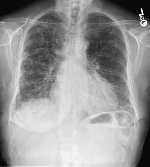
The chest radiograph is often normal in patients with EAA, with an estimated sensitivity of only 10%. In acute EAA, radiographs may show diffuse ground-glass or air space consolidation. Patients with subacute EAA may have a combination of nodular or reticulonodular opacities with ground-glass attenuation. Chronic fibrotic EAA usually has the appearance of reticular opacities with honeycombing on chest radiographs (Figure 55-1).

High-resolution computed tomography (HRCT) of the chest is the most sensitive imaging study for the detection of subtle changes associated with EAA (Figures 55-2 and 55-3). Multiple HRCT abnormalities are seen in patients with EAA that are loosely correlated with histologic and pulmonary function abnormalities (Table 55-3). Up to 50% of patients will have mediastinal lymphadenopathy; the nodes are usually smaller than 20 mm and are not detectable on the chest radiograph.

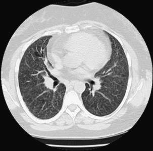
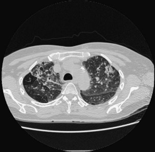
Table 55-3 Radiologic Abnormalities in EAA with Physiologic/Histologic Association

HRCT Abnormality Physiologic Correlate Histologic Correlate
Centrilobular nodules None Cellular bronchiolitis, alveolitis
Ground-glass opacities Restriction, decreased diffusion capacity Alveolitis, fine fibrosis, granulomas in alveolar septa
Mosaic attenuation Obstruction Bronchiolitis
Emphysema Obstruction, decreased diffusion capacity Emphysema, bronchiolar inflammation
Reticulation, honeycomb Restriction, decreased diffusion capacity Fibrotic change
Traction bronchiectasis

EAA, extrinsic allergic alveolitis; HRCT, high-resolution computed tomography.

Centrilobular nodules are the most frequent HRCT finding in EAA. The nodules are poorly defined, round, and less than 5 mm in diameter. Nodules predominate in the middle and lower lung zones, although this is not the rule, and are generally of ground-glass attenuation. Histologically, nodules correlate with polypoid intraluminal granulation tissue within the bronchioles (obliterative bronchiolitis [OB], or bronchiolitis obliterans) and reflect an active alveolitis. Centrilobular nodules do not correlate well with pulmonary function abnormalities.

Hazy opacity without obscuration of the underlying bronchovascular margins is most common in acute EAA but may also be seen in the subacute and chronic forms, especially with ongoing antigen exposure. Ground-glass opacities typically accompany other CT abnormalities such as centrilobular nodules and air trapping. Histologically, ground-glass opacities correlate with the presence of small granulomas within the alveolar septa, alveolitis, or fine fibrosis. Pulmonary function tests (PFTs) often reveals restrictive physiology with decreased diffusing capacity.

Mosaicism represents a patchwork of regions of differing attenuation caused by ground-glass opacities or air trapping, and often a combination of both in EAA. Air trapping reflects a failure of an area to increase in attenuation on expiratory imaging and suggests histologic bronchiolitis. It may also represent the presence of pulmonary hypertension in advanced cases. Obstructive physiology is often present on PFTs when mosaicism is apparent on imaging.

Multiple studies have shown that emphysema is more common than fibrosis in chronic farmer’s lung, even after adjusting for smoking status. The pattern of emphysema is similar to that caused by tobacco smoke and may be secondary to bronchiolar inflammation and obstruction.

In chronic EAA, the HRCT scan may show patterns typical for nonspecific interstitial pneumonitis (NSIP) or usual interstitial pneumonitis (UIP). Fibrotic EAA is associated with a reticular pattern, honeycombing, and traction bronchiectasis. Honeycomb change is seen in up to 50% of patients with chronic bird fancier’s lung and is less common in other forms of EAA. Fibrosis may predominate in the midlung, although a more diffuse distribution has been observed. Imaging features that favor a diagnosis of EAA over idiopathic pulmonary fibrosis (IPF) are upper-zone or midzone predominance, presence of ground-glass opacities, and less honeycomb change. Not surprisingly, the presence of fibrosis on HRCT is correlated with a poor prognosis.

Air space consolidation is rarely seen in EAA patients. Thin-walled cysts, resembling those in lymphocytic interstitial pneumonia, are found in up to 13% of patients with subacute EAA and have an uncertain pathogenesis.

Other Diagnostic Testing

Pulmonary Physiology

Classically, patients with EAA have a restrictive pattern with decreased forced vital capacity (FVC), total lung capacity (TLC), and diffusion capacity (DLCO). However, patients with farmer’s lung more often exhibit obstructive physiology than those with EAA caused by other antigens. About 40% of patients with farmer’s lung exhibited obstructive physiology in 6-year follow-up. Patients with subacute and chronic EAA may have a mixed pattern of obstruction and restriction. Methacholine challenge testing is often positive. Gas exchange abnormalities, if present, are characterized by hypoxemia that worsens with exercise. Exercise-induced hypoxemia is usually present early in disease.

In the setting of diagnostic uncertainty, an inhalational challenge using aerosolized material of the suspected causative antigen may be helpful. However, this is not recommended for most patients with suspected EAA. There are no purified, standardized, commercially available antigens to be used for this application, and most centers do not have experience with this technique. Patients may have an immediate reaction, but more frequently a delayed reaction occurs after several hours. In a study of patients with pigeon breeder’s disease, which also included normal subjects and those with ILD, all patients with EAA versus approximately 18% of those with ILD and 0% of healthy controls had an increase in body temperature and significant decreases in FVC, arterial oxygen tension (PaO2), and oxygen saturation (SaO2) after inhalational challenge with pigeon serum.

Histopathology

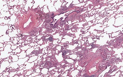
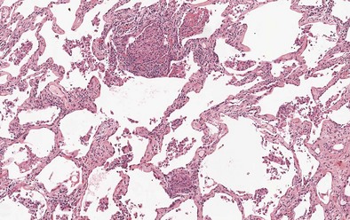
Findings on transbronchial biopsy are nonspecific and nondiagnostic in 50% of patients with EAA. Proceeding to surgical lung biopsy may be necessary when faced with diagnostic uncertainty, because features of EAA overlap with many other inflammatory lung diseases. The histologic findings vary depending on the stage of disease, although lung biopsies are rarely required in patients with acute EAA. Findings in acute EAA include neutrophilic and eosinophilic alveolar infiltration, small-vessel vasculitis, diffuse alveolar damage, and vascular immunoglobulin and complement deposition. A classic triad of histopathologic features is found in subacute EAA: lymphocytic alveolitis; small, loose non-necrotizing epithelioid cell granulomas; and cellular bronchiolitis (Figures 55-4 and 55-5). Foamy macrophages are present in air spaces, while lymphocytes are more prominent in the interstitium. Additionally, foci of obliterative bronchiolitis, intraalveolar fibrosis, and cellular NSIP may be found in subacute EAA.

In chronic EAA, variable stages of interstitial fibrosis may be found. Several patterns may be present, including NSIP, centrilobular and peribronchiolar fibrosis, bridging fibrosis (between centrilobular and perilobular areas), and a UIP-like pattern. The presence of mild to moderate lymphocytic infiltration, giant cells, poorly formed granulomas, and bridging fibrosis is more specific for EAA and can help differentiate EAA from other fibrotic lung diseases.

Treatment

Early diagnosis and elimination of antigen exposure are key elements in minimizing morbidity from EAA and treating the disease. Antigen elimination is the most effective approach. For example, maple bark stripper’s lung and bagassosis are now rare because of changes in handling organic substrates that minimize growth of microorganisms. Indoor sources of moisture that may lead to microbial contamination, such as humidifiers, leaking pipes, or appliances, and indoor hot tubs should be eliminated. A detailed environmental exposure history is important to identify potential causal antigens and remove the patient from exposure. However, in bird fancier’s lung associated with residential exposures, removal of birds from the home may not be sufficient; high levels of bird antigens may persist in the home and require extensive environmental cleanup. Efforts to ensure exposure abatement are often costly and difficult to assess for adequacy. It is therefore important for patients with EAA to receive regular clinical follow-up, PFTs, and imaging to monitor for progression and to direct further efforts at minimizing antigen exposure.

Oral corticosteroids are the first-line pharmacologic agents used at all stages of disease. Corticosteroids can shorten the duration of illness in acute EAA but do not improve long-term prognosis. Generally, patients should be started on high doses of prednisone, followed by gradual tapering once there is clinical improvement. Initiating prednisone at 0.5 mg/kg/day for 1 month, followed by a gradual taper until reaching a maintenance dose of 10 to 15 mg/day, is the recommended empiric regimen. PFTs should be performed within 2 months of therapy initiation, along with a clinical assessment of symptoms and possible steroid side effects. Prednisone should be discontinued when symptoms have resolved or there is no clear clinical or functional response. Inhaled corticosteroids can be useful adjuncts to steroid therapy, along with inhaled β-agonists if airway hyperresponsiveness is prominent. However, no randomized controlled clinical trials (RCTs) support the use of inhaled therapies in EAA. Supplemental oxygen is recommended in patients with hypoxemia.

In EAA patients taking corticosteroids without improvement or with severe steroid side effects, steroid-sparing agents such as azathioprine should be considered because of reported anecdotal success. In patients with progressive fibrotic EAA, early referral for lung transplantation should be considered.

Debate continues as to whether hot tub lung represents an infectious or hypersensitivity reaction to nontuberculous mycobacterial (NTM) aerosols and whether treatment with antimycobacterial antibiotics is necessary. The literature suggests that most patients improve with removal from exposure and treatment with corticosteroids. Thus, most immunocompetent patients with hot tub lung do not require prolonged courses of antimycobacterial therapy.

Prognosis

The natural history of EAA is variable and probably depends on the type and duration of antigen exposure and the host immune response. Acute EAA generally resolves within several weeks with corticosteroid therapy and removal from antigen exposure. However, continued symptoms and progressive lung impairment have been reported after recurrent acute attacks and even after a single acute attack. Additionally, persistent airway hyperresponsiveness and emphysema may impact long-term recovery.

Mortality estimates for patients with chronic EAA range from 1% to 10%. Patients with more severe lung fibrosis on biopsy specimens, honeycomb change on imaging, and digital clubbing have a worse prognosis. Moreover, patients with fibrotic EAA can present with acute exacerbations not triggered by recurrent antigen exposure. Unfortunately, they seem to have a similar poor prognosis as those with acute exacerbations of IPF. A recent study has shown that all-cause mortality in patients with EAA is three times higher than in the general population. The reason for this difference is unclear and needs further investigation.

Both prevention of disease and prevention of progression are key areas for intervention. Primary prevention is important in at-risk work environments and begins with informing workers of exposure risks and offering appropriate respiratory protection. Engineering controls and other workplace interventions are essential, such as improving ventilation systems, mechanizing feeding processes on farms, using additives to prevent the growth of mold in hay and silage, preventive maintenance on heating and cooling systems, enclosing selected metalworking fluid machining operations, and regular home cleaning to eliminate microbial colonization. Once EAA has been diagnosed, similar interventions can still be undertaken with a focus on elimination of antigen exposure to prevent disease progression.

Controversies and Pitfalls: A Focus on Treatment

Minimal evidence exists to guide treatment decisions for patients with EAA. When to start therapy and choice of pharmacologic agent are often challenging decisions. If, despite antigen elimination, the patient does not improve or even worsens oral corticosteroid therapy should be initiated. The recommended starting dose is 0.5 mg/kg of prednisone (usually 40-60 mg) once daily for 1 month, followed by a gradual taper. PFTs (e.g., DLCO) along with the patient’s symptoms should be assessed every 2 to 3 months to determine the clinical response to treatment. Prednisone may be tapered by 5-mg increments every 1 to 2 weeks, with close patient monitoring and adjustment as needed. Corticosteroid therapy should be tapered and discontinued if there is no clear response after 3 months.

Steroid-sparing agents (e.g., azathioprine) have been used with anecdotal success in patients who have failed prednisone or cannot tolerate steroids because of side effects. Some EAA patients require a steroid-sparing immunosuppressive agent for long-term preservation of lung function.

Again, antigen elimination, sometimes with several weeks of tapering oral corticosteroids, is effective for treatment of immunocompetent patients with hot tub lung, with no need for antibiotic therapy.

Long-acting inhaled steroids and β-agonists may be helpful in some EAA patients with airway hyperreactivity. Supplemental oxygen therapy is indicated in patients with hypoxemia. Early referral for lung transplantation should be considered in those with progressive fibrotic EAA.

Suggested Readings

Camarena A, Aquino-Galvez A, Falfan-Valencia R, et al. PSMB8 (LMP7) but not PSMB9 (LMP2) gene polymorphisms are associated with pigeon breeder’s hypersensitivity pneumonitis. Respir Med. 2010;104:889–894.

Camarena A, Juarez A, Mejia M, et al. Major histocompatibility complex and tumor necrosis factor-alpha polymorphisms in pigeon breeder’s disease. Am J Respir Crit Care Med. 2001;163:1528–1533.

Dakhama A, Hegele RG, Laflamme G, et al. Common respiratory viruses in lower airways of patients with acute hypersensitivity pneumonitis. Am J Respir Crit Care Med. 1999;159:1316–1322.

Lacasse Y, Assayag E, Cormier Y. Myths and controversies in hypersensitivity pneumonitis. Semin Respir Crit Care Med. 2008;29:631–642.

Lacasse Y, Selman M, Costabel U, et al. Clinical diagnosis of hypersensitivity pneumonitis. Am J Respir Crit Care Med. 2003;168:952–958.

Lacasse Y, Selman M, Costabel U, et al. Classification of hypersensitivity pneumonitis: a hypothesis. Int Arch Allergy Immunol. 2009;149:161–166.

Lynch DA, Rose CS, Way D, et al. Hypersensitivity pneumonitis: sensitivity of high-resolution CT in a population-based study. AJR Am J Roentgenol. 1992;159:469–472.

Rose CS, Lara AR Hypersensitivity pneumonitis, Mason RJ, et al. Murray and Nadel’s textbook of respiratory medicine, ed 5, Philadelphia: Saunders-Elsevier, 2010.

Selman M. Hypersensitivity pneumonitis: a multifaceted deceiving disorder. Clin Chest Med. 2004;25:531–547.

Selman M. Hypersensitivity pneumonitis. In: Schwarz M, King TEJ. Interstitial lung disease. Shelton, Conn: People’s Medical Publishing House; 2011:597–635.