Chapter 54 Extremity Injuries
Development
1 How do the pediatric musculoskeletal system and its response to stress differ from those in the adult?


2 What unique categories of fractures are commonly seen in pediatrics as a result of these differences?


3 Describe the Salter-Harris classification of fractures.
 Salter I: A fracture within the growth plate. The fracture line itself is not visible on radiographs, but a widening of the physis or displacement of the epiphysis may be suggestive of such a fracture.
 Salter II: A fracture that extends through the growth plate and metaphysis
 Salter III: An intra-articular fracture that extends through the growth plate and the epiphysis
 Salter IV: An intra-articular fracture that involves the metaphysis, growth plate, and epiphysis

5 What is the Thurston-Holland sign?
This is the triangular fracture segment of the metaphysis found in Salter-Harris type II fractures.
6 What are the four zones in the physis? Through which zone do fractures commonly occur?
9 In what order do the growth centers in the elbow ossify? Why is this clinically important?
The mnemonic CRITOE is a useful reminder of the order in which these ossified growth centers appear:


Radiology cases in pediatric medicine: elbow ossification centers in a child: www.hawaii.edu/medicine/pediatrics/pemxray/v1c11.html
Diagnosis


14 How do you evaluate distal nerve function in an uncooperative or preverbal child following extremity or digit injury?
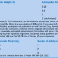
16 What name is associated with a Salter III fracture of the distal tibia? How is this fracture related to the age and growth of a child?
A Salter III fracture of the distal tibia is called a Tillaux fracture (Fig. 54-1). It classically occurs in teenagers shortly before growth plate closure. The medial segment of the distal tibial physis fuses last, and the medial aspect of the distal epiphysis remains anchored to the fibula by the anterior tibiofibular ligament. External rotation of the foot with sufficient force produces an avulsion fracture through the unfused medial segment of growth plate and down through the epiphysis.
19 What is a toddler’s fracture?
Tenenbein M, Reed MH, Black GB: The toddler’s fracture revisited. Am J Emerg Med 8:208–211, 1990.
21 How do you differentiate an avulsion fracture at the proximal fifth metatarsal from the normal secondary site of ossification?
Management
23 Describe the initial approach to the trauma victim with a significantly deformed extremity fracture.
24 What factors determine the need for fracture reduction?


25 What pediatric extremity injuries require emergent orthopedic consultation?


27 Describe the appropriate acute management for the vast majority of extremity injuries.


Complications
29 What clinical findings suggest a compartment syndrome?
In addition to a tense, swollen area at the injury site, clinical findings may include the 5 Ps:


30 What extremity injuries are at the highest risk for compartment syndrome?


Abuse Injuries
33 Describe the diagnosis and management of metaphyseal chip fractures.
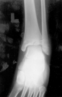
The radiograph in Fig. 54-2 depicts metaphyseal chip or “corner” fractures. The finding is highly suggestive of child abuse. The likely mechanism is vigorous pulling or shaking of the limbs. Corner fractures are most common in the distal femur and proximal tibia. Treatment should consist of splinting and pain management as well as a search for other evidence of abuse, including a skeletal survey. Social work services and child welfare should be involved.
34 What extremity fractures are associated with abuse?


Elbow Injuries
41 What are the three classifications of supracondylar fractures?
The three Gartland classifications of supracondylar fractures are:

 Type II: Mild displacement with intact posterior cortex
 Type III: Displaced fracture with no anterior and posterior cortical contact
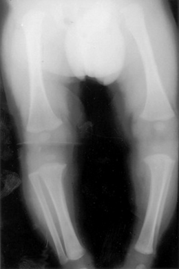
All type III fractures and most type II supracondylar fractures (Fig. 54-3) require percutaneous pinning to obtain appropriate reduction and to reduce complications.
Pediatric supracondylar fractures of the humerus. Wheeless’ Online Textbook of Orthopedic Surgery: www.wheelessonline.com/ortho/pediatric_supracondylar_fractures_of_the_humerus
1 The most common mechanism of pediatric elbow injury is a fall on an outstretched hand.
2 The presence of a posterior fat pad or elevated anterior fat pad is evidence of a supracondylar fracture until proven otherwise.
3 Type II and type III Gartland fractures typically require surgical pinning for adequate reduction.
4 Secondary ossification centers in the elbow appear in a predictable order.