133
Extramammary Paget disease
Extramammary Paget disease is a rare intraepidermal adenocarcinoma that presents on the scrotum in men and vulva in women. It may be misdiagnosed as eczema or tinea.
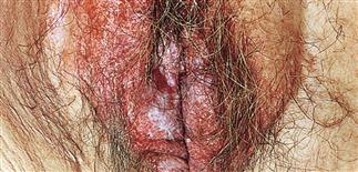
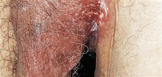
The disease appears as white to red, scaling or macerated, infiltrated, eroded or ulcerated plaque most frequently observed on the labia majora and scrotum.
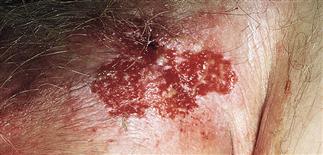
Three biopsies were taken before malignant cells were demonstrated at the periphery of this chronic ulcer at the base of the scrotum.
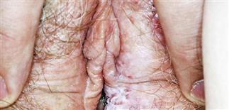
White eroded plaque with ill-defined borders on the labia are features of extramammary Paget disease that can be confused with lichen sclerosis.
DESCRIPTION
Intraepidermal adenocarcinoma involving anogenital or axillary skin. Occurs in areas where apocrine glands are found. May be divided into two groups based on source of underlying primary adenocarcinoma. • Most cases represent adenocarcinoma in situ with extension of primary adenocarcinoma in situ from adnexal structures. Apocrine gland carcinoma is the most common associated malignancy. • A minority of cases reflect an intraepidermal spread of tumor cells from non-cutaneous adenocarcinomas, via local or lymphatic spread. Urogenital and rectal carcinomas are the most common associated non-cutaneous adenocarcinoma. Local contiguous or regional lymphatic spread of these carcinomas leads to intraepidermal invasion.
HISTORY
• Rare before age 40. • More common in women than in men. • On vulva and perineum in older women. In men, scrotum, penis, anal and perianal skin most commonly affected. • May extend to involve lower abdomen, inguinal folds, buttocks, thighs. • Lesions slowly and relentlessly increase in size.
PHYSICAL FINDINGS
• A red to white-gray plaque with a velvety or scaly surface is typical. The plaque is sharply demarcated and has irregular borders. The lesion may appear eczematous or lichenified. Scaling, erosion, and serous exudate may occur. • Most often unilateral. • Differential diagnosis includes eczema, psoriasis, intertrigo, tinea, Candida, lichen simplex chronicus, and Bowen disease. • Depending on site of origin, the non-cutaneous primary adenocarcinoma may be visible and palpable. Regional lymph nodes are usually not palpable early in the course of the disease. • Unlike Bowen disease, dermal invasion and regional metastases appear to occur earlier in the disease course. • Fewer than 25% of all patients with extramammary Paget disease have an underlying non-cutaneous malignancy. Of those patients with underlying malignancy, 25% eventually die from the underlying malignancy. • Most common sites of metastases are the inguinal and pelvic lymph nodes, followed by liver, bone, lungs, brain, bladder, prostate, and adrenal glands. Regional and widespread metastases may develop from any one of the primary sites.
TREATMENT
• Local excision with obvious clear margins of the involved areas is standard. • Although lesions appear sharply defined clinically, histologic confirmation of margins is vital. Surrounding, clinically normal-appearing skin may also be involved. • High recurrence rate, even after excision with apparently appropriate margins. • May be benefit from Mohs micrographic excision as initial procedure. • Dissection of palpable regional lymph nodes may be warranted. • Radiotherapy also an option for difficult cases, recurrent disease.


