External skull

Published on 17/03/2015 by admin

Filed under Basic Science

Last modified 17/03/2015

Print this page

rate 1 star rate 2 star rate 3 star rate 4 star rate 5 star
Your rating: none, Average: 0 (0 votes)

This article have been viewed 10472 times

CHAPTER 26 External skull

The skull is the bony skeleton of the head. It shields the brain, the organs of special sense and the cranial parts of the respiratory and digestive systems, and provides attachments for many of the muscles of the head and neck. Movement of the lower jaw (mandible) occurs at the temporomandibular joint.

The skull without the mandible is called the cranium, and may be subdivided into two regions. The cranial vault or neurocranium encloses the brain, cranial meninges and cerebrospinal fluid, while the facial skeleton or viscerocranium hangs down from the front of the neurocranium and houses the organs of special sense.

The skull is the most complex osseous structure in the body: the young adult skull is composed of an average of 28 separate bones, many of which are paired, although some in the median plane are single. Most of the vault bones are flat, and consist of two tables or plates of compact bone enclosing a narrow layer of relatively dense cancellous marrow (diploic bone). The marrow within the skull bones is a site of haemopoiesis, at least in the young individual. These bones form by intramembranous ossification of a highly vascular connective tissue membrane and have often been referred to as ‘dermal’ in deference to their alleged ancient phylogenetic origin. The inner table is thinner and more brittle while the outer tends to be thicker and more resilient: this is important to remember when examining fractures to the skull caused by either blunt or sharp trauma. The skull bones vary in thickness in different regions, tending to be thinner where they are covered by muscles, e.g. in the temporal region, and thicker where muscles attach, e.g. in the occipital region. The thinner regions are more prone to fracture.

The majority of bones in the skull are held together firmly by fibrous joints termed sutures: in the developing skull sutures allow for growth. The three main sutural morphologies are dependent upon the magnitude of strains placed upon them. Thus the margins of adjacent bones of a suture may be smooth and meet end-to-end, giving a simple (butt-end) suture (e.g. median palatine suture); bevelled, so that the border of one bone overlaps the other (e.g. parietotemporal suture); or present numerous projections that interlock, giving a serrated appearance (e.g. sagittal suture). The complexity of serrated sutures increases from the inner to the outer surface. Fusion across sutures (synostosis) can start early in the third decade, although its variability precludes using this information to assess age at death in skulls with any degree of accuracy. The process of fusion starts on the internal surface of the cranium first and proceeds externally; the sagittal suture is often one of the first affected. By middle age, many of the larger sutures will show evidence of synostoses, although there are some which rarely show fusion, e.g. the zygomaticofacial. Premature fusion of sutures during the early growth phase of the skull will result in various abnormalities.

The bones forming the base of the skull develop mainly via endochondral ossification, and also play an important part in the overall growth of the face and the neurocranium. The joints between bones in the skull base are primary cartilaginous. One of the most important is the spheno-occipital synchondrosis between the body of the sphenoid anteriorly and the basilar part of the occipital bone posteriorly: fusion is completed between 13 and 18 years of age.

There are only two sites of synovial articulation associated with the exterior of the skull – the temporomandibular joint with the mandible and the atlanto-occipital joint between the condyles of the occipital bone and the superior articular facets of the atlas. Rotation of the skull does not directly involve any joints of the skull, but occurs at the atlanto-axial joint between the first and second cervical vertebrae.

Many important nerves and vessels pass in and out of the skull via openings (foramina). The skull is a prime site for fractures resulting from trauma, which means that these structures can be damaged as a result of head injury. In addition to main foramina, irregular emissary foramina allow veins situated externally on the face and scalp to communicate with those lying intracranially. The spread of infection along these routes can have serious clinical consequences.

In the account of the skull that follows, only generalized standard views of the skull will be considered. A more detailed account of each individual bone will be found associated with the relevant regional text.

FRONTAL (ANTERIOR) VIEW

Viewed from the front, the skull is generally ovoid in shape and is wider above than below (Fig. 26.1). The upper part is formed by the frontal bone which underlies the forehead region above the orbits. Superomedial to each orbit is a rounded superciliary arch (more pronounced in males) between which there may be a median elevation, the glabella. The glabella may show the remains of the inter-frontal (metopic) suture, which is present in about 9% of adult skulls. Above each superciliary arch is a slightly elevated frontal tuber or eminence which is generally more obvious in the female. Below, where the nasal bones meet the frontal bone, is a depression marking the root of the nose. The frontal bone articulates with the two nasal bones at the frontonasal sutures; the point at which the frontonasal and internasal sutures meet is the anthropometric landmark known as the nasion.
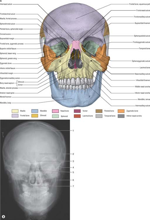
The upper part of the face is occupied by the orbits and the bridge of the nose. Each orbital opening is approximately quadrangular (see Ch. 39). The upper, supraorbital, margin is formed entirely by the frontal bone, interrupted at the junction of its sharp lateral two-thirds and rounded medial third by the supraorbital notch or foramen, which transmits the supraorbital vessels and nerve. The lateral margin of the orbit is formed largely by the frontal process of the zygomatic bone and is completed above by the zygomatic process of the frontal bone; the suture between them lies in a palpable depression. The infraorbital margin is formed by the zygomatic bone laterally and maxilla medially. Both lateral and infraorbital margins are sharp and palpable. The medial margin of the orbit is formed above by the frontal bone and below by the lacrimal crest of the frontal process of the maxilla.

The central part of the face is occupied mainly by the two maxillae, separated by the anterior nasal aperture. Each maxilla contributes to the upper jaw, the floor of the orbital cavity, the lateral wall of the nose, the floor of the nasal aperture and the bone of the cheek. Medially, the maxilla forms the nasal notch which is the floor and inferolateral border of the anterior nasal aperture. The prominent anterior nasal spine surmounts the intermaxillary suture at the lower margin of the anterior nasal aperture and is palpable in the nasal septum. The infraorbital foramen, which transmits the infraorbital vessels and nerve, lies about 1 cm below the infraorbital margin. The maxillary alveolar process bears the upper teeth. The short, thick zygomatic process of the maxilla has an oblique upper surface that articulates with the zygomatic bone at the zygomaticomaxillary suture. The frontal process of the maxilla ascends posterolateral to the nasal bone to articulate with the frontal bone.

The anterior nasal aperture is piriform in shape, and is wider below than above and bounded by the nasal bones and maxillae. The upper boundary of the aperture is formed by the nasal bones while the remainder is formed by the maxillae. In life, various cartilages (septal, lateral nasal, major and minor alar) help to delineate two nasal cavities (see Ch. 32). The defleshed skull presents a single anterior nasal aperture because these cartilages are lost during preparation or decomposition. However, the shape of these bones can be used quite successfully to predict the shape of the cartilaginous nose.

The lower part of the face, below the nose, is formed from the alveolar arch of the maxillae and the upper dentition, and the body of the mandible, the alveolar process of the mandible and the lower dentition. In the midline the mental protuberance produces the characteristic prominence of the chin. The mental foramen, which transmits the mental nerve and accompanying vessels, lies in the same vertical plane as the supraorbital and infraorbital foramina.

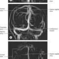
AP radiographs of the skull clearly show the central location of the paranasal air sinuses in the frontal bone, the maxilla and the ethmoid. These can be particularly useful indicators of identity when postmortem images are compared with premortem clinical films.

POSTERIOR VIEW

The parietal, temporal and occipital bones form the entirety of the posterior view (Fig. 26.2). The superolateral region is occupied by the parietal bones, the mastoid part of the temporal bones comprises the inferolateral regions and the central portion is occupied by the occipital bone, which is why this aspect is also referred to as the occipital view. The parietal bones articulate with the occipital bone at the lambdoid suture and extend inferiorly into the occipitomastoid and the parietomastoid sutures behind and above the mastoid process respectively. Sutural or wormian bones are islands of bone that are commonly found within the lambdoid suture. They may arise from separate centres of ossification and they appear to have no clinical significance, being of genetic rather than pathological aetiology. A large interparietal bone is not uncommon and is sometimes referred to as an Inca bone (Fig. 26.2).

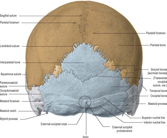
The external occipital protuberance is a midline ridge or a distinct process on the occipital bone which can become particularly well developed in the male. Superior nuchal lines extend laterally from the protuberance and represent the boundary between the scalp and the neck. Inferior nuchal lines run parallel to, and below, the superior nuchal lines; a set of highest nuchal lines may sometimes occur above the superior lines. The external occipital protuberance, nuchal lines and roughened external surface of the occipital bone between the nuchal lines, all afford attachment to muscles of the neck.
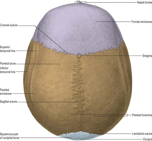
SUPERIOR VIEW

Seen from above, the contour of the cranial vault varies greatly but is usually ellipsoid, or more strictly, a modified ovoid with its greatest width lying nearer to the occipital pole (Fig. 26.3). Four bones constitute this view and articulate via three well-defined sutures. The squamous part of the frontal bone is anterior, the squamous part of the occipital bone is posterior and the two parietal bones meet in the midline and separate the frontal from the occipital bone. The maximal parietal convexity on each site is palpable at the parietal tuber or eminence: it is most conspicuous in the female who ostensibly retains a paedomorphic appearance. The superior and inferior temporal lines run close to the parietal eminence but are best seen in a lateral view.
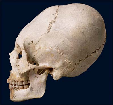
The coronal suture marks the articulation between the posterior margin of the frontal bone and the anterior margins of the two parietal bones. It descends across the cranial vault and projects inferiorly until it meets the junction between the greater wing of the sphenoid and the squamous temporal bone at the pterion. The sagittal suture runs in the midline between the two parietal bones and extends from the bregma anteriorly to the lambda posteriorly. The lambdoid suture delineates the articulation between the posterior borders of the right and left parietal bones and the superior border of the occipital bone.

The bregma represents the position of the fetal anterior fontanelle and is the junction between the coronal and sagittal sutures. The anterior fontanelle is a diamond-shaped membrane-filled space located between the two frontal and two parietal bones of the developing fetal skull and persists until approximately 18 months after birth. The lambda, at the junction of the sagittal and lambdoid sutures, is the site of the posterior fontanelle, which persists for the first 2–3 months after birth.

A parietal foramen may pierce either or both parietal bones near the sagittal suture about 3.5 cm anterior to the lambda. It transmits a small emissary vein from the superior sagittal sinus. The vertex is the highest point on the skull and it usually occupies a position in the middle third of the sagittal suture.

LATERAL VIEW

The skull, viewed from the side, can be subdivided into three zones: face (anterior); temporal region (middle); occipital region (posterior) (Fig. 26.4). The face has been considered in the section on the anterior view of the skull.
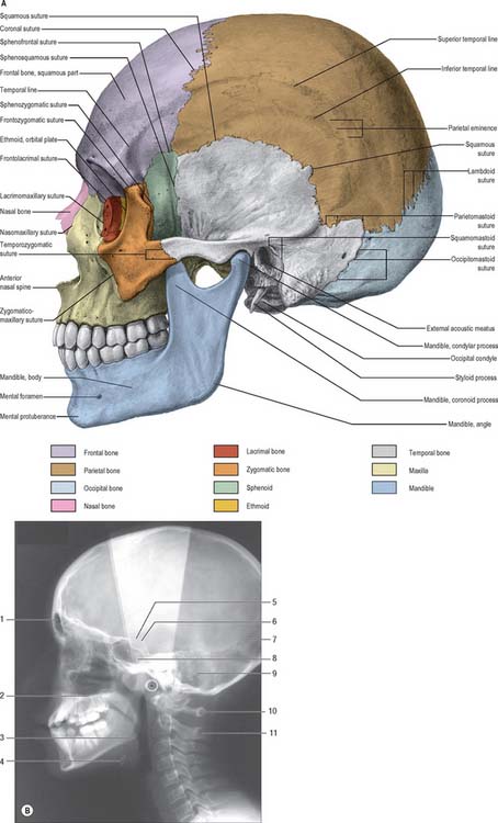
The temporal region can be divided into an upper temporal fossa and a lower infratemporal fossa, separated by the zygomatic arch. The temporal fossa is bounded inferiorly by the zygomatic arch, superiorly and posteriorly by the temporal lines and anteriorly by the frontal process of the zygomatic bone, and is continuous inferiorly with the infratemporal fossa deep to the zygomatic arch. The temporal lines often present anteriorly as distinct ridges, but become much less prominent as they arch posteriorly across the parietal bone. Indeed, the superior line may become quite indistinct posteriorly. The inferior temporal line becomes more prominent as it curves down the posterior part of the squamous temporal bone, forming a supramastoid crest at the base of the mastoid process. The superior temporal line gives attachment to the temporal fascia while the inferior temporal line provides attachment for temporalis.

The floor of the temporal fossa is formed by the frontal and parietal bones superiorly and the greater wing of the sphenoid and squamous temporal inferiorly. All four bones of one side meet at an H-shaped sutural junction termed the pterion. This is an important anthropometric landmark as it overlies both the anterior branch of the middle meningeal artery and the lateral fissure of the cerebral hemisphere. The pterion corresponds to the site of the anterolateral (sphenoidal) fontanelle of the neonatal skull which closes in the third month after birth.

The vertical suture between the sphenoid and temporal bones, the sphenosquamosal suture, is formed by articulation between the posterior border of the greater wing of the sphenoid and the anterior border of the squamous part of the temporal bone.

The lateral surface of the ramus of the mandible will be described briefly here as its position lies within the middle region of this view of the skull (see Ch. 31). The ramus is a plate of bone projecting upwards from the back of the body of the mandible; its lateral surface gives attachment to masseter. The ramus bears two prominent processes superiorly, the coronoid process anteriorly and condylar process posteriorly, separated by the mandibular notch. The coronoid process is the site of insertion of temporalis and the condylar process articulates with the mandibular fossa of the temporal bone at the temporomandibular joint. The inferior and posterior borders of the mandible meet at the angle, which is often splayed in the male because of the larger site of muscle attachment for medial pterygoid on the internal surface.

The zygomatic arch stands clear of the rest of the skull, and the temporal and infratemporal fossae communicate via the gap thus created. In life, this space is largely filled by temporalis. The zygomatic bone is the principal bone of the cheek together with the zygomatic processes of the maxillae and temporal bones. The term ‘zygomatic arch’ is generally restricted to the temporal process of the zygomatic bone and the zygomatic process of the temporal bone, which articulate at the zygomaticotemporal suture. The suture between the zygomatic process of the frontal bone and the frontal process of the zygomatic bone is the frontozygomatic suture, that between the maxillary margin of the zygomatic bone and the zygomatic process of the maxillary bone is the zygomaticomaxillary suture, and between the sphenoid and zygomatic is the sphenozygomatic suture. As the zygomatic process of the temporal bone passes posteriorly, it becomes associated with the articular tubercle of the mandibular fossa anteriorly and the supramastoid crest posteriorly.

The temporal bone is a prominent structure on the lateral aspect of the skull. Its squamous part lies in the floor of the temporal fossa and its zygomatic process contributes to the structure of the cheek. Additional components visible in the lateral view of the skull are the mandibular fossa and its articular eminence (tubercle), the tympanic plate, the external acoustic meatus, and the mastoid and styloid processes.

The mandibular (glenoid) fossa is the part of the temporomandibular joint with which the condylar process of the mandible articulates. It is bounded in front by the articular eminence and behind by the tympanic plate. The articular eminence provides a surface over which the mandibular condyle glides during mandibular movements (see Ch. 31).

The tympanic plate of the temporal bone contributes most of the margin of the external acoustic (auditory) meatus, and the squamous part forms the posterosuperior region (see Ch. 36). The external margin is roughened to provide an attachment for the cartilaginous part of the meatus. A small depression, the suprameatal triangle, lies above and behind the meatus and is related to the lateral wall of the mastoid antrum.

The mastoid process is a small inferior projection of the temporal bone, lying posteroinferior to the external acoustic meatus and is the site of attachment of sternocleidomastoid. It is in contact behind with the posteroinferior angle of the parietal bone at the parietomastoid suture and with the squamous part of the occipital bone at the occipitomastoid suture. These two sutures meet the lateral end of the lambdoid suture at the asterion, which coincides with the site of the posterolateral fontanelle in the neonatal skull and which closes during the second year. A mastoid foramen may be found near or in the occipitomastoid suture and transmits an emissary vein from the sigmoid sinus. Sutural bones may appear in the parietomastoid suture.

The styloid process lies anterior and medial to the mastoid process and gives attachment to several muscles and ligaments. Its base is partly ensheathed by the tympanic plate and it descends anteromedially, its tip usually reaching a point medial to the posterior margin of the mandibular ramus. However, the styloid process is very variably developed, and ranges in length from a few millimetres to a few centimetres.

The infratemporal fossa is an irregular postmaxillary space located deep to the ramus of the mandible; it communicates with the temporal fossa deep to the zygomatic arch (see Ch. 31). It is best visualized, therefore, when the mandible is removed but, for completeness, is considered here. Its roof is the infratemporal surface of the greater wing of the sphenoid. The lateral pterygoid plate lies medial to the fossa and the ramus of the mandible and styloid process lie laterally and posteriorly respectively. The infratemporal fossa has no anatomical floor. Its anterior and medial walls are separated above by the pterygomaxillary fissure lying between the lateral pterygoid plate and the posterior wall of the maxilla. The infratemporal fossa communicates with the pterygopalatine fossa through the pterygomaxillary fissure.

INFERIOR (BASAL) VIEW

The inferior surface of the skull, the base of the cranium, is complex: it extends from the upper incisor teeth anteriorly to the superior nuchal lines of the occipital bone posteriorly (Fig. 26.5). The region contains many of the foramina through which structures enter and exit the cranial cavity. The inferior surface can be conveniently subdivided into anterior, middle, posterior and lateral parts. The anterior part contains the hard palate and the dentition of the upper jaw and lies at a lower level than the rest of the cranial base. The middle and posterior parts can be arbitrarily divided by a transverse plane passing through the anterior margin of the foramen magnum. The middle part is occupied mainly by the base of the sphenoid bone, the apices of the petrous processes of the temporal bones and the basilar part of the occipital bone. The lateral part contains the zygomatic arches, mandibular fossa, tympanic plate and the styloid and mastoid processes. The posterior part lies in the midline and is exclusively formed from the occipital bone. Whereas the middle and posterior parts are directly related to the cranial cavity (the middle and posterior cranial fossae), the anterior part (the palate) is some distance from the anterior cranial fossa, being separated from it by the nasal cavities.

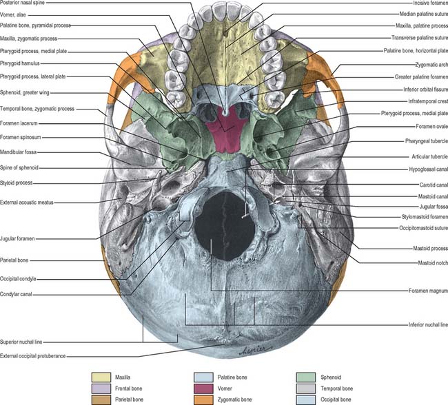
ANTERIOR PART OF CRANIAL BASE

The bony palate within the superior alveolar arch is formed by the palatine processes of the maxillae anteriorly and the horizontal plates of the palatine bones posteriorly, all meeting at a cruciform system of sutures (Fig. 26.5). The median palatine suture runs anteroposteriorly and divides the palate into right and left halves. This suture is continuous with the intermaxillary suture between the maxillary central incisor teeth. The transverse palatine (palatomaxillary) sutures run transversely across the palate between the maxillae and the palatine bones. The palate is arched sagittally and transversely: its depth and breadth are variable but are always greatest in the molar region. The incisive fossa lies behind the central incisor teeth, and the lateral incisive foramina, through which incisive canals pass to the nasal cavity, lie in its lateral walls. Median incisive foramina are present in some skulls and open onto the anterior and posterior walls of the fossa. The incisive fossa transmits the nasopalatine nerve and the termination of the greater palatine vessels. When median incisive foramina occur, the left nasopalatine nerve usually traverses the anterior foramen and the right nerve traverses the posterior foramen. The greater palatine foramen lies near the lateral palatal border of the transverse palatine suture; a vascular groove, deeper behind and shallower in front, leads forwards from the foramen. The lesser palatine foramina, usually two, lie behind the greater palatine foramen and pierce the pyramidal process of the palatine bone which is wedged between the lower ends of the medial and lateral pterygoid plates. The palate is pierced by many other small foramina and is marked by pits for palatine glands. Variably prominent palatine crests extend medially from behind the greater palatine foramina. The posterior border projects back as the posterior nasal spine. In the adult, the alveolar arch bears a maximum of 16 sockets or alveoli for the teeth: the sockets vary in size and depth, some are single and some are subdivided by septa, according to the morphology of the dental roots.

The nasal fossae lie above the hard palate and are separated by the nasal septum in the midline. The nasal septum is formed from the perpendicular plate of the ethmoid superiorly and the vomer inferiorly. The upper border of the vomer approximates to the inferior aspect of the body of the sphenoid, where it expands into an ala on each side. The two posterior nasal apertures (choanae) are located where the nasal fossae end: they are separated by the free posterior border of the vomer, and bounded below by the posterior border of the horizontal plate of the palatine bones, above by the sphenoid and laterally by the medial pterygoid plates.

MIDDLE PART OF CRANIAL BASE

The middle part of the cranial base is made up from the body of the sphenoid, the petrous temporal bones and the basiocciput (Fig. 26.5). It extends from the posterior nares anteriorly to an artificial line drawn transversely through the anterior margin of the foramen magnum posteriorly. In the adult, the body of the sphenoid fuses with the basiocciput to form a midline bar of bone that extends posteriorly to the foramen magnum. The basilar part of the occipital bone bears a small midline pharyngeal tubercle, which gives attachment to the pharyngeal raphe and the highest attachment of the superior pharyngeal constrictor.

The middle part of the cranial base is completed by the petrous processes of the two temporal bones, which pass from the lateral sides of the base of the skull and fill the triangular space between the greater wing of the sphenoid anteriorly and the lateral margins of the basiocciput posteriorly. Each petrous process meets the basilar part of the occipital bone at a petro-occipital suture, which is deficient posteriorly at the jugular foramen. The petrosphenoidal suture and the groove for the pharyngotympanic tube lie between the petrous process and the infratemporal surface of the greater wing of the sphenoid. The apex of the petrous process does not meet the spheno-occipital suture, and the deficit so produced forms the foramen lacerum.

Pterygoid processes descend from the junction between the greater wing and body of the sphenoid bone. Each separates into two laminae, the medial and lateral pterygoid plates, which are separated by a pterygoid fossa. Anteriorly the plates are fused, except inferiorly, where they are separated by the pyramidal process of the palatine bone. Sutures are usually discernible at this site. Laterally the pterygoid plates are separated from the posterior maxillary surface by the pterygomaxillary fissure, which leads into the pterygopalatine fossa. The posterior border of the medial pterygoid plate is sharp, and bears a small projection near the midpoint, above which it is curved and attached to the pharyngeal end of the pharyngotympanic tube. Above, the medial pterygoid plate divides to enclose the scaphoid fossa, while below it projects as a slender pterygoid hamulus, which curves laterally and is grooved anteriorly by the tendon of tensor veli palatini. The pterygoid hamulus gives origin to the pterygomandibular raphe. The lateral pterygoid plate projects posterolaterally and its lateral surface forms the medial wall of the infratemporal fossa. Superiorly and laterally the pterygoid process is continuous with the infratemporal surface of the greater wing of the sphenoid bone which forms part of the roof of the infratemporal fossa. This surface forms the posterolateral border of the inferior orbital fissure and bears an infratemporal crest associated with the origin of the upper part of lateral pterygoid. The infraorbital and zygomatic branches of the maxillary nerve and accompanying vessels pass through the inferior orbital fissure. Laterally the greater wing of the sphenoid articulates with the squamous part of the temporal bone.

The medial aspect of the greater wing of the sphenoid presents a crescent of foramina of which only the most posterior two, foramen ovale and foramen spinosum, can be viewed on the basal aspect. The foramen ovale lies medial to the foramen spinosum and lateral to the foramen lacerum on the infratemporal surface of the greater wing of the sphenoid bone. It transmits the mandibular division of the trigeminal nerve, the lesser petrosal nerve, the accessory meningeal branch of the maxillary artery and an emissary vein which connects the cavernous venous sinus to the pterygoid venous plexus in the infratemporal fossa. Posterolaterally, the smaller and rounder foramen spinosum transmits the middle meningeal artery and a recurrent meningeal branch of the mandibular nerve. The irregular spine of the sphenoid projects posterolateral to the foramen spinosum. The medial surface of the spine is flat and, with the adjoining posterior border of the greater wing of the sphenoid, forms the anterolateral wall of a groove that is completed posteromedially by the petrous part of the temporal bone. This groove contains the cartilaginous pharyngotympanic tube which leads posterolaterally into the bony portion of the tube that lies within the petrous part of the temporal bone. Occasionally the foramen ovale and foramen spinosum are confluent or the posterior edge of the foramen spinosum may be defective. A small foramen, the sphenoidal emissary foramen (of Vesalius), is sometimes found between the foramen ovale and scaphoid fossa. When present, it contains an emissary vein linking the pterygoid venous plexus in the infratemporal fossa with the cavernous sinus in the middle cranial fossa.

The foramen lacerum is bounded in front by the body and adjoining roots of the pterygoid process and greater wing of the sphenoid bone, posterolaterally by the apex of the petrous part of the temporal bone and medially by the basilar part of the occipital bone. Although it is nearly 1 cm long, it is not traversed by any major structure. The almost circular carotid canal lies behind and posterolateral to the foramen lacerum in the petrous part of the temporal bone. The internal carotid artery enters the skull through this foramen, ascends in the carotid canal, and turns anteromedially to reach the posterior wall of the foramen lacerum. It ascends through the upper end of the foramen lacerum accompanied by venous and sympathetic nerve plexuses. Meningeal branches of the ascending pharyngeal artery and emissary veins from the cavernous sinus also traverse the foramen lacerum. In life, the lower part of the foramen lacerum is partially occluded by cartilaginous remnants of the embryological chondrocranium. The pterygoid canal can be seen on the base of the skull at the anterior margin of the foramen lacerum, above and between the pterygoid plates of the sphenoid bone. It leads into the pterygopalatine fossa and transmits the nerve of the pterygoid canal and accompanying blood vessels.

POSTERIOR PART OF CRANIAL BASE

The posterior part of the cranial base is largely formed by the occipital bone (Fig. 26.5). Prominent features are the foramen magnum and associated occipital condyles, jugular foramen, mastoid notch and squamous part of the occipital bone up to the external occipital protuberance and the superior nuchal lines, hypoglossal canals (anterior condylar canals) and condylar canals (posterior condylar canals).

The foramen magnum lies in an anteromedian position and leads into the posterior cranial fossa. It is oval and wider behind, with its greatest diameter being anteroposterior. It contains the lower end of the medulla oblongata, meninges, vertebral arteries and spinal accessory nerve: the apical ligament of the dens and the tectorial membrane pass through it to attach to the internal basiocciput. Anteriorly, the margin of the foramen magnum is slightly overlapped by the occipital condyles which project down to articulate with the superior articular facets on the lateral masses of the atlas. Each occipital condyle is oval in outline and oriented obliquely so that its anterior end lies nearer the midline than its posterior end. It is markedly convex anteroposteriorly, less so transversely, and its medial aspect is roughened by ligamentous attachments. The hypoglossal canal, directed laterally and slightly forwards, traverses deep to each condyle and transmits the hypoglossal nerve, a meningeal branch of the ascending pharyngeal artery and an emissary vein from the basilar plexus. A depression, the condylar fossa, lies immediately posterior to the condyle and sometimes contains a (posterior) condylar canal for an emissary vein from the sigmoid sinus. This fossa accommodates the posterior margin of the atlas when the head is fully extended. A jugular process articulates with the petrous part of the temporal bone lateral to each condyle and its anterior free border forms the posterior boundary of the jugular foramen.

Laterally, the occipital bone articulates with the petrous part of the temporal bone anteriorly at the petro-occipital suture, and the mastoid process of the temporal bone more posteriorly at the petromastoid suture. The jugular foramen, a large irregular hiatus, lies at the posterior end of the petro-occipital suture between the jugular process of the occipital bone and the jugular fossa of the petrous part of the temporal bone. A number of important structures pass through this foramen: inferior petrosal sinus (anterior); glossopharyngeal, vagus and accessory cranial nerves (middle); internal jugular vein (posterior). A mastoid canaliculus runs through the lateral wall of the jugular fossa and transmits the auricular branch of the vagus nerve. The canaliculus for the tympanic nerve branch of the glossopharyngeal nerve lies on the ridge between the jugular fossa and the opening of the carotid canal. A small notch, related to the inferior glossopharyngeal ganglion, may be found medially, on the upper boundary of the jugular foramen (it is more easily identified internally). The orifice of the cochlear canaliculus may be found at the apex of the notch.

The squamous part of the occipital bone exhibits the external occipital protuberance, supreme, superior and inferior nuchal lines, and the external occipital crest, all of which lie in the midline, posterior to the foramen magnum. The region is roughened for the attachment of muscles whose primary function is extension of the skull (see Ch. 42).

LATERAL PART OF CRANIAL BASE

The lateral part of the cranial base consists of the zygomatic arch and infratemporal fossa anteriorly and the mandibular fossa, tympanic plate and styloid and mastoid processes posteriorly (Fig. 26.5) – the anterior structures have been considered earlier in this chapter.

A thin-walled depression in the temporal bone, the mandibular fossa, may be most easily inspected when the mandible is removed. The zygomatic arch extends laterally in front of the fossa and a distinct ridge, the articular eminence, lies anterior to the fossa. Three fissures can be distinguished behind the mandibular fossa. The squamotympanic fissure extends from the spine of the sphenoid, between the mandibular fossa and the tympanic plate of the temporal bone, and curves up the anterior margin of the external acoustic meatus. A thin wedge of bone forming the inferior margin of the tegmen tympani lies within the fissure and divides the squamotympanic fissure into petrotympanic and petrosquamous fissures. The petrotympanic fissure transmits the chorda tympani branch of the facial nerve from the intracranial cavity into the infratemporal fossa. The tympanic plate forms the floor of the external auditory meatus.

The stylomastoid foramen lies between the mastoid and styloid processes on the lateral aspect of the temporal bone. It transmits the facial nerve and the stylomastoid artery. A groove, the mastoid notch, lies medial to the mastoid process and gives origin to the posterior belly of digastric. A groove related to the occipital artery often lies medial to the mastoid notch. A mastoid foramen may be present near or in the occipitomastoid suture; when present, it transmits an emissary vein from the sigmoid sinus. The external acoustic meatus lies in front of the mastoid process. It is surrounded inferiorly by the tympanic plate which partly ensheathes the base of the styloid process as the vaginal process.

DISARTICULATED INDIVIDUAL BONES

Individual bones are described in appropriate chapters. The bones of the facial skeleton and cranial vault are described in the chapters on the face and scalp (29), nose and paranasal sinuses (32), external and middle ear (35) and orbit (39). The sphenoid and mandible are described in the chapter on the infratemporal fossa and the occipital bone is described in the chapter on the neck.

NEONATAL, PAEDIATRIC AND SENESCENT ANATOMY
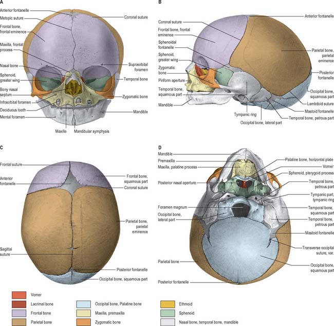
THE SKULL AT BIRTH

At birth the skull is large in proportion to other skeletal parts; the facial region is relatively small and constitutes only about one-eighth of the neonatal cranium, compared with half in adult life (Fig. 26.6). Smallness of the face at birth is largely due to the rudimentary stage of development of the mandible and maxillae because the teeth are unerupted. The nose lies almost entirely between the orbits, and the lower border of the nasal aperture is only slightly lower in position than the orbital floors. The large size of the calvaria, especially the neurocranium, reflects precocious cerebral maturation. Bones of the cranial vault are unilaminar and lack diploë. Frontal and parietal tuberosities are prominent; in the frontal view, the greatest width occurs between the parietal tuberosities. The glabella, superciliary arches and mastoid processes are not developed and the cranial base is relatively short and narrow.

Ossification is incomplete, and many bones are still in several elements united by fibrous tissue or cartilage. The ‘os incisivum’ is continuous with the maxilla; pre- and postsphenoids have just united, but the two halves of the frontal bone and mandible, and the squamous, lateral and basilar parts of the occipital bone are all separate. A second styloid centre (stylohyal) has not appeared, and parts of the temporal bones are separate except that fusion of the tympanic with the petrous and squamous parts has started. The fibrous membrane that forms the cranial vault before ossification is unossified at the angles of the parietal bones, producing six fontanelles: two median (anterior and posterior) and two lateral pairs (sphenoidal/anterolateral and mastoid/posterolateral). The anterior fontanelle is the largest and measures approximately 4 cm in anteroposterior and 2.5 cm in transverse dimensions. It occupies the junction between the sagittal, coronal and frontal sutures and is therefore rhomboid in shape. The posterior fontanelle lies at the junction between the sagittal and lambdoid sutures and is therefore triangular. The sphenoidal (anterolateral) and mastoid (posterolateral) fontanelles are small, irregular and occur at the sphenoidal and mastoid angles of the parietal bones respectively.

At birth the orbits appear relatively large. The developing tooth germs are generally contained within the alveolar crypts, although eruption of the upper central incisor teeth can occur prior to, or shortly after, birth. Temporal bones differ greatly from their adult form. The internal ear, tympanic cavity, auditory ossicles and mastoid antrum are all almost adult in size, the tympanic plate is an incomplete ring which has usually started to fuse with the squamous part, and the mastoid process is absent. The external acoustic meatus is short, straight and wholly cartilaginous. The external aspect of the tympanic membrane faces more inferiorly than laterally, in accord with the basal cranial contour. The stylomastoid foramen is exposed on the lateral surface of the skull, the styloid process has not fused with the temporal bone, the mandibular fossa is flat and more lateral, and its articular tubercle is undeveloped. The paranasal sinuses are rudimentary or absent and only the maxillary sinuses are usually identifiable.

During birth the skull is moulded by slow compression. That part of the scalp which is more central in the birth canal is often temporarily oedematous as a result of interference with venous return, and is called the caput succedaneum. Fontanelles and the openness and width of the sutures allow bones of the cranial vault some overlap. The skull is compressed in one plane with compensatory orthogonal elongation. These effects disappear within the first week after birth.

POSTNATAL GROWTH

Although postnatal growth of the vault and facial skeleton usually proceeds in a co-ordinated fashion, these regions exhibit different rates and temporal periods of expression. In general, early growth of the vault is largely influenced by cerebral maturation while growth of the facial skeleton responds to spatial requirement for the development of the teeth and the muscles of mastication. Growth of the cranial base is largely independent of both vault and facial development and therefore the three regions must be considered separately.

Growth of the vault

Growth of the vault is rapid during the first year and then continues at a slower rate until the seventh year, when it has reached almost adult dimensions. For most of this period, expansion is largely concentric; overall form is determined early in the first year and remains largely unaltered thereafter. However, the shape of the vault is not solely related to cerebral growth, but is also influenced by genetic factors that manifest in an extensive range of shapes and sizes that may be sufficient to allow a reliable evaluation of racial origin. During the first and early second year, growth of the vault occurs primarily through ossification at apposed margins of bones (which possess an osteogenic layer) accompanied by some accretion and absorption of bone at surfaces in order to adapt to continually altering curvatures. Growth in breadth is said to occur at the sagittal, sphenofrontal, sphenotemporal, occipitomastoid and petro-occipital junctions, while growth in height is said to occur at the frontozygomatic and squamosal sutures, pterion and asterion. During this period, fontanelles are closed by progressive ossification of the bones around them, but separate rogue centres of ossification may develop into sutural bones. The sphenoidal and posterior fontanelles ossify within 2 or 3 months of birth, the mastoid fontanelles usually ossify near the end of the first year and the anterior fontanelle ossifies around the middle of the second year. Those fontanelles which close first are most likely to show sutural bones.

Early in the first years the calvarial bones commence interlocking at sutural junctions. Further expansion of the vault is largely achieved by accretion and absorption on external and internal bone surfaces respectively. At the same time the bones also thicken, although this is not a uniform process. At birth the vault is unilaminar but the tabular structure with intervening diploë is generally apparent by about the fourth year. Thickening of the vault and development of external muscular markings reflect the influences of musculoskeletal maturation. The mastoid processes do not develop until the second year and they are invaded by air cells (pneumatized) in the sixth year.

Growth of the face

Growth of the facial skeleton occurs over a longer time period than is witnessed for the calvaria. The ethmoid and the orbital and upper nasal cavities have almost completed growth by the seventh year. Orbital and upper nasal growth is achieved by sutural accretion, with deposition of bone preferentially occurring on the facial aspects of the sutural junctions. The maxilla is carried downwards and forwards by expansion of the orbits and nasal septum and by sutural growth, especially at the fontanelles and zygomaticomaxillary and pterygomaxillary sutures. In the first year, growth in width occurs at the symphysis menti and midpalatal, internasal and frontal sutures: such growth diminishes or even ceases when the symphysis menti and frontal suture close during the first few years, even though the midpalatal suture persists until mature years. Facial growth continues up to puberty and shows a period of expansion that is linked to the growth spurt and hormonal influences of secondary sexual alteration. After sutural growth, near the end of the second year, expansion of the facial skeleton occurs by surface accretion on the face, alveolar processes and palate, and resorption in the walls of the maxillary sinuses, the upper surface of the hard palate and the labial aspect of the alveolar process. Co-ordinated growth and divergence of the pterygoid processes reflects deposition and resorption of bone on appropriate surfaces.

Obliteration of the calvarial sutures progresses with age, starting between 20 and 30 years internally, and somewhat later on the exterior. Closure times vary greatly. Obliteration usually begins in the coronal suture and then extends into the sagittal and lambdoid sutures, in that order. The most striking senile feature is diminution in size of the mandible and maxillae following the loss of teeth and absorption of alveolar bone; this reduces the vertical depth of the face and increases the mandibular angles.

Sutural bones

Additional ossification centres may occur in or near sutures, giving rise to isolated sutural bones (also called Wormian bones, Fig. 26.2). Usually irregular in size and shape, and most frequent in the lambdoid suture, they frequently occur at fontanelles, especially the posterior fontanelle. They may represent a pre-interparietal element, a true interparietal, or some composite. An isolated bone at the lambda is sometimes referred to as an Inca bone or Goethe’s ossicle. One or more pterion ossicles or epipteric bones may appear between the sphenoidal angle of the parietal and the greater wing of the sphenoid; they vary greatly in size but are more or less symmetrical. Sutural bones usually have little morphological significance. However, they appear in great numbers in hydrocephalic skulls (Fig. 26.7), and they have therefore been linked with rapid cranial expansion. For a detailed analysis of these and other epigenetic variations in adult crania, consult Berry & Berry (1967) and Berry (1975).
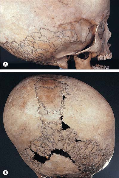
image

Fig. 26.7 Lateral (A) and posterior (B) views of the hydrocephalic skull of a 25-year-old male showing numerous sutural bones.

(By courtesy of the Museum of the Royal College of Surgeons of England. Photograph by Mr J Carr.)

Craniosynostosis

Sutural growth makes an important contribution to growth of the skull, especially during the first few years of life. The reasons for premature fusion of sutures (synostosis) are varied, but many occur as a result of small brain size or failure of the development of dural bands between the sutures. Premature fusion may occur in one or more of the cranial sutures: when the sutures around the skull base are involved, severe limitation of facial bone growth will occur. Metabolic disorders such as rickets and familial hypophosphatasia can also result in synostosis. Raised intracranial pressure with or without hydrocephalus, visual deterioration and mental retardation may result. Scaphocephaly (sagittal craniosynostosis) is the commonest and leads to lengthening of the vault in an anteroposterior direction. It also occurs in conjunction with other sutures, e.g. Crouzon’s syndrome. Coronal synostosis, either unilateral (plagiocephaly) or bilateral (brachycephaly/oxycephaly) is the next most frequently seen and results in reduced anteroposterior development with marked supraorbital recession. When it is unilateral the face develops asymmetrically and is rotated away from the side with premature fusion. Metopic craniosynostosis (trigonocephaly) and pansynostosis (turricephaly, where both the coronal and sphenofrontal sutures are involved) are much less common.

Treatment of these premature sutural fusions is critical to prevent abnormalities of skull growth and to relieve raised intracranial pressure. It is usually carried out between 3–6 months of age. Treatment consists of release of the sutures involved and the prevention of refusion, which is usually achieved by covering the bone edges with silastic sheeting after the radical removal of bone from either side of the suture line. Simultaneous expansion and reshaping of the skull is often required, particularly if diagnosis has been delayed. It is usually necessary to advance the supraorbital ridge and to straighten it if there is asymmetrical growth (as seen in plagiocephaly).

The craniofacial dysostosis syndromes such as Crouzon’s, Apert’s, Saethre–Chotzen, Pfeiffer’s and Carpenter’s, show varying degrees of calvarial synostoses which are usually accompanied by a significant lack of growth in the mid-face. Early release of the calvarial synostoses does not result in normal facial growth, and a midfacial osteotomy at the Le Fort III level is usually required later in life. When significant orbital hypertelorism develops, a transcranial bipartitioning procedure is needed in order to bring the two orbits together.

Skull deformities can also be derived deliberately by affecting sutural growth using binding and other pressure, as has been practiced in certain cultures of the world (Fig. 26.8).

image

Fig. 26.8 Skull binding. Egyptian Copts.

(By courtesy of John Langdon.)

Congenital anomalies affecting the skull

A large number of malformations and anomalies affect the bones and associated soft tissue structures of the skull and are the result of a localized error of morphogenesis during embryological development. Many are recognized patterns of malformation which are presumed to have the same aetiology. They do not arise as the result of just one isolated error in morphogenesis, and are described as syndromes.

Hemifacial microsomia (Goldenhar syndrome)

These patients present with unilateral microtia, macrostomia and varying degrees of failure of formation of the mandibular ramus and condyle. Vertebral anomalies and epibulbar dermoids are common. Most cases are sporadic, but familial instances have been described. The facies are strikingly asymmetric as a result of hypoplasia or displacement of the ear. The ear deformities vary from complete aplasia to minor distortions of the pinna, which is displaced anteriorly and inferiorly. Absence of the external auditory meatus is common, as are middle ear deficiencies, which result in conduction deafness. Supernumerary ear tags are present and occur anywhere along a line from the tragus to the angle of the mouth.

The maxilla, temporal and zygomatic bones show varying degrees of hypoplasia on the affected side. The mandible shows varying degrees of hypoplasia ranging from mild asymmetry to major failure of development of the ramus and condyle. The mastoid process also shows degrees of hypoplasia. Often there is frontal bossing. Ten per cent of cases are bilateral, but invariably one side is more severely affected.

There is concomitant hypoplasia of the main masticatory muscles on the affected side, and occasionally the muscles of facial expression are involved. In 10% of cases there is a lower motor neurone weakness of the facial nerve. Epibulbar dermoids occur and are usually located at the limbus or lower outer quadrant. A coloboma of the upper lid is present in most cases. Unilateral microphthalmia or anophthalmia can occur and is associated with mental retardation, however mental retardation is unusual. Vertebral anomalies are common and include occipitalization of the atlas, cuneiform vertebrae, and fusion of several adjacent vertebrae. A variety of cardiac anomalies have been described ranging from ventricular septal defects to Fallot’s tetralogy. Pulmonary hypoplasia and renal anomalies have also been recorded.

Mandibulofacial dysostosis (Treacher Collins syndrome)

Mandibulofacial dysostosis mainly involves structures derived from the first branchial arch, groove and pouch. It is inherited as an autosomal dominant trait with variable penetration. The facial appearance is characteristic: downward sloping palpebral fissures, depressed cheek prominences, deformed ears, mandibular hypoplasia and a large fishlike mouth. The hairline often shows a tongue-shaped extension toward the cheek.

Clinically the skull vault appears normal, but on imaging it is seen that the supraorbital ridges are poorly developed and despite normal sutural development there may be increased digital markings (copper beating) on the inner table. The zygomatic bones may be totally absent, or more frequently are grossly deficient in a symmetrical manner with failure of fusion of the zygomatic arches. The mastoid processes are not pneumatized and may be sclerotic. The paranasal sinuses are frequently abnormally small or even absent. The infraorbital rims are also poorly developed.

There may be various eye anomalies, including colobomas affecting the lateral third of the lower eyelid (75% of cases) and microphthalmia. The ears are usually severely deformed, have a crumpled appearance and are often wrongly positioned. In a third of patients the external auditory meatus is absent, and there may be ossicular defects which result in conduction deafness. Additional ear tags and blind fistulae may be present anywhere between the tragus and the angle of the mouth. The nasofrontal angle is usually obliterated and the bridge of the nose elevated. The alar cartilages are hypoplastic and there may be choanal atresia. The mandible is almost always hypoplastic, the angle is obtuse and the ramus deficient. The coronoid and condylar processes may also be hypoplastic. A cleft palate is present in 30% of cases and there is a high arched palate. Mental retardation is common.

Distraction osteogenesis

The pioneering Russian orthopaedic surgeon Ilizarov demonstrated that long bones could be lengthened by performing osteotomies in the axial plane and then slowly separating (distracting) the two ends of the bone. This stimulates release of bone morphogenetic proteins and new bone is formed between the sectioned bone ends: when the desired length has been achieved, the long bone is immobilized in its desired position. After a period of a few weeks the initial woven bone is replaced by normal mature bone and the resulting lengthened bone is stable and functional.

These techniques are now applied to the bones of the skull. Mandibular distraction is commonly used, particularly to treat asymmetry of the mandible, e.g. in hemifacial microsomia. By performing the osteotomies at the angles of the mandible, and using carefully adjusted distraction devices, the mandible can be lengthened in the vertical and anteroposterior planes. Distraction is usually performed at a rate of one millimetre per day; the jaw can be lengthened indefinitely until the distraction is stopped and the callus allowed to mature and unite.

Distraction techniques have now been applied to the middle third of the facial skeleton, using complicated frames bolted onto the skull vault. The technique is particularly useful for the management of the craniofacial syndromes such as Crouzon’s, where the facial bones which articulate with the sphenoid fail to develop normally. If the facial bones can be released at an early age, and the mobilized bones distracted, the facial profile can develop normally.

IDENTIFICATION FROM THE SKULL

There are many ways of identifying an individual: in physical and forensic anthropology the most important concern biological and personal identity. Biological identity pertains to those features that allow an individual to be classified in relation to features present in other individuals e.g. sex, age, race and stature, whereas personal identity establishes criteria that are characteristic and discriminatory for a particular individual e.g. DNA, fingerprints and dental information. The skull is a useful source of information for the establishment of both biological and personal identity. It is probably the most studied aspect of the skeleton. The foundation of this obsession has many historical roots, but fundamentally it has arisen from the importance that humans place in the concept that the skull is the repository of ‘self’, and that it is the means by which interpersonal communication is effected. Our face is our primary means of recognition and communication and therefore it plays a pivotal role in establishing and reconstructing the identity of an individual.

SEX

The determination of sex from a juvenile skull is notoriously unreliable. While sexual differences have been detected in measurements of the mandible, orbits, tooth size and pattern of dental eruption, they do not reach a level of discrimination that will allow accurate and reliable assessment. It is equally difficult to assign sex to the face of a child because the faces of prepubertal boys and girls are comparable: perceivable sexual dimorphism is not manifest until secondary sexual changes are completed. Growth in the female face ceases in advance of the male, and consequently female sex-related characteristics are more paedomorphic. The defining characteristics of sex in an adult skull are therefore male in orientation and reflect the effects of the increased mass of the muscles of mastication, which attach to the mandible, and the muscles associated with maintaining the erect head. It is reported that using the skull alone, sex can be predicted with over 80% accuracy in the adult. This is extremely encouraging because research has shown that the correct sex can usually be predicted from the adult living face with around 96% accuracy.

Generally speaking, the male skull is more robust and the female more gracile, although there are obvious genetic, and therefore racial, variations which must be considered when attempting to assign sex from a skull. The female forehead is generally higher, more vertical and more rounded than the male, and there is a clear retention of the frontal eminences in the female. The male mandible is more robust and larger than that of the female: it generally displays a greater height in the region of the symphysis menti, the chin is more square, the condyles are larger, the muscle attachments are more pronounced and the gonial angle is generally less than 125°. A male skull has thicker and more rounded orbital margins, pronounced supra-orbital ridges, and often a well-defined glabella that occupies the midline above the root of the nose. The temporal lines are more pronounced in the male and the supramastoid crest generally extends posterior to the external auditory meatus.

Other sites of muscle attachment on the skull reflect the biomechanical requirement to keep the more robust male head erect. They include the mastoid process for sternocleidomastoid, which is generally more robust in the male and more gracile in the female, and the nuchal lines, especially the external occipital protuberance for the attachment of the ligamentum nuchae. The cranial base in the male is generally more robust and the bone is thicker, which means that this area of the skull survives inhumation particularly well and is therefore of great value in sex identification from fragmentary remains.

AGE DETERMINATION

Age is a continuous variable: to establish chronological age from a skull requires that structures change with age at a relatively constant and predictable rate. The relationship between chronological age and skeletal maturity is closest in the juvenile years, and therefore greater accuracy is achieved in the prediction of age from the juvenile than from the adult skull.

The neonatal skull has been described above. Examples of additional features that allow reliable age determination throughout the subadult years are: development of the nasal spine (by year 3), completion of the hypoglossal canal (by year 4), formation of the foramen of Huschke (by year 5), ossification of the dorsum sellae (by year 5) and fusion of the different parts of the occipital bone (by year 7). The fontanelles are usually all closed by the middle of the second year: the posterolateral is the first to close in the first two months after birth, and the anterior fontanelle is the last to close around the middle of the second year. The mastoid process appears in the second year and the metopic suture between the two frontal bones will close by year 4. The spheno-occipital synchondrosis will fuse between 11 and 16 years in the female and 13 and 18 years in the male, while the vomer and the ethmoid will fuse between 20 and 30 years of age. The last part of the skull to show active age-related growth is the jugular growth plate, a small triangular area sited posterolateral to the jugular foramen in the occipitotemporal suture. Fusion here does not begin until 22 years of age, and bilateral fusion may not be completed before 34 years; in a small proportion of individuals, the plate may remain unfused beyond 50 years. Closure of the cranial sutures is age-related but the correlation is not strong and displays strong genetic variation. Thus, while it can be said that suture closure may begin in the early part of the third decade, and it is likely that many of the sutures will be obliterated with advancing age, it is not a reliable means of establishing the age of an individual.

The most accurate means for determining age from the skull (of both a living and a deceased individual) is by assessment of dental maturation. Tooth development can be studied throughout the entire juvenile age span (from the early embryo to the adolescent), and importantly dental age and chronological age have been shown to exhibit a stronger correlation than skeletal and chronological age. Further, the teeth tend to survive inhumation successfully and are remarkably resilient to fire and explosion, ensuring their value in forensic investigations.

The chronological pattern of dental maturation is well documented and is an extremely important tool for age evaluation. Tooth development can be separated into a number of well defined stages: deciduous mineralization (crown and root), deciduous emergence and maturation, deciduous root resorption, shedding of deciduous teeth, mineralization (crown and root) of permanent dentition, emergence and maturation of deciduous dentition and attrition of permanent crowns (see Ch. 30). These stages do not occur in a linear fashion: while some of the deciduous teeth are emerging, permanent teeth are already being formed. For example, mineralization of the deciduous central incisor commences around the 15th week post fertilization, and this is the first tooth to emerge within the first 5 months after birth. All deciduous teeth are in occlusion by around 3 years of age. The first deciduous teeth to be shed are generally the central and lateral incisors around 7 years of age, when the permanent incisors emerge. The last deciduous tooth to be shed is generally the second molar in the 10th year. The first permanent tooth to show mineralization is the first molar, which occurs around the time of birth (sometimes earlier and sometimes later): it is also the first permanent tooth to emerge at around 6 years of age, and it will reach occlusion by the end of the 7th year. The last permanent tooth to emerge is the third molar: the variability of this occurrence makes it of restricted value for age prediction.

While the patterns of mineralization, emergence and shedding are extremely useful, there are other methods available for determining age from the dentition. These include tooth length, cementum apposition, secondary dentine formation, incremental enamel lines, attrition rates, root translucency and dentine transparency.

RACE DETERMINATION

The determination of racial or genetic origin is particularly difficult to achieve although it is something that both physical and forensic anthropologists insist on trying to do. The traditional view of race is that it is ‘one of the major zoological subdivisions of mankind, regarded as having a common origin and exhibiting a relatively constant set of physical traits’ (Bamshad & Olson 2003). Classifying groups on this basis is rather restrictive and, in our migrant modern world, somewhat artificial. It is still useful to be able to attempt to assign a ‘most likely’ racial group, especially when dealing with unidentified forensic remains, but there are enormous areas of overlap between the characteristics, and within any racial group there is often a full spectrum of representation. Yet we persist in classification on the basis of visual characteristics, and the area of the body that is most often analysed in this way is the skull.

Early anthropologists classified man largely through geographical origins and recognized physical traits. The four traditional races of man were: (1) Caucasoid – geographically from Europe, North Africa, Middle East, Indian subcontinent and parts of Central Asia. Classically the Caucasoid skull has a rounded to long shape (dolicocephalic) with a narrow nasal aperture, moderately developed supraorbital ridging, a prominent nasal spine, a steeple shaped nasal root, little prognathism and a narrow interorbital distance. The forehead is steep, the chin is prominent, the palate is long and narrow, the cheek bones are not overly prominent and there is a tendency to maxillary protrusion or mandibular retrusion. (2) Negroid – geographically represented by sub-Saharan and West African groups. The Negroid skull is also long with a wide nasal aperture, strong alveolar prognathism, low nasal root, guttering of the nasal aperture and a wide interorbital distance. The forehead is rounder, the palate is wider and the teeth are larger. (3) Mongoloid – geographically represented by groups in East Asia, South East Asia, Central Asia, Americas, Greenland, Inuit, Polynesia, South Asia and Eastern Europe. The Mongoloid skull is generally described as round with a nasal aperture of medium width, well developed and high cheek bones, moderate prognathism, a tented nasal root, short nasal spine and shovel shaped incisors. The palate is foreshortened, the forehead is vertical, the nasal bridge is low and there is a tendency for a forward rotation of the mandible. (4) Australoid – geographically represented by Australian Aborigines, Maori, Pacific Islanders, Fijians and Papuans. This skull of this rather heterogeneous group is generally represented by a broad nasal aperture, well developed supraorbital ridging and glabella, and a wide palate with large teeth.

There is a current resurgence in research into racial determination which is largely centred on genetic markers: skeletal indicators now play a significantly reduced role in this task.

FACIAL APPROXIMATION (RECONSTRUCTION)

Once the biological identity of an individual has been established (i.e. sex, age, stature and race) an attempt to establish personal identity may be necessary, particularly in relation to a forensic investigation. Achieving a possible name for a victim is necessary before comparison of antemortem and postmortem data can be achieved and a positive identity established. One of the accepted ways to achieve this is to reconstruct the face from the skull, thereby producing a facial approximation that can be released to the community or the public at large in an attempt to identify the victim. Experts in this discipline utilize the many variations in the skull to reconstruct individual possible representations of the face from the material available. Biological variation will dictate the appropriate data used for tissue thickness that are applied to the skeletal scaffold.

There are fundamentally two approaches to facial reconstruction: (1) Computer reconstruction – the skull is usually scanned by a laser 3-D scanner and an ‘average’ virtual face is wrapped around the skeletal scaffold. This approach is largely automated and requires limited training and expertise. It is rapid to achieve and relatively inexpensive, but relies on a large data set to ensure that the ‘average’ face utilized is appropriate. (2) Modelled reconstruction – the skull is usually cast, and pegs are inserted into the cast at the appropriate tissue depth requirements. In the ‘American’ approach a skin of clay is then moulded over the pegs to approximate the face. In the ‘Manchester’ method each sequential muscle and soft tissue layer is built up around the pegs before a clay skin is moulded over the underlying structures. The modelling approach is clearly more dependent upon experience, takes longer to achieve and is more costly.

There is great debate over which approach is the most accurate and, as yet, no agreement has been reached. Most practitioners state that the process achieves an approximation of one of the potential faces for the deceased and does not purport to reconstruct the actual face. The rationale is to produce an image that will jog the memory of the public and provide some possible names that will allow comparison of antemortem and postmortem identity data.

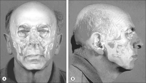
FACIAL SUPERIMPOSITION

Once a possible name has been derived it may be necessary to compare the skull with photographs of the suspected individual. In these circumstances an image of the skull is superimposed onto an image of the face of the missing person (Fig. 26.9). This relies on achieving a live capture image of the skull so that it can be rotated and manipulated into an identical position and to an identical size as the photograph. Features that do not change are lined up: a photograph that shows teeth is ideal because teeth can be lined up with the dentition on the skull. The image of the skull and photograph can then be faded in and out: if this is undertaken at speed, any discrepancies will show up on the image as distortion.

image

Fig. 26.9 Facial superimposition: a live capture image of the skull has been manipulated into the identical position and size of the face in the photograph.

(By courtesy of Dr Caroline Wilkinson and Professor Sue Black, Centre for Anatomy and Human Identification, College of Life Sciences, University of Dundee.)

REFERENCES

Bamshad M, Olson S. Does race exist? Sci Am. 2003;289:78-85.

Berkovitz BKB, Moxham BJ. Color Atlas of the Skull. London: Mosby-Wolfe, 1994.

Berry AC. Factors affecting the incidence of non-metrical skeletal variants. J Anat. 1975;120:519-535.

Berry AC, Berry RJ. Epigenetic variation in the human cranium. J Anat. 1967;101:361-380.

Bruce V, Burton AM, Hanna E, Healey P, Mason O, Coombes A, Fright R, Linney A. Sex discrimination: how do we tell the difference between male and female faces? Perception. 1993;22:131-152.

Gill GW, Rhine S. Skeletal Attribution of Race: Methods for Forensic Anthropology. New Mexico: Maxwell Museum of Anthropology, 2004.

Ilizarov GA, Ledyaev VI. The replacement of long tubular bone defects by lengthening distraction osteotomy of one of the fragments. Clin Orthopaed Relat Res. 1992;280:7-10.

Lahr MM. The Evolution of Modern Human Diversity: A Study of Cranial Variation. Cambridge: Cambridge University Press, 1996.

Lele S, Richtsmeier JT. Euclidean distance matrix analysis: a coordinate-free approach for comparing biological shapes using landmark data. Am J Phys Anthropol. 1991;86:415-427.

Lieberman DE, McCarthy RC. The ontogeny of cranial base angulation in humans and chimpanzees and its implications for reconstructing pharyngeal dimensions. J Hum Evol. 1999;36:487-517.

Lieberman DE, Pearson OM, Mowbray KM. Basicranial influence on overall cranial shape. J Hum Evol. 2000;38:291-315.

Liversidge H. The dentition. In: Scheuer L, Black S. The Juvenile Skeleton. London: Elsevier, 2004.

Maat GJR, Mastwijk RW. Ossification status of the jugular growth plate. An aid for age at death determination. Int J Osteoarch. 1995;5:163-168.

Scheuer L, Black S. Developmental Juvenile Osteology. London: Academic Press, 2000.

Sperber GH. Craniofacial Development, 4th edn. Ontario: BC Decker, 2001.

Vidarsdottir US, O’Higgins P, Stringer C. A geometric morphometric study of regional differences in the ontogeny of the modern human facial skeleton. J Anat. 2002;201:211-229.

Whittaker DK, MacDonald DG. A Colour Atlas of Forensic Dentistry. London: Wolfe, 1989.

Wilkinson C. Forensic Facial Reconstruction. Cambridge: Cambridge University Press, 2004.