Examination of the Cervical Spine

Published on 23/05/2015 by admin

Filed under Physical Medicine and Rehabilitation

Last modified 22/04/2025

Print this page

rate 1 star rate 2 star rate 3 star rate 4 star rate 5 star
Your rating: none, Average: 1 (1 votes)

This article have been viewed 1969 times

CHAPTER 48 Examination of the Cervical Spine

SCOPE AND LIMITATIONS

Perhaps there is no better example of the ‘art’ of medicine than in a review of the various physical examination techniques used in clinical diagnosis. More than any other organ system, the musculoskeletal system is the purview of a diverse group of medical specialists and subspecialists who care for persons with spinal and spine-related pain.

In the field of medicine alone, the responsibility for physical diagnosis is undertaken by many disciplines including neurology, neurosurgery, occupational medicine, orthopedic surgery, physical medicine and rehabilitation, sports medicine, etc. Beyond allopathic medicine there are osteopathic physicians, chiropractors, and physical therapists among others who examine the spine. While there are some elements common to all groups, each craft presents its own unique nomenclature and terms of reference and stakes a claim to the role of gatekeeper for musculoskeletal pathology.

This diversity leads to a heterogeneous approach to the physical diagnosis of spinal pain. Examination techniques vary widely both across and within disciplines. The algorithm used to make a diagnosis by examination is derived primarily from a particular practitioner’s own experience and education and promulgated via authoritative opinion. These techniques are passed down from generation to generation as one clinician incorporates it as part of his own routine, or copies it from other sources. Not surprisingly, then, the evidence base supporting the diagnostic utility of any particular clinical maneuver varies widely. The cervical spinal examination is no exception to this dilemma.

For example, cervical spinal range of motion is perhaps the most commonly performed assessment, common to most if not all practitioners, in the assessment of the patient presenting with neck pain. Notwithstanding the popularity of this examination component, there is a wide range of intra-subject variability depending upon the time of day it is measured. In addition, within a given individual, motion in a particular plane differs according to where the starting point of the motion is measured. Accordingly, establishing normal ranges for spinal motion is challenging.1,2

This has led to the development of various devices and technologies that can measure spinal range of motion more precisely.37 While certain devices have shown improved reliability as compared to manual physical examination techniques, the use of such devices is not commonplace in clinical practice, leaving the practitioner to rely upon manual techniques to guide clinical decision-making.

Compounding these reliability issues is the notion that impaired spinal range of motion correlates with impaired spinal function. In spite of evidence demonstrating a lack of correlation between loss of range of spinal motion and spinal dysfunction,8,9 the use of range of motion as a diagnostic tool remains well engrained in medical culture. Accordingly, range of motion models are regularly used as the basis upon which spinal impairment is rated.

Even more problematic is the attempt to establish whether a particular spinal motion segment, within the multiarticulated spine, has an abnormally restricted or lax range of motion. Despite numerous descriptions of techniques for the palpation of spinal structures, the inter-rater reliability and validity for motion palpation are both lacking in literature support.1013

Consequently, there is no evidence-based normative database that represents a gold standard against which any single range of motion data set can be compared to determine whether it is ‘normal’ or not. Moreover, there is no hard and fast rule to determine whether a particular patient’s range of motion values are likely to be clinically relevant, even if they do fall outside the range of ‘normal.’ This example will be discussed in more detail later in this chapter.

The quest for sources of anatomic pain generators has led examiners to teach provocative maneuvers that are designed to provoke local or referred pain.14 As a result, many syndromic diagnoses have included reproduction of a patient’s habitual pain as an essential diagnostic element. Nevertheless, the combination of the subjectivity of the pain response to palpation and the inherent biases of both examiner and examinee, have limited the predictive value of this aspect of the examination.15,16

WHY EXAMINE THE SPINE AT ALL?

The goal of the spinal physical examination is to provide the clinician with clues that support or refute the possible origin of spinal pain and dysfunction. This information is used in conjunction with the history to direct diagnostic testing and treatment to form a clinical report card. When the results of each of these components are concordant, the clinician can be more confident about the certainty of the clinical diagnosis.

In addition, cervical spinal pain can be broadly classified into one of three descriptive diagnostic categories: ominous, neurogenic, and non-specific.

Ominous spinal pain implies a diagnosis with potentially severe adverse health consequences including spinal tumors, infections, fractures, etc. Physical examination often detects focal abnormalities, but is generally insufficient to render a tissue-specific clinical diagnosis.

Neurogenic spinal pain implies a referral from the neural elements including the spinal nerves or spinal cord. In general, these presentations are associated with objective findings more readily and reliably demonstrable on physical examination, including altered spinal reflexes, diminished sensation, weakness on manual muscle testing, and possibly adverse dural tension.

Finally, non-specific spinal pain implies pain of non-ominous and non-neurogenic origin. In general, this diagnostic category is consistent with a more benign prognosis than the other two categories. The anatomic origin, if detectable by alternate means, may be discrete or diffuse. The spinal examination is often non-specific and exhibits wide variability among patients in this descriptive category.

Using this diagnostic frame of reference, the anatomic sources in both ominous and neurogenic spinal pain conditions are readily diagnosed using the clinical report card. In these two categories (neurogenic spinal pain in particular), the physical examination is a very powerful tool in the development of management decisions and in the determination of clinical progress.

However, the same cannot be said for non-specific pain disorders. Here, the physical examination is often insufficiently specific to render an anatomic diagnosis. The reader is referred elsewhere in this text for the specific techniques used to diagnose anatomic pain generators (disc, zygapophyseal joints, etc.).

Nevertheless, there is a role for physical examination, even in this category. Even in the absence of a discrete anatomic diagnosis, function can be assessed either quantitatively or qualitatively by physical examination. In this way the clinician has a measuring stick to use to gauge clinical progress, including response to treatment.

Therefore, the utility of the physical examination of the cervical spine is to provide the clinician with an overall impression of the patient. While individual physical examination maneuvers may be lacking in either sensitivity or specificity, the combination of clinical tests used, in the context of the clinical report card, is meaningful in providing the clinician with evidence to generate clinical hypotheses and working diagnosis upon which to base initial treatments.

Spinal examination is a dynamic process where findings may change over time, either by chance, as a response to intervention (sometimes even despite intervention) or as a reflection of the condition’s natural history. Repeated examinations over time, in the context of the clinical report card, may prove to be the best way to compensate for the deficiencies inherent in the examination process.

In the end, the clinician using physical examination techniques still has to rely somewhat on the art of medicine. The following physical examination protocol is but one of many approaches to clinical examination, and is a synthesis of techniques derived from many sources in the context of the authors’ collective clinical experiences.

PHYSICAL EXAMINATION

Since there is often an overlap in the symptoms associated with upper limb conditions and neck conditions referring to the upper limb, evaluation of the cervical spine should not only include a detailed neurological evaluation of the upper limbs, but also an examination of the upper limb joints that could potentially be the source of the presenting symptoms. Symptoms referable to the shoulder often mirror presenting complaints frequently seen in the cervical spine. The examination of the upper limb joints is outside the scope of the present chapter, so the focus will be on the remaining elements of the cervical spinal evaluation. For further information on the shoulder, please refer to Chapter 49.

The examination of the cervical spine is tailored to the clinical impression presented by the patient’s history. The presence of local or referred pain, motion restriction, guarding, posturing, etc. will influence the emphasis placed by the examiner on various examination techniques.

The various components of the examination should include inspection of posture and musculoskeletal symmetry, evaluation of range of motion in both sitting and supine positions, palpation of the bony and soft tissue structures, and special neural tests. In addition, a detailed neurological examination of the upper limbs must be performed.

Inspection

Inspection should be performed with the patient both standing and sitting. This provides the examiner with the opportunity to evaluate the patient’s habitual posture to look for signs of asymmetry and potential sources of soft tissue overload.

The skin should be inspected for scars, subcutaneous masses, and for lesions that may be consistent with herpes zoster (shingles). Since the thoracic spine is the base of support for the cervical spine, it too should be assessed for scoliosis, kyphosis, and scapular winging. Lastly, station and gait should be screened, specifically looking for signs of spasticity or altered motor tone. Any gait or balance abnormalities may direct further investigation of the central nervous system.

Poor posture, in particular, poor sitting posture, is considered to be a significant contributor in back and neck pain.17 When sitting in a slumped or unsupported position, there is a loss of lumbar lordosis, which results in a compensatory thoracic kyphosis. Consequently, there is a resultant compensatory forward inclination of the head with flexion of the lower cervical spine and hyperextension of the upper cervical pole to level the head horizontally.

Postural imbalances such as these may impart significant mechanical stress to the cervical spine during sitting. Therefore, it is important to evaluate the patient in both the seated and standing position for compensatory changes in cervical lordosis based on the posture of the lumbar spine.

The head-forward posture is typically associated with ‘rounded shoulders,’ with protracted scapulae and internally rotated shoulders. This is often associated with scapulothoracic muscle imbalance with weakness of the mid and lower trapezius, rhomboids, and serratus anterior, and tightness of the anterior shoulder girdle muscles (pectoralis and latissimus dorsi), upper trapezius, and levator scapulae.

Since many of these muscles have spinal attachments or are innervated by cervical nerves, abnormal scapulothoracic muscle mechanics can alter the mechanics of cervical paraspinal muscles. Over time, substitution patterns develop to compensate for these pathomechanical features, resulting in a vicious cycle of tissue overload often present in local or referred pain.

Diminished cervical lordosis is often the result of paravertebral muscle guarding. This is often a ‘reflex’ response to a variety of possible conditions including muscle overload or a structural abnormality. Altered cervical lordosis should be investigated with a detailed clinical examination and diagnostic imaging, if indicated.

Changes in cervical lordosis may also result from compensation for thoracic or lumbar spine posture. Thoracic kyphosis may arise from weak posterior and tight anterior shoulder and chest wall musculature. However, underlying congenital or developmental structural abnormalities must be excluded. These include Scheuermann’s kyphosis, a hemivertebrae, and scoliosis. Acquired thoracic kyphosis may be a result of fractures, both pathologic and traumatic, as well as infections such as tuberculosis.

In addition, the examiner should look for signs of muscle atrophy and side-to-side asymmetry that may provide clues of the underlying clinical diagnosis. Normal asymmetry such as depressed dominant shoulder needs to be recognized, and, unless extreme in nature, need not raise concern.

Muscle wasting is generally not seen in the neck itself, but rather in the upper limbs, periscapular region, or shoulder girdle. In particular, neurogenic cervical conditions may reveal a myotomal pattern of wasting that provides valuable clues as to the source of anatomic dysfunction.

Muscle atrophy, however, is not always indicative of a neurogenic source. Pain is a powerful inhibitor of function, and therefore disuse atrophy should also be considered when muscle asymmetry is noted.

Range of motion

The spine specialist must be able to assess active and passive ranges of motion (ROM) of the cervical spine and understand the potential clinical implications of ‘abnormal’ findings. With the patient seated, the examiner should stand behind the patient in order to assess spinal range of motion. From behind, the examiner can best control motion and optimally view the response of the trunk muscles and adjacent spinal regions to motion.

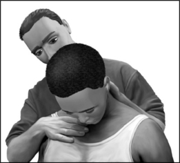
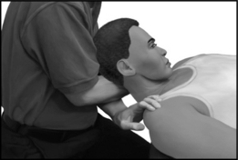
The patient should be given simple commands such as, ‘Touch your chin to your chest,’ ‘Look up to the sky,’ etc. By placing one hand on the head or chest, the examiner can also provide proprioceptive cues to guide the patient through his active motion (Fig. 48.1). In this way the unrehearsed patient is easily able to perform the requested active range of motion with confidence, and without the need for the examiner to demonstrate the required maneuver.

Notwithstanding the controversies regarding quantitative spinal range of motion mentioned previously, there is a wide range of published values that are considered ‘normal’ for cervical range of motion. The valid quantification of cervical range of motion demands that the examiner be able to isolate cervical motion from that occurring below. In addition, since the aggregate motion observed is as a result of the sum of spinal segmental motion, it is difficult to determine, especially by manual techniques, whether a particular segment or segments are the source of the limitation.

With this in mind, certain parameters can be applied to the evaluation of cervical range of motion to depict the normal population.

Cervical flexion is limited by contact of the chin upon the chest. Generally, up to two finger-widths between the chin and chest with a closed mouth is considered full. This may also be envisioned by the arc formed by a point at the vertex with a maximum range of 90°. Extension is limited by the approximation of the posterior zygapophyseal joints. A point on the vertex may pass approximately 70° in extension.

Guidelines for cervical side-bending are more difficult to establish since the range of motion is dependent of position (sitting versus supine), neck muscle bulk and tone. All of these factors may affect the particular observed range of motion in a particular individual. In sitting, the passage of a point on the vertex through an arc of approximately 20–45° in either direction is often considered as normal. This is often equivalent to approximately 2–4 finger-breadths between the ear and the shoulder tip.

Lateral rotation may also be subject to the variation affecting side-bending. In sitting, the arc passed by a point of the subject’s nose is generally 70–90° in either direction.

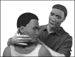
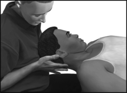
At the end of the patient’s active range, gentle over-pressure can be applied to determine the end-feel and the extent of the passive range (Fig. 48.2). This overpressure also allows the examiner to differentiate between the anatomical versus physiological end range. Palpation will be discussed below.

In general, passive ROM (PROM) is greater than the active (AROM), and PROM is increased in the supine versus sitting position. This positional ROM increase is due to the relaxation of resting muscular tone that normally holds the head against gravity. A primary example is the relative ease with which the patient is able to approximate his ear to his shoulder with side-bending when supine as compared to sitting (Fig. 48.3).

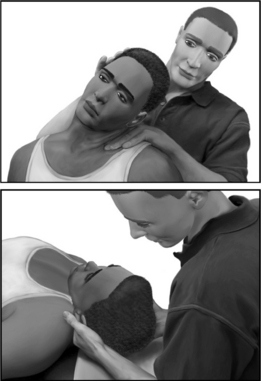
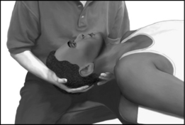
In the supine examination, cervical extension may be evaluated with the head and cervical spine off the end of the table. Cervical extension can then be performed with passive assistance with one of the examiner’s hands guiding motion and the other blocking motion at the lower, mid and upper cervical spine (Fig. 48.4). Evaluation of flexion is then assessed by resting the examinee’s head against the examiner’s crossed arms and trunk (Fig. 48.5).

Cervical range of motion is the summation of movements of the entire cervical spine. Nevertheless, cervical spinal ROM is often reported as ‘flexion is 90 degrees’ leaving the reader the impression that the observed motion occurred at ‘the neck joint.’ To some extent overall ROM also takes into account disc flexibility, inclination of the articular processes, and laxity of the supporting ligaments.

With the aforementioned wide variability in cervical ROM, and the many associated factors that influence an examiner’s ability to validly measure range of motion, it is perhaps even more important to assess and document range of motion qualitatively. In this regard, it is essential to understand that the different regions of cervical spine serve different functions.

For example, the upper cervical segments, C0–C1–C2 function quite differently than the lower segments C3–7. The primary observed upper cervical spine motions are nodding (C0–C1) and rotation (C1–2). The latter motion contributes approximately 50% of rotation with the lower cervical segments contributing the balance.

In general, the upper cervical segments function to orient the head on the neck, while the lower segments orient the neck on the trunk. For example, consider the execution of two qualitatively different motions: right rotation and right side-bending. In both cases, the motion at C3–7 is qualitatively similar with ipsilateral coupled side-bend/rotation occurring at each segment. However, the upper cervical motion differs markedly between the two. In the first instance, there is ipsilateral C1–2 rotation, whereas contralateral C1–2 rotation occurs in the latter instance.

It is frequently helpful, when AROM restriction is suspected, to assess the motion separately, thus determining to what degree the blockage is due to the upper and lower segments, respectively.

For example, when observing sagittal plane motion (flexion–extension), it is helpful to note whether the subject is primarily nodding (indicating lower c-spine restriction) or chin thrusting (indicating upper c-spine restriction). Rotational restrictions can be evaluated by comparing the degree of rotation achievable with the cervical spine in neutral flexion–extension as compared to full flexion. In the latter position, almost all the rotation observed is occurring at the C1–2 joint. By relatively limiting lower cervical spine rotation through flexion, the examiner can estimate the degree to which the restriction is arising from the upper or the lower cervical segments.

The examiner should be mindful that both age and possibly gender affect cervical ROM. Chen and colleagues performed a meta-analysis of 13 reports looking at these correlations. The majority of these studies found that ROM generally decreases with age. Most found that women had greater range of motion than men, although the gender differences were not statistically significant.1

As discussed previously, spine ROM is a major determinant of impairment in many disability rating schedules. A study by Lowery et al. suggests that impairment ratings based solely on decreased ROM produces less than accurate results.8 In this study, 81 healthy subjects were evaluated using a double inclinometer method. They concluded the current method of impairment determination, based on spinal motion criteria, overestimates impairment by up to 38%. Accordingly, impairment guides have moved away from a range of motion model of assessment of spinal impairment to one based on diagnosis-related estimates.

Range of motion evaluation, however, should not be dismissed entirely in correlating pathology. Dall’Alba et al. demonstrated that ROM alone allowed blinded evaluators the ability to discriminate between asymptomatic persons and those with persistent whiplash-associated disorders in 90% of their subjects, with a sensitivity of 86.2% and specificity of 95.3%.18 Perhaps other methods for impairment evaluation should be developed and incorporated that are more specific for individuals with true functional impairment and that account for age-related differences in spinal motion.

Palpation

In palpating the soft tissues, the examiner should be cognizant of the three-dimensional anatomy of the underlying tissues. In this way the examiner may better appreciate superficial from deep structures and may direct palpation intentionally either parallel or tangential to the structure’s orientation.

While both tenderness to palpation and tissue texture are frequently used tests, they are both lacking in reliability and validity. Nevertheless, these techniques may be useful clinical adjuncts when interpreted in the context of the examination as a whole as part of the clinical report card. Focal tenderness or perceived alteration of tissue texture may be more meaningfully interpreted if accompanied by other focal signs of anatomic dysfunction (e.g. myotomal weakness, crepitus, etc.).

Where possible, bony palpation should be performed on each spinal segment. While this may be non-specific as to anatomic cause (and occasionally falsely positive) it may provide the examiner with clues as to the region of the cervical spine (e.g. upper, mid or lower) that is dysfunctional.

In general, bony or segmental palpation of the spine is best performed with the patient supine since the postural muscles are better relaxed in this position, providing better access to bony landmarks. In this position, longitudinal friction can be applied to the posterior articular pillars at almost each cervical segment. Pressure can be applied along the groove located between the paraspinal muscles and the trapezius. This can occasionally be facilitated by slightly rotating the examinee’s neck away from the side of palpation (Fig. 48.6).

The examiner should also assess the cervical soft tissues including skin texture and muscle tone about the neck. Although non-specific, alterations is texture and tone are often a result of an underlying condition.

There must be a familiarity with bony landmarks, including the external occipital protuberance, the spinous processes of each cervical vertebrae, and regions overlying the Z-joints. The occipital protuberance is found at the posterior skull in the midline. The spinous processes of C2, C6, and C7 are most obvious. The first prominence palpated caudal to the occiput is C2. The next most obvious spinous processes distally are C6 and C7.

Vertebra prominens, also known as the nuchal tubercle, is the most prominent spinous process in the cervicothoracic region. It is the spinous process of the seventh cervical vertebra in 70% of the cases, sixth in 20%, and the first thoracic vertebra in 10%. When the examiner is uncertain which vertebra is the most prominent, flexing and extending the patient’s neck can typically discriminate between the C6 and C7 prominences. Whereas C7 remains fairly stationary with this maneuver, C6 will translate anteriorly on extension and then posteriorly on flexion.

In the lateral aspect of the cervical spine, the transverse processes can be palpated. The C1 transverse process is located slightly inferior and anterior to the mastoid process. Moving caudally, the subsequent transverse processes follow the lordotic path of the cervical vertebrae under the sternocleidomastoid muscle. Lymph nodes, carotid pulse, and parotid glands are also palpated in the lateral aspect of the cervical spine. Lymph nodes are readily palpable if swollen. They are located along the anterior border of the sternocleidomastoid muscles. The carotid pulse should be examined at the midportion of the lateral neck to assess whether the pulse is normal and symmetric bilaterally. The parotid gland lies superior to the angle of the mandible and may feel boggy if swollen.

Other anterior structures important to identify are the trachea, thyroid cartilage, and thyroid gland. The trachea must be in midline, and any deviation is cause for concern. The thyroid cartilage lies anterior to the C4 and C5 vertebrae. Superficial to the cartilage is the thyroid gland. The gland should be palpated for tenderness or enlargement, which also mandates further investigation.

Special neural tests

After completing an evaluation of motion in the cardinal planes, the examiner can evaluate the effect of complex motions by combining various degrees of sagittal, frontal, and transverse planar motion. In 1944, Spurling and Scoville, first described the ‘neck compression test’ stating that it is almost pathognomonic of a cervical intraspinal lesion.19 They performed the test by side-bending the neck towards the painful side and applying axial pressure on the top of the head to reproduce the patient’s characteristic pain and radicular symptoms. They did not indicate how long this position was to be held or with how much axial loading. Side-bending the neck away from the lesion usually provides relief.

Tong et al. performed a cross-sectional study to determine the sensitivity and specificity of Spurling’s maneuver for cervical radiculopathy.20 The test was performed as in Spurling’s original description, and was considered positive if it reproduced pain or paresthesias that began in the shoulder and radiated distally to the elbow. They concluded that Spurling’s maneuver is not very sensitive (30%) but is specific (93%) for cervical radiculopathy diagnosed by electromyography. Therefore, it appears not as useful as a screening test, but more clinically useful in confirming a cervical radiculopathy.

Bradley and colleagues suggested a three-stage protocol to the test.21 If symptoms are reproduced, subsequent stages need not be performed. The first stage consists of axial compression with the cervical spine in neutral position. The second stage involves compression with the neck in extension, and the third stage places the neck in extension and lateral rotation to the unaffected side first and then to the symptomatic side, both with axial compression. Radicular pain into the arm is considered a positive test and indicates irritability of a nerve root. The dermatomal distribution of the symptoms suggests involvement of a specific nerve root. The neck positions of each stage of the test progressively narrows the intervertebral foramen, which may be seen in conjunction with uncovertebral and zygapophyseal hypertrophy and disc herniations.

A similar test, called the maximal compression test, incorporates side-bending and rotation towards the symptomatic side, along with extension and axial compression (Fig. 48.7). This combination of positions causes maximal neuroforaminal compression and is positive if pain radiates into the arm. Most clinicians perform the maximal compression test but erroneously refer to it as the Spurling’s maneuver, when in fact it is a ‘modified’ Spurling’s compression test.

Another provocative maneuver used in patients with cervical radiculopathy is the application of lateral pressure against the vertebrae. This maneuver, also known as the doorbell sign, is considered positive when pressure to the anterolateral cervical spine reproduces referral of pain into the ipsilateral upper limb (Fig. 48.8). A positive sign is considered to be consistent with spinal nerve irritability or other unspecified spinal segmental dysfunction.

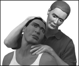
The distraction test is used to diagnoses spinal nerve irritation by alleviating radicular symptoms. The examiner places one hand on the patient’s chin and the other hand around the occiput and slowly applies upward traction to the patient’s head. The test is positive if the patient reports a decrease in radicular symptoms. The shoulder abduction relief sign, also called Bakody’s sign, is observed when the patient abducts the arm and places the hand or forearm on the top of the head. This arm placement, which may be performed subconsciously by the patient for symptomatic relief, is also indicative of presence of cervical nerve root traction that is being alleviated as a result of this maneuver.

Thoracic outlet syndrome (TOS) should be tested as part of the comprehensive cervical examination. Classic, or neurogenic, TOS occurs when the lower trunk of the brachial plexus becomes compressed within the scalene triangle by a cervical rib, fibrous band of tissue, or from an elongated C7 transverse process. In TOS, neck pain is rarely a prominent symptom.22 The patient may complain of deep, aching pain in the ulnar side of the forearm, occasionally involving the hand. Subjective hand weakness and clumsiness may be present. Muscle wasting may be seen in the thenar eminence, particularly involving the abductor pollicis brevis. This is also referred to as the Gilliatt-Sumner hand.23 There is sometimes decreased sensation on the ulnar side of the hand. Roos describes several methods to reproduce symptoms of classic TOS.24 The more well known of the tests, also referred to as Roos’ test, involves a 3-minute elevated arm stress test. The shoulder is abducted to 90° and externally rotated, and the elbow is flexed at 90°. Additional stress is incorporated by asking the patient to open and close the hands for the duration of the test. The test is considered positive if typical upper limb symptoms are reproduced. Another less-known TOS test was also described by Roos. Percussion or light pressure held for 30 seconds over the supraclavicular fossa may reproduce pain, thereby distinguishing these symptoms from those emanating from the cervical spine.

Compression of the subclavian vessels has been termed vascular TOS. Adson’s maneuver evaluates for obliteration of the radial pulse when the arm is placed at the side in a slightly extended and externally rotated position with the head laterally rotated to the contralateral side. However, the false-positive rate is high.25 Furthermore, Wright’s test is positive when the radial pulse is diminished when the arm is placed in a horizontally abducted position with the head turned to either side. This indicates only positional compression of the subclavian artery, and is not necessarily indicative of vascular TOS. In Wright’s series of 150 asymptomatic normal subjects, 92.6% had reduction of the radial pulse in this position, calling into question the clinical utility of this test.26

Upper limb neural tension tests, also known as dural tension tests, place stress on the neural elements of the upper limb. They are analogous to the straight leg raise test performed in the lower extremities for reproduction of lumbar radicular pain. The upper limb neural tension tests may be performed in ulnar, median, and radial bias positions. In all testing positions, it is important to keep the ipsilateral shoulder depressed.

For upper limb neural tension testing with median nerve bias, the shoulder is first abducted to 90°. The arm is externally rotated, the forearm is supinated and the elbow, wrist, and fingers are extended.

For ulnar nerve bias testing, the arm is abducted to 90°, the elbow is flexed to 90°, the forearm is maximally pronated, the wrist is extended and radially deviated, and the fingers are extended.

The radial nerve bias dural tension is performed with the shoulder abducted to approximately 10°, the elbow extended, the forearm fully pronated, the wrist flexed with ulnar deviation, and the fingers held in flexion.

With all of the above neural tension tests, the neck may be positioned in contralateral side-bending to further increase tension on the nerves analogous to slumping or neck flexion in the straight leg raise. Upper limb neural tension tests are considered positive only if the patient’s typical symptoms are reproduced, and if there is a side-to-side difference, assuming the contralateral side is asymptomatic.

Coppieters et al. tested the reliability of neural provocation tests in both the laboratory and clinical setting.27 Their study, focusing on median bias dural tension, demonstrated that pain provocation during neurodynamic testing is a stable phenomenon with good inter-tester and intra-tester reliability with intra-class correlation coefficient of 0.98.

The neurologic examination

Neurogenic neck pain should be suspected when a patient presents with a complaint of neck and upper limb pain. Shoulder girdle pain, while often interpreted to be of thoracic origin, is most commonly a sign of either intrinsic shoulder pathology or referred pain from the cervical spine.

The regional neurological examination should screen for special neural tests, altered reflexes (either augmented or diminished), weakness in a myotomal pattern, and sensory loss in a dermatomal pattern.

Key reflexes to be evaluated include:

Key muscle groups to be evaluated include:

Key sensory areas to be evaluated correspond to regions of minimal dermatomal overlap including:

The regional neurological evaluation may be performed in either sitting or supine position, provided that the patient is relaxed. Once in position, it is desirable not to move the patient’s limbs too much in order to avoid altering tone or guarding.

The biceps reflex is performed with the forearm in neutral pronation/supination. Firm pressure is applied with the thumb over the biceps tendon at the elbow. The reflex hammer is applied directly to the examiner’s thumb, and the muscle contraction is either observed or palpated. If the reflex is not evoked, the forearm may be supinated to increase tension and facilitate the reflex.

The brachioradialis reflex is applied directly to the tendon at the distal forearm. Augmentation can be induced with slight stretch applied as ulnar wrist deviation.

The pronator reflex is performed with the patient’s wrist in supination. The examiner’s thumb is firmly applied to the distal radius. The reflex hammer strikes the thumb in an effort to induce further supination, thus stretching the pronator. The response is forearm pronation or elbow flexion. The reflex may be augmented by applying more supination or slightly decreased elbow flexion.

The triceps reflex is performed with patient’s forearm supported comfortably by the examiner. The reflex may be augmented by slightly increasing the elbow flexion, or by asking the patient to very gently push away to activate the triceps.

Manual muscle testing requires that the examiner be in a position where mechanical advantage is obtained. In this way a mismatch between a small examiner and large patient is minimized and the interpretation of weakness is more sensitive.

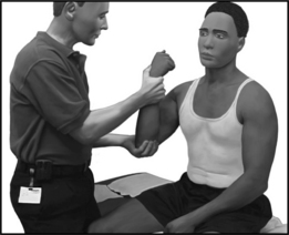
Simple commands should be given to the patient in order to evoke the desired patient response. Generally, the examiner should position the limb in the position to be tested (Fig. 48.9). The patient should be told, ‘Don’t let me move it.’ Prior to resisting, the examiner can apply a few brief jerks in the direction of the resistance in order to give proprioceptive cues to guide the patient through their resisted motion.

Manual muscle testing should always be performed bilaterally, one muscle at a time. This allows for a more valid side-to-side comparison. The unaffected side should be tested first in order to maximize the learning effect prior to assessing the side where weakness is suspected. This helps ensure better effort on the affected side, since the process has been learned by the patient.

Sensory function may be best screened with a single modality such as pinprick. The key sensory areas noted previously from C4 to T2 should be screened bilaterally one level at a time. The patient should be asked to report any side-to-side differences. A positive response may include hypoesthesia or hyperesthesia.

Tests for upper motor neuron (UMN) signs can direct the physician to consider abnormalities occurring at the level of the spinal cord or above. A widely practiced UMN response is the Babinski reflex, or plantar response. This reflex is elicited by stroking the undersurface of the foot from the heel to the great toe. Extension and fanning of the toes is considered a positive, abnormal response. The Hoffman reflex is the UMN sign analog in the hand. It is elicited by quick flexion of the distal interphalangeal joint of the long finger. Abnormal reflex is noted if there is immediate interphalangeal joint flexion of the ipsilateral thumb. This reflex can be indicative of an upper motor neuron lesion. However, the reflex is normal in some, especially young female individuals, and may be seen in the overly anxious patient. Lhermitte’s sign is elicited by briskly flexing the patient’s neck. Shock-like sensations radiating down the patient’s thoracic spine often indicate spinal cord pathology, although this can also occur in some patients with herniated cervical discs.

Other findings on inspection warranting further work-up include muscle fasciculations, which may be benign in nature or can indicate underlying UMN pathology. Gait dysfunction, such as spastic or circumducted gait, raises clinical suspicion for cervical myelopathy.

References

1 Chen J, Solinger AB, Poncet JF, et al. Meta-analysis of normative cervical motion. Spine. 1999;24(15):1571-1578.

2 Chiu TT, Sing KL. Evaluation of cervical range of motion and isometric neck muscle strength: reliability and validity [see comment]. Clin Rehabil. 2002;16(8):851-858.

3 Dvir Z, Prushansky T. Reproducibility and instrument validity of a new ultrasonography-based system for measuring cervical spine kinematics. Clin Biomech. 2000;15(9):658-664.

4 Lantz CA, Chen J, Buch D. Clinical validity and stability of active and passive cervical range of motion with regard to total and unilateral uniplanar motion. Spine. 1999;24(11):1082-1089.

5 Schaufele MK, Boden SD. Physical function measurements in neck pain. Phys Med Rehabil Clin N Am. 2003;14(3):569-588.

6 Tousignant M, de Bellefeuille L, O’Donoughue S, et al. Criterion validity of the cervical range of motion (CROM) goniometer for cervical flexion and extension. Spine. 2000;25(3):324-330.

7 Tousignant M, Duclos E, Lafleche S, et al. Validity study for the cervical range of motion device used for lateral flexion in patients with neck pain. Spine. 2002;27(8):812-817.

8 Lowery WDJr, Horn TJ, Boden SD, et al. Impairment evaluation based on spinal range of motion in normal subjects. J Spinal Dis. 1992;5(4):398-402.

9 Parks KA, Crichton KS, Goldford RJ, et al. A comparison of lumbar range of motion and functional ability scores in patients with low back pain: assessment for range of motion validity. Spine. 2003;28(4):380-384.

10 Najm WI, Seffinger MA, Mishra SI, et al. Content validity of manual spinal palpatory exams – a systematic review. BMC Complement Altern Med. 2003;3(1):7.

11 Pool JJ, Hoving JL, de Vet HC, et al. The interexaminer reproducibility of physical examination of the cervical spine. J Manip Physiol Ther. 2004;27(2):84-90.

12 Strender LE, Lundin M, Nell K. Interexaminer reliability in physical examination of the neck. J Manip Physiol Ther. 1997;20(8):516-520.

13 van Mameren H, Sanches H, Beursgens J, et al. Cervical spine motion in the sagittal plane. II. Position of segmental averaged instantaneous centers of rotation – a cineradiographic study. Spine. 1992;17(5):467-474.

14 Dvorak J. Epidemiology, physical examination, and neurodiagnostics. Spine. 1998;23(24):2663-2673.

15 Ohrbach R, Gale EN. Pressure pain thresholds, clinical assessment, and differential diagnosis: reliability and validity in patients with myogenic pain. Pain. 1989;39(2):157-169.

16 Sandmark H, Nisell R. Validity of five common manual neck pain provoking tests. Scand J Rehabil Med. 1995;27(3):131-136.

17 Black KM, McClure P, Polansky M. The influence of different sitting positions on cervical and lumbar posture. Spine. 1996;21(1):65-70.

18 Dall’Alba PT, Sterling MM, Treleaven JM, et al. Cervical range of motion discriminates between asymptomatic persons and those with whiplash [see comment]. Spine. 2001;26(19):2090-2094.

19 Spurling RG, Scoville WB. Lateral rupture of the cervical intervertebral disc. Surg Gynec Obstet. 1944;78:350-358.

20 Tong HC, Haig AJ, Yamakawa K. The Spurling test and cervical radiculopathy. Spine. 2002;27(2):156-159.

21 Bradley JP, Tibone JE, Watkins RG. History, physical examination, and diagnostic tests for neck and upper extremity problems. In: Watkins RG, editor. The spine in sports. St Louis: Mosby-Year Book, 1994.

22 McGillicuddy JE. Cervical radiculopathy, entrapment neuropathy, and thoracic outlet syndrome: how to differentiate? Invited submission from the Joint Section Meeting on Disorders of the Spine and Peripheral Nerves, March 2004. J Neurosurg Spine. 2004;1(2):179-187.

23 Huang JH, Zager EL. Thoracic outlet syndrome. Neurosurgery. 2004;55(4):897-902.

24 Roos DB. Congenital anomalies associated with thoracic outlet syndrome. Anatomy, symptoms, diagnosis, and treatment. Am J Surg. 1976;132(6):771-778.

25 Adson W, Coffey RJJr. Cervical rib: a method of anterior approach for relief of symptoms by division of the scalenus anticus. Ann Surg. 1927;85:839-857.

26 Wright IS. The neurovascular syndrome produced by hyperabduction of the arms. Am Heart J. 1945;157:1-19.

27 Coppieters M, Stappaerts K, Janssens K, et al. Reliability of detecting ‘onset of pain’ and ‘submaximal pain’ during neural provocation testing of the upper quadrant [erratum appears in Physiother Res Int 2002; 7(4):following 250]. Physiother Res Int. 2002;7(3):146-156.