Essential thrombocythaemia and myelofibrosis

Published on 03/04/2015 by admin

Filed under Hematology, Oncology and Palliative Medicine

Last modified 22/04/2025

Print this page

rate 1 star rate 2 star rate 3 star rate 4 star rate 5 star
Your rating: none, Average: 0 (0 votes)

This article have been viewed 1304 times

33

Essential thrombocythaemia and myelofibrosis

Essential thrombocythaemia

Essential thrombocythaemia (ET) is a chronic myeloproliferative neoplasm characterised by a persistent increase in platelet count. It is thought to be a clonal stem cell disorder although recent studies suggest that it is heterogeneous. Almost half of ET patients are positive for the JAK2 V617F mutation (see p. 64). They appear to have distinct clinical features including a closer link to polycythaemia vera and a higher incidence of thrombosis. ET may be associated with either thrombotic or haemorrhagic complications, the latter caused by abnormal platelet function. The average age of presentation is around 60 years. The prognosis is generally good although there is a risk of transformation to myelofibrosis, polycythaemia and acute myeloid leukaemia.

Diagnosis

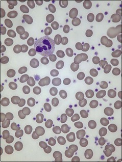
Platelet counts can be as high as 2000 × 109/L and usually exceed 450 × 109/L (the normal range is 150–400 × 109/L) (Fig 33.1). In practice, there is no single test to specifically identify ET – diagnosis is often a process of exclusion. As thrombocytosis may accompany a wide range of disorders including infections, inflammatory conditions, malignancy and iron deficiency, a thorough history and examination is mandatory. The lack of a measurable ‘acute phase response’ (i.e. normal erythrocyte sedimentation rate, plasma viscosity and fibrinogen) increases the likelihood of ET as opposed to a ‘reactive’ thrombocytosis. Even where there is an acquired JAK2 gene mutation, other myeloproliferative disorders must be excluded. Bone marrow examination is worthwhile to exclude chronic myeloid leukaemia (absence of Philadelphia chromosome), myelofibrosis or myelodysplasia, and to check iron stores. Patients with polycythaemia vera may have thrombocytosis, while patients with ET can have an increased red cell mass. In practice such patients are better diagnosed as having myeloproliferative neoplasms rather than forced into either category. Only about 5% of all raised platelet counts are due to ET, but persistence of the count above 1000, particularly with coexistence of thrombosis or haemorrhage, makes it the likely diagnosis. Abnormal platelet function tests suggest ET rather than a reactive thrombocytosis.

Management

Management is not straightforward. The decision whether to treat at all must follow consideration of the patient’s age, the degree of thrombocytosis and the presence or perceived risk of significant thrombotic or haemorrhagic events. Any clinical benefit must be weighed against potential toxicity of cytotoxic drugs. In a patient of more advanced age (>60 years) or with a very high platelet count (>1500) or a history of thromboembolic disease, the treatment of choice is hydroxycarbamide and low-dose aspirin. Anagrelide or interferon alfa may be preferred where hydroxycarbamide is not tolerated. The objective of treatment is to maintain the platelet count in the normal range and prevent thrombosis and haemorrhage. Low-dose aspirin alone is a reasonable option in patients at lower risk of these complications. Interferon is the usual drug of choice in pregnancy.

Myelofibrosis

Primary myelofibrosis is a myeloproliferative neoplasm characterised by bone marrow fibrosis and splenomegaly. It may develop de novo or in the setting of polycythaemia vera or essential thrombocythaemia. Most patients are over 50 years.

Myelofibrosis is a neoplastic clonal disorder originating in a single pluripotential stem cell. Abnormal megakaryocytes are produced in increased numbers and it is these cells which release cytokines such as platelet-derived growth factor (PDGF) and transforming growth factor-β, which stimulate fibroblast proliferation and build-up of collagen in the bone marrow. The scarred marrow is unable to function normally and haematopoietic stem cells move to the spleen and liver (extramedullary haematopoiesis).

Clinical features

The disease is often insidious in onset with fatigue and weight loss. Splenomegaly is present in all cases and massive in 10% (Fig 33.4). Splenic pain is common and a bulky spleen may lead to portal hypertension, bleeding varices and ascites. Hepatomegaly is seen in two-thirds of cases.

Diagnosis

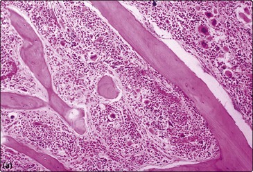
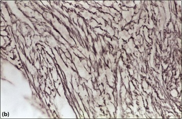
Anaemia is almost universal and the blood film shows tear-drop poikilocytes and a ‘leucoerythroblastic’ picture (Fig 33.2). In the early stages, thrombocytosis and neutrophilia may occur but in more advanced disease low counts are the rule. Bone marrow aspiration characteristically results in a ‘dry tap’ (i.e. only peripheral blood aspirated), and a marrow trephine showing dense reticulin fibres on silver staining, fibrosis and osteosclerosis is needed for diagnosis (Fig 33.3). There is usually megakaryocytic hyperplasia. The JAK2 gene mutation is present in approximately 50% of cases. X-rays often show bone sclerosis. The major differential diagnosis is from other myeloproliferative disorders and myelodysplastic syndromes which may be associated with a degree of marrow fibrosis. Systemic causes of marrow fibrosis such as marrow infiltration by carcinoma or lymphoma and disseminated tuberculosis should also be considered.
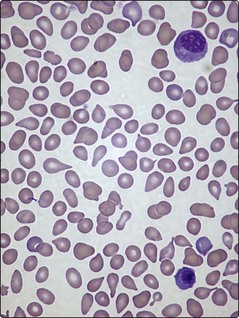
Prognosis and management

Average survival is 4–7 years but this is very variable. Management is increasingly guided by prognostic scoring systems. Leukaemic transformation occurs in about 15% of patients. Asymptomatic patients may require no treatment. For anaemia a trial of a corticosteroid, androgen or erythropoietin is worthwhile but regular transfusion is usually needed. Oral chemotherapeutic agents such as hydroxycarbamide may improve quality of life by reducing systemic upset and shrinking the spleen. There is abnormal bone marrow angiogenesis in myelofibrosis and the anti-angiogenic agents thalidomide and lenalidomide can improve blood counts and reduce splenomegaly with some durable responses. The JAK2 inhibitor ruxolitinib is a promising new agent. Its major benefits are reduced splenomegaly and constitutional symptoms. Other JAK2 inhibitors are under investigation.

Splenic irradiation can alleviate splenic pain. Splenectomy must not be undertaken lightly as it is associated with considerable mortality (around 5–10%). However, it is considered for painful splenomegaly, unacceptable transfusion requirements, life-threatening thrombocytopenia, profound cachexia or complications of portal hypertension.

Allogeneic stem cell transplantation is the only potentially curative procedure. Use of reduced intensity conditioning regimens (see p. 57) may allow its wider application.