[level-membership-for-radiology-category] Lacks muscle layer (unlike distal esophageal A and B rings)
IMAGING
• Full column barium esophagram with rapid-sequence filming or have patient swallow small marshmallow followed by barium
• Appearance: 1-2 mm wide, shelf-like filling defect
Usually along anterior wall of cervical esophagus
Circumferential, radiolucent ring in some cases
May occur at other sites in esophagus
• Mild, moderate, or severe luminal narrowing
• Partial obstruction suggested by
Jet phenomenon: Barium spurting through ring
Dilatation of esophagus or pharynx proximal to web
TOP DIFFERENTIAL DIAGNOSES
• Esophageal strictures
Longer length of luminal narrowing of esophagus
• Schatzki ring
Lower (GE junction) esophageal or Β ring
PATHOLOGY
• Esophageal webs may be associated with
Plummer-Vinson (Paterson-Kelly) syndrome
Epidermolysis, pemphigoid
Eosinophilic esophagitis
Celiac-sprue disease
Chronic GE reflux
Graft vs. host disease
CLINICAL ISSUES
• Usually asymptomatic
• Dysphagia with impaction of food or pills above web
• Respond to balloon or bougie dilation, but often recur
(Left) Rapid-sequence barium esophagram in a 45-year-old woman with dysphagia shows a thin, shelf-like indentation from the anterior and lateral walls of the pharyngoesophageal junction.
(Right) Frontal esophagram in the same patient shows abnormal distension of the pharynx above this web , confirming that it is causing partial obstruction and luminal narrowing.
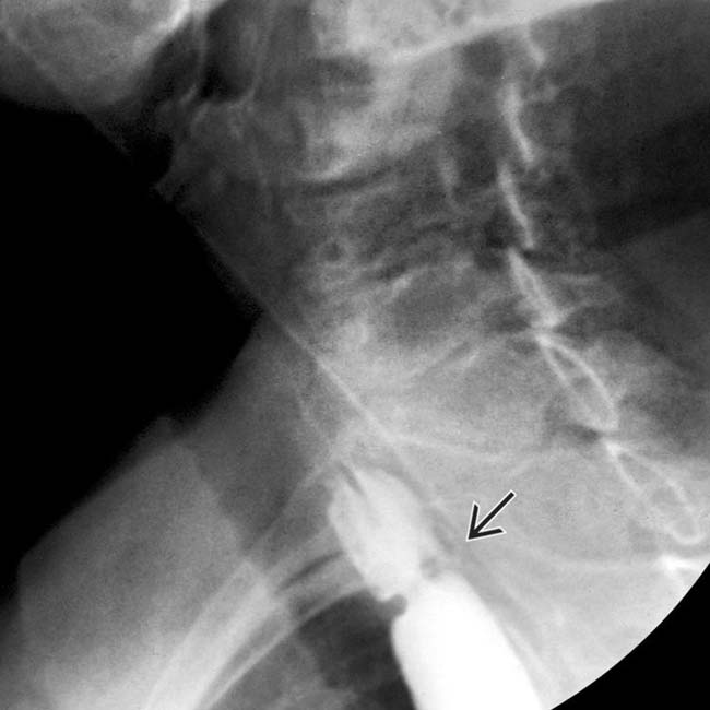
(Left) Barium esophagram in a 50-year-old woman with solid food dysphagia shows a shelf-like narrowing of the proximal esophageal lumen. This web is unusually thick and circumferential.
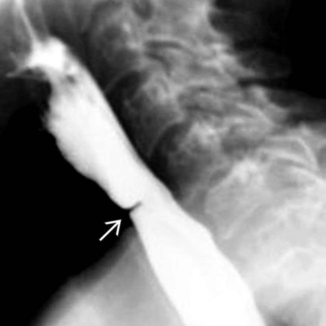
(Right) Lateral esophagram in a young patient with epidermolysis bullosa shows a web-like stricture near the pharyngoesophageal junction, representing a stricture due to repeated episodes of mucosal ulceration, typical of patients with epidermolysis.
Lateral esophagram shows a tight, web-like narrowing at the pharyngoesophageal junction due to epidermolysis bullosa.
Lateral esophagram shows a circumferential and relatively thick web causing significant luminal narrowing.
Frontal esophagram in a patient with Plummer-Vinson syndrome with glossitis, iron deficiency anemia, and dysphagia shows esophageal web and stricture .
Lateral barium esophagram shows a thin, shelf-like indentation of the esophagus.


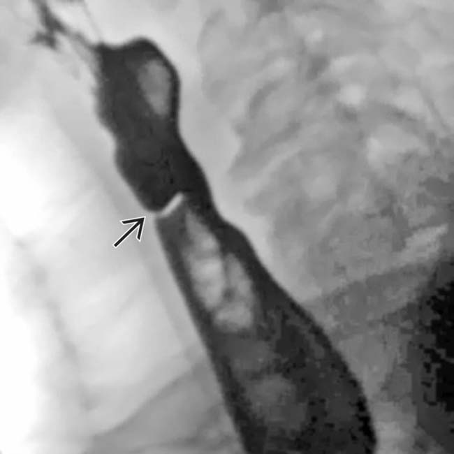
 from the anterior and lateral walls of the pharyngoesophageal junction.
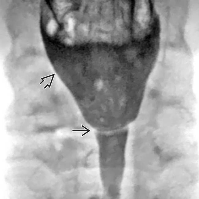
 above this web
  , confirming that it is causing partial obstruction and luminal narrowing.
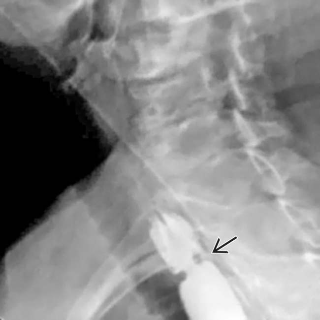
 of the proximal esophageal lumen. This web is unusually thick and circumferential.
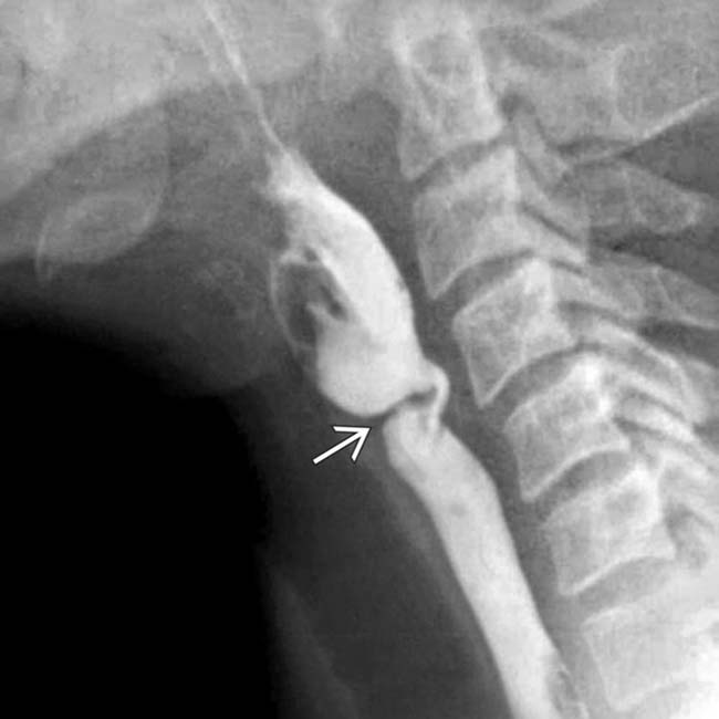
 near the pharyngoesophageal junction, representing a stricture due to repeated episodes of mucosal ulceration, typical of patients with epidermolysis.
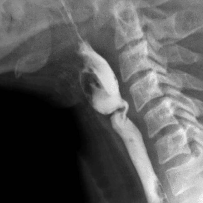
 causing significant luminal narrowing.
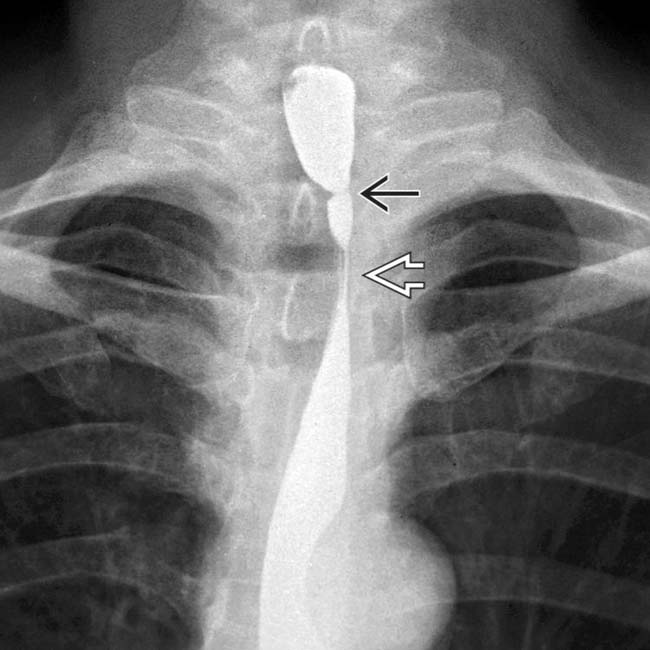
 and stricture
  .
.
 of the esophagus.