5 Esophageal Diseases
Anatomy of the Esophagus
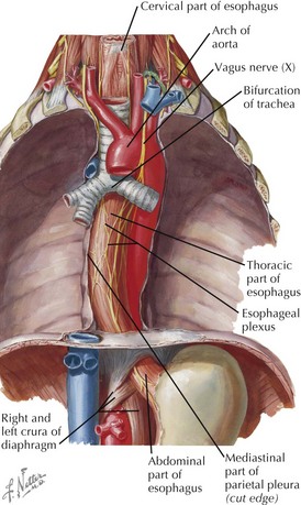
• Fibromuscular tube (~25 cm) running from the pharynx in the neck, through the thorax and diaphragm, to the stomach in the abdomen
• Runs just posterior to the trachea and anterior to vertebral bodies in the neck and superior mediastinum
• Tends to run to the left below T4 but is pushed to the center by the arch of the aorta and the root of the left lung
• Upper esophageal sphincter: circular muscle of the superior esophagus, including the cricopharyngeus, the first region of anatomical constriction


• Retropharyngeal danger space: possibility of infection spreading retroesophageally into the thorax
Microscopic Anatomy
Mucosa


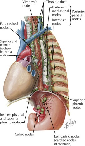
Vessels and Lymphatics
• Extensive submucosal vascular plexuses allow mobilization of large extents of the esophagus with reduced risk of ischemia.

Arterial Supply
• Cervical portion is supplied by branches of the inferior thyroid arteries from thyrocervical trunks of the right and left subclavian arteries.
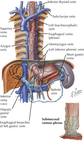
Venous Drainage
• Esophageal venous plexus has multiple connections.


• Because of portal and systemic (azygos, etc) connections of the submucosal veins, they can become enlarged (varices) in portal hypertension.
Clinical Correlates
Achalasia
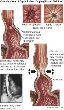
Gastroesophageal Reflux Disease (GERD)
• Failure of normal anatomical mechanisms: lower sphincter competence, normal esophageal structure, normal gastric reservoir