87
Erythema nodosum
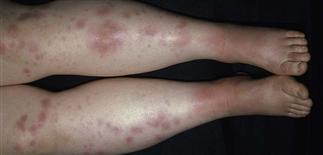
Pink, dusky, firm, painful nodules classically occur on the pretibial surfaces.
Erythema nodosum nodules have ill-defined borders.
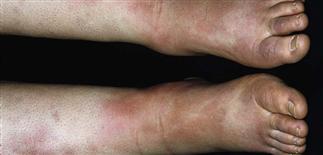
Erythema nodosum can be accompanied by edema.
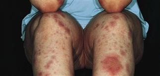
Although the legs are the typical location, erythema nodosum lesions can occur with varying shapes on the arms, head, neck, and torso.
DESCRIPTION
Panniculitis characterized by tender pink nodules on extensor surface of lower legs.
HISTORY
• In adults, five to six times more common in women, with a peak onset at 20–30 years. In children, boys and girls affected equally. Caused by a hypersensitivity reaction to various antigenic stimuli: bacteria, viruses, fungi, parasites, drugs, malignancies, connective tissue diseases. Streptococcal infections and sarcoidosis commonly associated with erythema nodosum. Half of cases are idiopathic. • Can be associated with fever, malaise, diarrhea, headache, conjunctivitis, cough. New crops of lesions usually develop for 3–6 weeks, although rarely erythema nodosum persists for months to years.
PHYSICAL FINDINGS
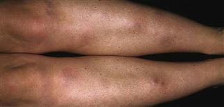
• Pink to dusky-red firm nodules with indistinct edges occur symmetrically on the pretibial surfaces. • Ankle and leg pain are common. Erythema nodosum occurs on head, neck, torso, arms, thighs. • Fades in 1–2 weeks without scars.
TREATMENT
• Associated diseases and infections should be identified and treated. • Discontinue precipitating medications. Symptomatic treatment with rest, compressive bandages, and non-steroidal anti-inflammatory drugs (indometacin, Naprosyn). • Supersaturated potassium iodide and systemic corticosteroids have been helpful for chronic recurrent erythema nodosum.


