164 Erythema multiforme
Salient features
History
Examination
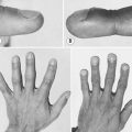
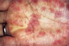
• Target-shaped lesions (Fig. 164.1), usually over the palms, soles and skin. A classical target lesion consists of three concentric zones of colour change: typically there is a central, dark, purple area or blister surrounded by a pale, oedematous round zone; this, in turn, is surrounded by a peripheral rim of erythema
Questions
Advanced-level questions
How would you investigate this patient?
What is the histology of erythema multiforme?
Early lesions. Superficial perivascular lymphocytic infiltrate with oedema of the dermis, accompanied by degeneration and necrosis of keratinocytes and margination of lymphocytes at the dermoepidermal junction.
Late lesions. Upward migration of lymphocytes into the epidermis; discrete and confluent portions of the epidermis necrose, resulting in blister formation and subsequently erosions (as a result of sloughing of the epidermis).
Target lesions. Characterized by central necrosis surrounded by perivenular inflammation.
How would you manage a patient with erythema multiforme and Stevens–Johnson syndrome?
Erythema multiforme is a self-limiting condition, but Stevens–Johnson syndrome may be fatal:
• Symptomatic treatment with antipyretics, intravenous fluids and antibiotics.
• Systemic corticosteroids, although the role of steroids is controversial.
• Other immunosuppressive drugs used in recurrent erythema multiforme and Stevens–Johnson syndrome include levamisole, azathioprine, dapsone, thalidomide, high-dose intravenous immunoglobulin and ciclosporin.
• Aciclovir is recommended by some authors as a therapeutic trial in any patient with severe recurrent erythema multiforme, even if no preceding herpes simplex infection has been documented.