85
Erythema multiforme
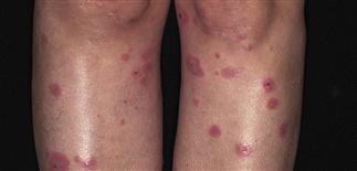
Erythema multiforme is commonly seen on the extremities, especially palms and soles. Iris or target lesions are classically seen along with fixed urticaria-like papules.
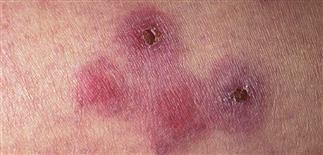
The central portion of the target lesion can appear dark red, blister, and heal with scale crust.
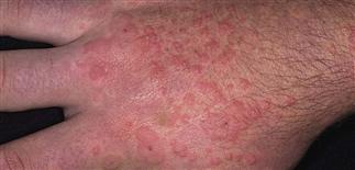
Erythema multiforme shows numerous lesion morphologies, urticaria-like papules, vesicles, bullae, and target-shaped papules.
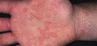
Target lesions on the palms can mimic a drug hypersensitivity reaction. Itching with erythema multiforme lesions is usually mild.
DESCRIPTION
Relatively common, acute—often recurrent—inflammatory disease characterized by target-shaped skin lesions.
HISTORY
• Commonly associated with herpes simplex, Mycoplasma pneumoniae, and upper respiratory tract infections. • Can be seen with contact allergens, drugs, connective tissue diseases, physical agents, X-ray therapy, pregnancy, internal malignancies. • Reactivation of herpes simplex can produce recurrent erythema multiforme due to cytotoxic immune response directed against keratinocyte-expressing foreign viral or drug antigens. • Usually resolves in 1 month.
PHYSICAL FINDINGS
• Numerous lesion morphologies: target lesions, erythematous macules and papules, urticaria-like lesions, vesicles, bullae. • Target lesions begin as dusky red, round macules and papules and are 1–3 cm. Can see symmetric pattern on palms, soles, backs of hands and feet, and extensor aspect of forearms and legs. • The center of the iris can appear dark red, purpuric, or vesicular, and is due to acute epidermal injury. Surrounding the central area is a pale area of edematous skin, which is in turn surrounded by a sharp, discrete ring of erythema. • Lesions appear in crops, resolving in 1–2 weeks without scarring. • Postinflammatory pigment changes are common (hypopigmentation and/or hyperpigmentation). • Bullae and erosions may be present in the oral cavity. • Lesions are fixed and may occur in areas of trauma (Koebner phenomenon). • Skin biopsy: interface reaction with necrotic keratinocytes.
TREATMENT
• Ruptured blisters and eroded skin respond to topical antibiotics (bacitracin, Bactroban). • Widespread erythema multiforme responds rapidly to 1–3 weeks of systemic corticosteroids. • Prednisone 40–80 mg q.d. initially until lesions resolve, followed by taper. •Triamcinolone acetonide (40 mg) i.m. can be effective • Can prevent recurrent herpes-associated erythema multiforme with oral acyclovir (200 mg b.i.d. or t.i.d. or 400 mg b.i.d.), valacyclovir (Valtrex) 500 mg q.d., or famciclovir (Famvir) 125 mg b.i.d. as continuous suppressive therapy.


