[level-membership-for-opthalmology-category]10.3
Epiretinal Membrane
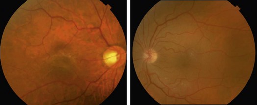
Clinical Features:
Patients may be asymptomatic or complain of metamorphopsia and blurring of varying severity. On examination, the epiretinal layer can be seen as a glistening membrane overlying the fovea, often associated with retinal striae in a radial fashion and macular thickening (Fig. 10.3.1). Contraction of the membranes can also cause retinal vascular distortion. More severe epiretinal membranes can cause loss of the normal foveal reflex and ‘pseudohole’ formation.
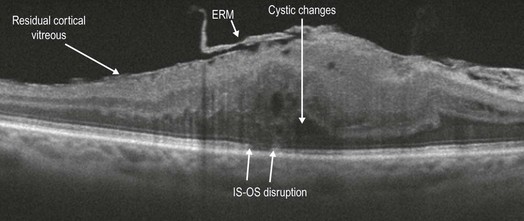
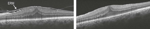
OCT Features:
ERM appears as a highly reflective layer overlying the inner retina, which may be adherent to the retina throughout the length of the scan, or adherent for only a portion of the macular region. Depending on the severity of the ERM, distortion of the inner retina, loss of foveal contour, retinal thickening and irregularity of the retinal surface, subretinal fluid and foveal schisis may occur (Figs 10.3.2 and 10.3.3). Cystic changes may also be seen within the retina. Occasionally, ERM may be associated with a pseudohole configuration, with disruption of the inner retina in the region of the ‘hole’ and separation between the outer plexiform layer and the outer nuclear layer seen adjacent to the site of the ‘hole’. However, the outer retinal structures are intact including the inner and outer segments of the photoreceptors.
[/level-membership-for-opthalmology-category][not-level-membership-for-opthalmology-category]10.3
Epiretinal Membrane
Clinical Features:
Patients may be asymptomatic or complain of metamorphopsia and blurring of varying severity. On examination, the epiretinal layer can be seen as a glistening membrane overlying the fovea, often associated with retinal striae in a radial fashion and macular thickening (Fig. 10.3.1). Contraction of the membranes can also cause retinal vascular distortion. More severe epiretinal membranes can cause loss of the normal foveal reflex and ‘pseudohole’ formation.
[/not-level-membership-for-opthalmology-category]
