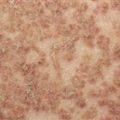
Epidermolysis bullosa simplex, Weber-Cockayne type.
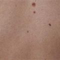
Epidermolysis bullosa simplex, Koebner type. Discrete blisters on a toddler’s foot.
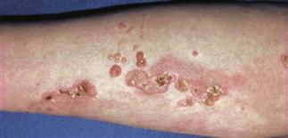
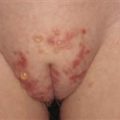
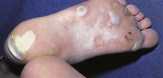
Epidermolysis bullosa simplex, Dowling-Meara type. Grouped blisters on the leg of a child.

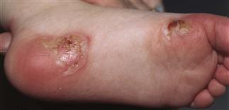
Epidermolysis bullosa simplex, Dowling-Meara type. Grouped blisters on the arm of a child.
CLINICAL FEATURES
Epidermolysis bullosa simplex (EBS) is characterized by blister formation within the epidermis, most commonly in the basal layer, and in rarer forms in the suprabasillar layer. There are mutations in six different genes that cause EBS. These are keratin 5, keratin 14, plectin, a6b4 integrin, plakophillin and desmoplakin. A new form of EBS caused by mutations in the dystonin gene has recently been described. The defects in the most common types of EBS are in keratins 5 or 14. There are multiple other rare variants.
TREATMENT
Treatment is symptomatic and supportive. Drain blisters with a sterile needle or blade. Do not remove the blister roof. If the blister roof is gone, apply Vaseline and a non-stick bandage. Secure the bandage with kerlex and netting. Avoid strenuous activity in warm weather. Antibiotics may be necessary if the blisters become infected.