122
Epidermal cyst
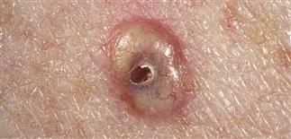
Epidermal cysts are yellow to gray, smooth, dome-shaped nodules with a central punctum and contain keratin. Epidermal cysts vary in size. Lesions of this size will show little tendency to rupture.
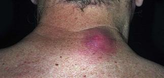
Spontaneous rupture of the wall results in discharge of a soft yellow keratin into the dermis. A tremendous inflammatory response ensues and the cyst becomes red, swollen and tender.
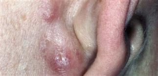
Epidermal cysts occur commonly on the back of the ear and may become clustered. Posterior neck and back are also common sites.
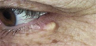
Smaller epidermal cysts can occur about the eyelids and in areas of scar trauma.
DESCRIPTION
An epidermal cyst is a firm, subcutaneous, keratin-filled cyst originating from squamous epithelium, most often from a hair follicle infundibulum.
HISTORY
• Epidermal cysts arise spontaneously. • They occur most commonly on the trunk, postauricular fold, and posterior neck. Cysts frequently develop in areas of friction and are usually solitary. • Most epidermal cysts arise from the squamous epithelium of the hair follicle. • Unlike pilar cysts, the epidermal cyst wall is fairly delicate and thus prone to rupture. Rupture is followed by a foreign body reaction to keratin extruded into the dermis and acute inflammation. Such lesions are tender and appear to be infected. However, cultures are usually sterile.
PHYSICAL FINDINGS
• The firm, dome-shaped, pale-yellowish, intradermal or subcutaneous cystic nodules range from 0.5 to 5.0 cm in size. • Cysts are somewhat mobile but are tethered to the overlying skin through a small punctum that often appears as a comedo. This punctum represents the follicle from which the cyst developed. • Inflamed epidermal cysts are warm, red, boggy, and tender on palpation. Furuncles have a similar appearance. Sterile, purulent material and keratin debris drain to the surface. • If the inflammatory response is brisk enough to destroy the cyst wall, then it is unlikely the cyst will recur. More often, the inflammation subsides and the cyst recurs. • Scarring often follows rupture, which makes the cyst more difficult to remove. • Multiple epidermal cysts occurring on the face, scalp, and back should raise suspicion of Gardner syndrome in the appropriate clinical setting. This very rare, autosomal dominant condition is associated with colonic polyposis and early malignant degeneration into adenocarcinoma of the colon.
TREATMENT
• Epidermal cysts on the face may rupture and lead to scarring. • The cosmesis of elective surgical excision must be weighed against a scar from potential rupture. Such lesions are far more difficult to remove once they have ruptured. • Asymptomatic epidermal cysts occurring elsewhere do not require treatment. Symptomatic or recurrent ruptured epidermal cysts should be removed. • Ruptured, inflamed epidermal cysts should be incised and drained under local anesthesia. • Attempts should be made to remove the cyst lining, by either curettage or blunt dissection. • Recurrent epidermal cysts that have previously ruptured and scarred are best excised along with the surrounding scar once the inflammation has subsided.


