9 Epiaortic Ultrasonography and Epicardial Echocardiography
Surface Imaging
Probe Orientation
Probe orientation marker is important for surface imaging techniques.
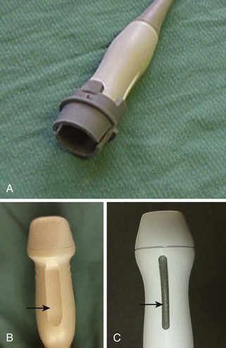
Probe Options
Image Acquisition and Optimization
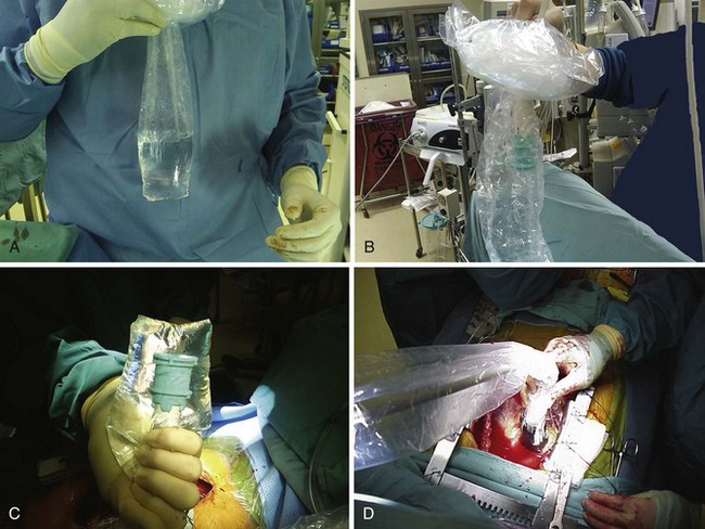
Maintaining a Sterile Field
Epiaortic Ultrasonography
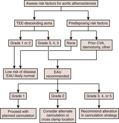
Prevention of Stroke
Key Points
Standard Views and 12-Segment Nomenclature
Key Points
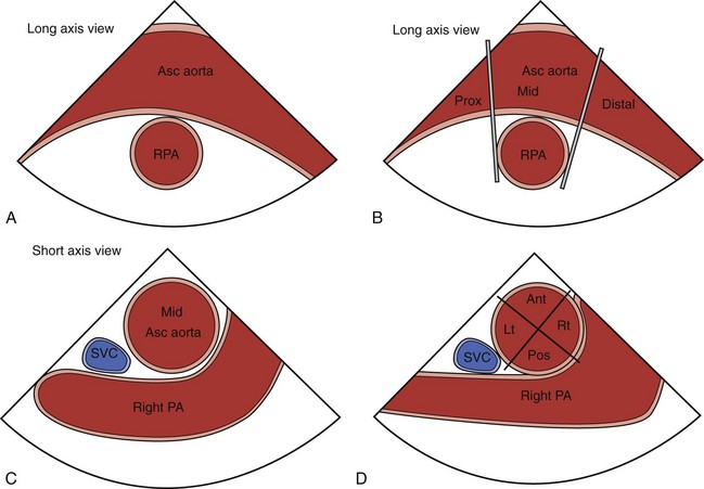
Performing an Epiaortic Ultrasonography Examination
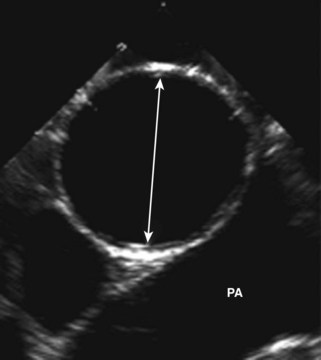
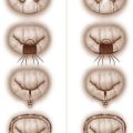
Atherosclerosis Grading (Figure 9-7)
| Grade | Severity | Description |
|---|---|---|
| Grade 1 | Normal | Normal, no intimal thickening |
| Grade 2 | Mild | Intimal thickening ≤ 3 mm |
| Grade 3 | Moderate | Sessile atheroma > 3 mm but < 5 mm |
| Grade 4 | Severe | Sessile atheroma ≥ 5 mm |
| Grade 5 | Mobile | Protruding atheroma with mobile components |
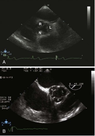
Epicardial Echocardiography
When Transesophageal Echocardiography Is Not an Option
ECE predates TEE for surgical diagnosis and evaluation and, currently, is most often used when a TEE probe cannot be placed or is contraindicated. Table 9-2 lists the recommended ECE views and the corresponding TTE views.
TABLE 9-2 EPICARDIAL AND TRANSESOPHAGEAL ECHOCARDIOGRAPHY VIEW NOMENCLATURE
| Epicardial Views | Corresponding TTE View |
|---|---|
| AV SAX | Parasternal AV SAX |
| AV LAX | Suprasternal AV LAX |
| LV basal SAX | Modified parasternal MV basal SAX |
| LV mid SAX | Parasternal LV mid SAX |
| LV LAX | Parasternal LAX |
| Two-chamber | Modified parasternal LAX |
| RVOT | Parasternal SAX |
AV, aortic valve; LAX, long axis; LV, left ventricle; MV, mitral valve; RVOT, right ventricular outflow tract; SAX, short axis.
Seven Standard Epicardial Echocardiographic Views
Image Acquisition
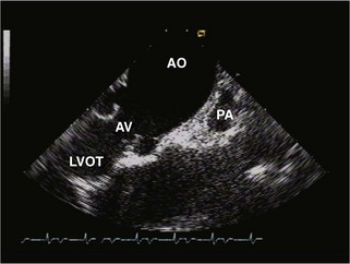
Step 1: Aortic Valve Short Axis (see Figure 9-3)
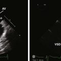
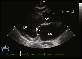
Step 2: Aortic Valve Long Axis (Figure 9-8)
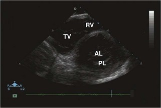
Step 3: Left Ventricle Basal Short Axis (Figure 9-9)
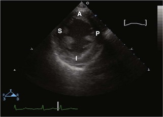
Step 4: Left Ventricle Mid Short Axis (Figure 9-10)
Step 5: Left Ventricle Long Axis (Figure 9-11)
Step 6: Two-Chamber (Figure 9-12)
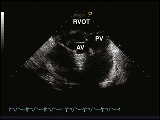
Step 7: Right Ventricular Outflow Tract (Figure 9-13)
1 Glas KE, Swaminathan M, Reeves ST, et al. Council for Intraoperative Echocardiography of the American Society of Echocardiography. Society of Cardiovascular Anesthesiologists. Society of Thoracic Surgeons. Guidelines for the performance of a comprehensive intraoperative epiaortic ultrasonographic examination: recommendations of the American Society of Echocardiography and the Society of Cardiovascular Anesthesiologists; endorsed by the Society of Thoracic Surgeons. Anesth Analg. 2008;106:1376-1378.
2 Davila-Roman VG, Phillips KJ, Daily BB, et al. Intraoperative transesophageal echocardiography and epiaortic ultrasound for assessment of atherosclerosis of the thoracic aorta. J Am Coll Cardiol. 1996;28:942-947.
3 Bucerius J, Gummert JF, Borger MA, et al. Stroke after cardiac surgery: A risk factor analysis of 16,184 consecutive adult patients. Ann Thorac Surg. 2003;75:472-478.
4 van der Linden J, Hadjinikolaou L, Bergman P, Lindblom D. Postoperative stroke in cardiac surgery is related to the location and extent of atherosclerotic disease in the ascending aorta. J Am Coll Cardiol. 2001;38:131-135.
5 Katz ES, Tunick PA, Rusinek H, et al. Protruding aortic atheromas predict stroke in elderly patients undergoing cardiopulmonary bypass: Experience with intraoperative transesophageal echocardiography. J Am Coll Cardiol. 1992;20:70-77.
6 Reeves ST, Glas KE, Eltzschig H, Shernan SK. Guidelines for performing a comprehensive epicardial echocardiography examination: Recommendations for the American Society of Echocardiography Council for Intraoperative Echocardiography and the Society of Cardiovascular Anesthesiologists. Anesth Analg. 2008;105:22-28.
7 Rosenberger P, Shernan SK, Loffler M, et al. The influence of epiaortic ultrasonography on intraoperative surgical management in 6051 cardiac surgical patients. Ann Thorac Surg. 2008;85:548-553.
8 Eltzschig HK, Kallmeyer IJ, Mihaljevic T, et al. A practical approach to a comprehensive epicardial and epiaortic echocardiographic examination. J Cardiothorac Vasc Anesth. 2003;17:422-429.
9 Hilberath JN, Shernan SK, Segal S, et al. The feasibility of epicardial echocardiography for measuring aortic valve area by the continuity equation. Anesth Analg. 2009;108:17-22.