Atrophic Endometrium
Synonyms/Description
Endometrial atrophy
Etiology
Endometrial atrophy is most often the result of postmenopausal status, although it may also occur in premenopausal women with lack of estrogen from other etiologies.
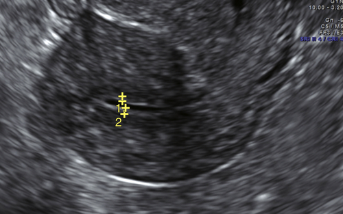
Ultrasound Findings
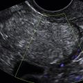
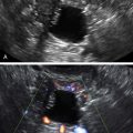
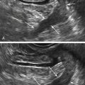
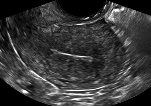
A very thin endometrial echo is characteristic of atrophic endometrium. This occurs primarily in postmenopausal or anovulatory women. Often the endometrium is so thin it is reflective of sound waves, such that it looks like a continuous line. An atrophic endometrium measures less than 4 mm, but is more commonly 1 to 2 mm in width. In postmenopausal women there can occasionally be a small amount of fluid in the cavity outlining a paper-thin endometrial lining. This is usually transudate associated with cervical stenosis and not associated with pathology.
Differential Diagnosis
If the endometrial lining is very thin in a patient who is hypoestrogenic, the diagnosis is likely to be atrophic endometrium.
Clinical Aspects and Recommendations
Postmenopausal bleeding is often caused by atrophy of the endometrium.
In the absence of estrogen, the functional layer atrophies, leaving only the basalis layer. In patients with postmenopausal bleeding, a workup is necessary to rule out uterine cancer, hyperplasia, and polyps before the cause can be attributed to atrophic endometrium, especially because endometrial cancer can coexist with atrophic endometrium.
Suggested Reading
Davidson K.G., Dubinsky T.J. Ultrasonographic evaluation of the endometrium in postmenopausal vaginal bleeding. Radiol Clin North Am.. 2003;41:769–780.
Goldstein S.R. Sonography in postmenopausal bleeding. J Ultrasound Med. 2012;31:333–336.
Tsai M.C., Goldstein S.R. Office diagnosis and management of abnormal uterine bleeding. Clin Obstet Gynecol. 2012;55(3):635–650.