Chapter 162 Endometriosis
 Diagnostic Summary
• Triad of symptoms: dysmenorrhea, dyspareunia, and infertility
• Physical examination reveals one or more of the following: tenderness of the pelvic area, cul-de-sac, or both; enlarged or tender ovaries; a uterus that tips backward and lacks mobility; fixed pelvic structures; adhesions
• Pelvic ultrasound: detection and consistency of endometriomas
• Definitive diagnosis: a laparoscopy or laparotomy visualizing endometrial implants within the pelvic cavity
 General Considerations
Etiologic Theories
Retrograde flow, the predominant theory first proposed in 1927, suggests that during menses blood flows backward and provides seeds of implants in the pelvic cavity. The problem with this theory is that more than 90% of menstruating women without endometriosis have retrograde flow; thus, this theory was challenged and questions were raised as to the biochemical and immunologic factors causing the implantation of endometrial tissue within the pelvic environment.1 Some theories are based on findings that endometrial implants from women with endometriosis are biochemically different from those from normal women.2
Other findings suggest that cells may implant only in women with altered cell immunity.1 Implants found in odd locations, such as the nose and lungs, suggest the transportation of endometrial tissue through lymphatic channels and blood vessels. Some researchers believe the implants to be of embryologic origin, when pieces of the uterus were left behind during development, and that these implants, when activated, secrete a chemical causing the nearby capillaries to bleed.3
Other research on endometriosis in baboons suggests activation by environmental toxins that mimic estrogens.4 Exposure to radiation and dioxin has been associated with a higher frequency of endometriosis.5,6 The highest incidence of endometriosis appears to be in Belgium, which is also the home to the highest level of dioxin pollution in the world.7
Two studies, however, one in Belgium, found no significantly increased risk with dioxins or polychlorinated biphenyls.8 The other study, in Italy, found no significantly increased risk of endometriosis in women who had high levels of dioxin in their blood.9 Although there is currently no definitive epidemiologic evidence linking any one class of chemicals to the risk of endometriosis, there does appear to be some suggestion of a link with estrogen-like compounds in the environment,10 called xenoestrogens, which can disrupt estrogen and estrogen metabolism. Substances that have been shown to have estrogenic effects in the body include PCBs, weed killers, liners in tin cans, plastics, detergents, and household cleaners.11
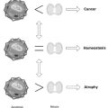
Immunologic alterations may exist in women with endometriosis. Increasingly, we are finding evidence that a lack of proper immune surveillance in the pelvic area is the cause of endometriosis and that impairment in other aspects of the immune system function are involved in the progression of the disease.12
Women with endometriosis have been found to have suppressed natural killer cell activity in their peritoneal fluid,13,14 high levels of immunoglobulins IgG and IgM,15 and high levels of autoantibodies against ovary and endometrial cells.16,17
Both types of immunity, cell-mediated and humoral, have been implicated in endometriosis, with immunologic defects present even in the mildest forms of the disease.18 Macrophages are found in greater numbers in the early stages of endometriosis.19
Cytokines, macrophages, T lymphocytes, and tumor necrosis factors have all been found to be increased in the peritoneal fluid in women with endometriosis,20–22 and their increase correlates with the severity of the disease. Growth factors, angiogenic factors, and lipid peroxidation in the peritoneal fluid may stimulate endometrial cell growth. Targeting of these proinflammatory compounds and blocking their action with antioxidants and other compounds provide a good rationale for new treatment strategies, both conventional and with natural compounds.
Genetic influences the area also likely involved in endometriosis. Grouping of endometriosis within families has been found in clinical studies,23,24 populations-based studies,25 and even studies of twins.26–28 Several abnormalities in detoxification enzymes, tumor suppressor genes, and other genetic factors occurring in multistep fashion may be involved both in the development and the progression of the disease.29
 Diagnosis
In some cases symptoms begin with the onset of menstruation, but in most symptoms begin later and worsen progressively over time. The triad of symptoms is dysmenorrhea (pain with menses), dyspareunia (pain with vaginal intercourse), and infertility. Acute pain occurs before the onset of menses; it can last for a day or two during the menses or throughout the month. In some women, vomiting, diarrhea, and fainting can occur concurrently with the intense pelvic/abdominal cramping and even labor-like pains. Other pains can involve a chronic bearing-down pain and pressure in the lower back, with pain sometimes radiating down the legs. There is not necessarily any correlation between pain and the extent of disease. Women with fixed ovaries and large endometriomas may report only mild discomfort, whereas those with visibly smaller lesions may report severe chronic pain. Upon surgery, these lesions are found to extend more deeply; they are possibly more influenced by circulating estrogens.30 Research has found that the severity of symptoms is correlated with the depth of the lesions rather than the number of lesions.31
 Therapeutic Considerations
Diet
Several dietary principles are key in a natural medicine approach to endometriosis:
• Reduce inflammatory foods and increase antiinflammatory foods.
• Enhance detoxification mechanisms.
• Increase dietary fiber to promote optimal transit time and optimal gut flora.
The consumption of trans fats appears to increase the risk of endometriosis, whereas long-chain omega-3 fats appear to be protective. Twelve years of prospective data from the Nurses’ Health Study II, which began in 1989, weres analyzed for dietary fat and its association with many health problems, including endometriosis.32 Although there was no association with total fat consumption and endometriosis risk, those women with the highest consumption of long-chain omega-3 fatty acids were 22% less likely to be diagnosed with endometriosis compared with women who had the lowest intake of these fats. Those women who had the highest intake of trans unsaturated fats were 48% more likely to be diagnosed with endometriosis.
Foods high in fiber are associated with optimal transit time in the intestines and an optimal balance of friendly microorganisms within the large intestines (see Chapter 52). These microorganisms, better known as gut flora, crowd out the other types of flora that deconjugate estrogens and allow estrogens to recycle back through the body. Studies suggest that a predominantly vegetarian diet emphasizing less protein and more fiber can lead to a decrease of biologically active unconjugated estrogens in the blood plasma.33 Although higher-protein diets are found to provide enzymes for the detoxification pathways of estradiol,34 vegetarian diets are of greater value owing to their lower fat content. Diets containing large amounts of animal protein, especially in the form of red meat, contain large amounts of arachidonic acid, which promotes inflammatory prostaglandins and thus inflammation and pain. By adding vegetable protein, soy, nut butters such as almond, and salmon to our diets, we tip the inflammatory pathway toward antiinflammatory prostaglandins that inhibit tumor growth—and possibly endometrial growth.
By increasing the intake of vegetables, specifically those that enhance liver function, cell damage from toxins and metabolites is prevented. “Liver foods” to be increased include carrots, beets, and cabbage-family vegetables, owing to their role in aiding phase 2 of the liver’s detoxification pathway. Indole-3-carbinol—found in broccoli, Brussels sprouts, cabbage, and cauliflower—favors the less active metabolite of estrogen.35 Other liver-cleansing foods are carrots, artichokes, lemons, dandelion greens, watercress, and burdock root. Onions, garlic, and leeks contain organosulfur compounds, which enhance the immune system and induce enzymes that detoxify in the liver. In addition, they contain the bioflavonoid quercetin, which is known to stimulate the immune response, protect against oxidation, block the inflammatory response, and inhibit tumor growth.36 Organically grown vegetables are expected to contain lower levels of pesticides that may also mimic estrogen.
The isoflavones in soy products and the lignans in flax seeds may also be important in a dietary approach to endometriosis. Many people might claim that these phytoestrogen compounds should be avoided, thinking that they would stimulate the growth of endometriotic implants, much as estrogen might. However, the evidence supports the inclusion of soy in the diets of women with endometriosis. A Japanese study demonstrated that a moderate intake of soy isoflavones was associated with a decreased risk of premenopausal hysterectomy. Because some of the surgeries involved endometriosis, these results led the authors to conclude that moderate soy intake may decrease the risk for endometriosis.37
Seasonings such as turmeric (curcumin) protect against environmental carcinogens, decrease inflammation, and increase bile secretion. Adding a tablespoon of milk thistle seeds that have been soaked and ground can also help with liver function. Fresh flax seeds increase antiinflammatory fatty acids. Seasoning with Fucus (a seaweed) helps to stimulate T-cell production and absorb toxins.38
Foods to decrease include sugar, caffeine, dairy, red meat, and alcohol. Sugar is known to increase estrogen levels in males.39 A study of 50 women with endometriosis examined the effect of the reduction of higher glycemic carbohydrates, the addition of omega-3 and omega-9 fatty acids, and the elimination of foods containing caffeine and tyramine; after 8 weeks, there was a significant reduction in symptoms.40 Endometriosis is found to be specifically correlated with caffeine consumption. Women consuming 5 to 7 g of caffeine monthly had a 1.2-times greater incidence of endometriosis, whereas those consuming more than 7 g had a 1.6-times greater increase.41 The Environmental Protection Agency estimates that 90% of human dioxin exposure is through food, primarily meat and dairy products.42 Cheese, particularly cottage cheese, and milk cause the lipid pathway to be tipped toward prostaglandins and leukotrienes, which cause inflammation, smooth muscle contraction, and vascular constriction. Alcohol consumption depletes stores of B vitamins in the liver and slows the metabolism of estrogen.
Nutritional Supplements
Vitamin C
Studies with the use of vitamin C show increases in cellular immunity, decreases in autoimmune progression, and decreases in fatigue.43 In addition, vitamin C enhances immunity and decreases capillary fragility and tumor growth, all of which are evident at varying levels in women with endometriosis. Studies on autoimmune progression point to the effectiveness of high levels of vitamin C.44
Beta-Carotene
Beta-carotene helps enhance immunity. Studies show that the use of beta-carotene increased T-cell levels after 7 days.45 In addition, beta-carotene was shown to be protective against early stages of tumor growth.46 Retinoids can moderate the effects of interleukin-6, an inflammatory mediator, which has been implicated in endometriosis.47 Impairment of phagocytosis is seen in vitamin A–deficient states.48 Although vitamin A was used in the latter study, one third of beta-carotene is converted to the active form of vitamin A, that of retinol. Additional studies suggest that immune function is affected more by carotenoids than by vitamin A.49 However, a significant portion of the population poorly converts beta-carotene to vitamin A, so supplemental vitamin A is still often needed (see Chapter 69) to ensure proper immune system function.
Vitamin E
Vitamin E helps to correct abnormal progesterone:estradiol ratios in patients with mammary dysplasia (increased growth of cells).50 Because parallels have been found between abnormal tumor growth in cancer and abnormal growth of lesions in endometriosis, vitamin E supplementation may be advantageous. Although secondary dysmenorrhea is usually involved with endometriosis, studies on the use of vitamin E with primary dysmenorrhea51 show benefit, perhaps through the inhibition of the arachidonic lipid pathway. Inhibition of the arachidonic pathway helps to prevent the release of chemicals that would normally cause edema, inflammation, and smooth muscle contraction.
Free radicals may contribute to the inflammation and excessive growth of endometrial tissue; vitamin E and N-acetyl cysteine, another antioxidant, can act to inhibit this proliferation.52,53
Pine Bark (Pycnogenol)
Pycnogenol is a special standardized extract from the bark of the French maritime pine. It is composed of polyphenols, several phenolic acids, catechins, taxifolin, and procyanidins. In laboratory research, Pycnogenol was found to selectively inhibit matrix metalloproteinases and other inflammatory cells; specifically, it inhibits cyclooxygenases 1 and 2. Its role in endometriosis was evaluated in a study of 58 women who were surgically diagnosed with endometriosis and then, after confirming regular menstruation and ovulation for 3 months, started on Pycnogenol within 6 months of surgery.54 These women were randomized to receive either Pycnogenol 30 mg twice daily for 48 weeks or a gonadotropin-releasing hormone agonist, leuprorelin acetate depot, 3.75 mg IM every 4 weeks for 24 weeks. After 4 weeks on Pycnogenol, patients slowly but steadily improved, reducing symptoms from severe to moderate. Overall, this group experienced a 33% reduction in symptoms of their endometriosis. The leuprorelin group had a greater response within the treatment period but relapsed after 24 weeks post treatment. The Pycnogenol group maintained regular menses and normal estrogen levels during treatment and, as expected, the leuprorelin group had suppressed menstruation and drastically lowered estrogen levels during treatment. In addition, five of the women taking Pycnogenol became pregnant.
Essential Fatty Acids
Gamma-linoleic acid (from borage, black currant, and evening primrose oil) and alpha-linolenic acid (from flax seed, canola, pumpkin, soy, and walnuts) help to decrease the inflammatory response on the tissue level through pathways that produce prostaglandins. Prostaglandins are potent hormone-like unsaturated fatty acids that act on target organs to increase or decrease inflammation depending on the kind of prostaglandins. Similarly, they can be pro-spasmodic or anti spasmodic, again depending on the prostaglandins that are being produced, as well as inhibit or promote tumor growth and other functions. Depending on one of three main pathways of prostaglandin production, the effects are helpful or harmful to the body. Animal fats lead to the production of prostaglandin products that increase inflammation, muscle constriction, and edema. However, when larger amounts of fats from the other two pathways, those of gamma-linoleic acid and alpha-linolenic acid, are taken, the opposite effects can occur. This means that these fatty acids can produce the prostaglandins involved in inhibiting tumor growth, dilating smooth muscle, and decreasing inflammation.55 Because endometriotic implants are thought to secrete chemicals that cause leakage from nearby capillary beds, decreasing the permeability of these vessels could help control tissue destruction and adhesions, thus decreasing irritation in the pelvis. (See Chapter 90 for a more complete discussion of this complex topic.)
An interesting illustrative study looked directly at the role of essential fatty acid ratios and the viability of endometrial cells, including their production of inflammatory cytokines.56 This in vitro study took endometrial cells from women with and without endometriosis attending an infertility clinic. In vitro cell cultures used culture media supplemented with various ratios of omega-3 polyunsaturated fatty acids and omega-6 polyunsaturated fatty acids. As a result, the investigators found that the higher the ratio of omega-6 to omega-3 fatty acids, the longer the survival time and secretion of interleukin-8 (IL-8) in cells from both women with and without endometriosis. Of particular significance was their observation that the cells from women with endometriosis secreted higher levels of IL-8 and that these levels were proportionately higher at higher ratios of omega-6 to omega-3.
B Vitamins
B vitamins help the liver to inactivate estrogen. Studies suggest that supplementation of B vitamins may cause the liver to become more efficient in processing estrogen.57
Selenium
Selenium aids in the synthesis of antioxidant enzymes responsible for detoxification reactions within the liver. In addition, selenium stimulates white blood cells and thymic function.58 Individuals with decreased selenium levels have suboptimal cell-mediated immunity, decreased T cells, and associated inflammation.59
Botanical Medicines
Vitex agnus castus
Vitex agnus castus, also known as chaste tree, has traditionally been used as a treatment for hormonal imbalances in women. Through its action on the pituitary gland, chaste tree increases progesterone production via an increase in luteinizing hormone. This herb is useful for fibroids, premenstrual syndrome, and perimenopausal times of increased estrogen. With its use, less estrogen is available to stimulate endometrial tissue.61
Taraxacum officinale
Taraxacum officinale, also known as dandelion root, is one of the most detoxifying herbs. It works principally on the liver and gallbladder to help remove waste products. By supporting the liver, excessive estrogens and toxins can be deactivated. Researchers in Japan have found a link between dandelion and antitumor activity.62 In addition, dandelion leaf contains vitamins A, C, and K; calcium; and choline.
Leonorus cardiaca
Leonorus, or motherwort, is antispasmodic and gently soothes the nerves. As women with endometriosis generally experience uterine cramps and pain, Motherwort is useful in promoting relaxation during times of extreme “bearing down” pain in the uterus and other regions.63 As a mild sedative, motherwort helps women get needed rest during their menstrual periods.
Turska’s Formula
Turska’s formula is a well-used old-time naturopathic treatment for decreasing aberrant cancer cell growth. A tincture of this formula is useful in endometriosis owing to the similarities in cell growth found in the pelvis. This formula contains Aconite napellus (monkshood), Gelsemium sempervirens (yellow jasmine), Bryonia alba (bryony), and Phytolacca americana (poke root). Monkshood and yellow jasmine contain various alkaloids that have been known to disrupt the assembly of microtubules, which eventually aid the formation of undifferentiated mesenchymal cells. Quite possibly, these herbal alkaloids interfere with the induction of abnormal ectopic lesions within the pelvis (consistent with the theory of cells left behind in embryonic development). Bryonia is also known to provide antitumor effects. Poke root contains glycoproteins, which are known to stimulate lymphocyte transformation for immune enhancement. In addition, Poke root also has antiinflammatory properties. Owing to its toxic properties, however, this tincture can be provided only by a licensed health professional.
 Therapeutic Approach
Supplements
• Vitamin C: 6 to 10 g in divided doses
• Beta-carotene: 50,000 to 150,000 IU a day
• Vitamin E: 400 to 800 IU a day
• Fish oil: 1000 mg EPA+DHA a day
• B complex: 50 to 100 mg B vitamin complex a day
• Selenium: 200 to 400 mcg a day
• Lipotropics: 1000 mg choline and 1000 mg methionine or cysteine three times a day
Topical
| Option 1 | Days 1 to 7 | No cream |
| Days 8 to 28 | ¼ to ½ tsp twice a day | |
| Option 2 | Days 1 to 14 | No cream |
| Days 15 to 28 | ¼ to ½ tsp twice a day | |
| Option 3 | Days 1 to 21 | No cream |
| Days 22 to 28 | ¼ to ½ tsp twice a day |
1. Dmowski W., Steele R., Baker G., et al. Deficient cellular immunity in endometriosis. Am J Obstet Gynecol. 1981 Oct;141:377–383.
2. Noble L., Simpson E., Johns A., et al. Aromatase expression in endometriosis. J Clin Endo Metab. 1996;81:174–179.
3. Peterson N., Hasselbring B. Endometriosis reconsidered. Med Self Care. 1987 May–June:52–55.
4. Rier S., Martin D., Bowman R., et al. Immunoresponsiveness in endometriosis: implications of estrogenic toxicants. Envir Health Perspect. 1995;103(suppl 7):151–156.
5. Ranton J., Golden J. Radiation-induced endometriosis in Maccaca mulatta. Radiat Res. 1991;126:141–146.
6. Rier S., Martin D., Bowman R., et al. Endometriosis in rhesus monkeys (Maccaca mulatta) following chronic exposure to 2,3,7,8 tetrachlorodibenzo-p-dioxin. Fundam Appl Toxicol. 1993;21:431–441.
7. Koninckx P., Braet P., Kennedy S., et al. Dioxin pollution and endometrisis in Belgium. Hum Reprod. 1994:91001–91002.
8. Pauwels A., Schepens P., D’Hooghe T., et al. The risks of endometriosis and exposure to dioxons and polychlorinated biphenyls: a case-controlled study of infertile women. Human Reprod. 2001;16:2050–2055.
9. Eskenazi B., Mocarelli P., Warner M., et al. Serum dioxin concentrations and endometriosis: a cohort sudy in Seveso, Italy. Environ Health Perspect. 2002;110:629–634.
10. Myers J., Guillette L., Jr., Palanza P., et al. The emerging science of endocrine disruption. Science and Culture Series. Erice, Italy: International Seminar on Nuclear War and Planetary Emergencies. 28th Session; 2003.
11. Davis D., Bradlow H. Can environmental estrogen cause breast cancer? Scient Amer. 1995 Oct:166–172.
12. Leibovic D., Mueller M., Taylor R. Immunobiology of endometriosis. Fertil Steril. 2001;75:1–10.
13. Wilson T., Hertzog P., Angus D., et al. Decreased natural killer cell activity in endometriosis patients: relationship to disease pathogenesis. Fertil Steril. 1994 Nov;62(5):1086–1088.
14. Oosterlynick D., Meuleman C., Waer M., et al. Immunosuppressive activity of peritoneal fluid in women with endometriosis. Obstet Gynecol. 1993 Aug;82(2):206–211.
15. Gleicher N., el-Roeiy A., Confino E., et al. Is endometriosis an autoimmune disease? Obstet Gynecol. 1987 July;70:115–122.
16. Gleicher N., Pratt D. Abnormal (auto)immunity and endometriosis. Int J Gynecol Obstet. 1993;40(suppl):S21–S27.
17. Mathur S., Peress M., Williamson H., et al. Autoimmunity to endometrium and ovary in endometriosis. Clin Exp Immunol. 1982;50:259–266.
18. Nomiyama M., Hachisuga T., Sou H., et al. Local immune response in infertile patients with minimal endometriosis. Gynecol and Obstet Invest. 1997;44:32–37.
19. Halme J., Becker S. Increased activation of pelvic macrophages in infertile women with mild endometriosis. Am J Obstet Gynecol. 1983 Feb;145:333–337.
20. Oosterlynck D., Cornillie F., Waer M., et al. Women with endometriosis show a defect in natural killer activity resulting in a decreased cytotocxicity to autologous endometrium. Fertil Steril. 1991;56:45–51.
21. Chegini N. Peritoneal molecular environment, adhesion formation, and clinical implication. Front Biosci. 2002;1:e91–e115.
22. Bazvani R. Templeton A. Peritoneal environment, cytokines and angiogenesis in the pathophysiology of endometriosis. Reproduction. 2002;123:217–226.
23. Simpson J., Elias S., Malinak L., et al. Heritable aspects of endometriosis. Am J Obstet Gynecol. 1980 June;137:327–331.
24. Kennedy S., Mardon H., Barlow D. Familial endometriosis. J Assist Reprod Genet. 1995;12:32–34.
25. Stefansson H., Geirsson R., Steinthorsdottir V., et al. Genetic factors contribute to the risk of developing endometriosis. Human Reprod. 2002;17:555–559.
26. Moen M. Endometriosis in monozygotic twins. Acta Obstet Gynecol Scand. 1994;73:59–62.
27. Hadfield R., Mardon H., Barlow D., et al. Endometriosis in monozygotic twins. Fertil Steril. 1997;68:941–942.
28. Treloar S., O’Connor D., O’Connor V., et al. Genetic influences on endometriosis in an Australian twin sample. Fertil Steril. 1997;71:701–710.
29. Wang Y., Guo S. Statistical methods for detecting genomic alterations through array-based comparative genomic hybridization (CGH). Front Biosci. 2004;9:540–549.
30. Koninckx P., Meuleman C., Mourman P., et al. Suggestive evidence that pelvic endometriosis is a progressive disease, whereas deeply infiltrating endometriosis is associated with pelvic pain. Fertil Steril. 1991 April;55(4):759–765.
31. Koninckx P., Martin D. Treatment of deeply infiltrating endometriosis. Curr Opin Obstet & Gynec. 1994 June;6(3):231–241.
32. Missmer S., Chavarro J.E., Malspeis S., et al. A prospective study of dietary fat consumption and endometriosis risk. Human Reprod. 2010 Jun;25(6):1528–1535.
33. Goldin B., Adlercreutz H., Dwyer J.T., et al. Effect of diet on excretion of estrogens in pre- and postmenopausal women. Cancer Res. 1981;41:3771–3773.
34. Kappas A., Anderson K., Conney A., et al. Nutrition-endocrine interactions: induction of reciprocal changes in the delta 4-5 alpha-reduction of testosterone and the cytochrome P-450-dependent oxidation of estradiol by dietary macronutrients in man. Proc Natl Acad Sci U S A. 1983;80:7646–7649.
35. Michnovicz J., Bradlow H. Altered estrogen metabolism and excretion in humans following consumption of indole-3-carbinol. Nutr Cancer. 1991;16:59–66.
36. Leibovitz B.E., Mueller J.A. Bioflavonoids and polyphenols: medical applications. J Opt Nutr. 1993;2:17–35.
37. Nagata C., Takatsuka N., Kawakami N., et al. Soy product intake and premenopausal hysterectomy in a follow-up study of Japanese women. Eur J Clin Nutr. 2001 Sep;55(9):773–777.
38. Leung A. Encyclopedia of common natural ingredients used in food, drugs and cosmetics. New York: Wiley & Sons; 1980. 17
39. Yudkin J., Elisa O. Dietary sucrose and oestradiol concentration in young men. Ann Nutr Metab. 1988;32:53–55.
40. Mathias J.R., Franklin R., Quast D.C., et al. Relation of endometriosis and neuromuscular disease of the gastrointestinal tract: new insights. Fertil Steril. 1998 Jul;70(1):81–88.
41. Grodstein F., Goldman M., Ryan L., et al. Relation of female infertility to consumption of caffeinated beverages. Am J Epidemiol. 1993;137:1353–1360.
42. Tremblay L. Reproductive Toxins Conference—Pollution Prevention Network. Endomet Assoc Newsletter. 1996;17(5–6):13–15.
43. Anderson R. The immunostimulatory, anti-inflammatory and anti-allergic properties of ascorbate. Adv Nut Res. 1984;6:19–45.
44. Leibovitz B., Siegel B. Ascorbic acid and the immune response. Adv Exp Med Biol. 1981;135:1–25.
45. Alexander M., Newmark H., Miller R.G. Oral beta-carotene can increase the number of okT4+ cells in human blood. Immunol Lett. 1985;9:221–224.
46. Gerster H. Anticarcinogenic effect of common carotenoids. Internat J Vit Nutr Res. 1993;63:93–121.
47. Sawatsri S., Desai N., Rock J.A., et al. Retinoic acid suppresses interleukin-6 production in human endometrial cells. Fertil Steril. 2000 May;73(5):1012–1019.
48. Ongsakul M., Sirisinha S., Lamb A. Impaired blood clearance of bacteria and phagocytic activity in vitamin A deficient rats. Proc Soc Exp Biol Med. 1985;178:204–208.
49. Watson R., Prabhala R., Plezia P., et al. Effect of beta-carotene on lymphoycyte subpopulations in elderly humans: evidence for a dose-response relationship. Am J Clin Nutr. 1991;53:90–94.
50. London R., Sundaram G., Schultz M., et al. Endocrine parameters and alpha-tocopherol therapy of patients with mammary dysplasia. Cancer Res. 1981;41:3811–3813.
51. Butler E.B., McKnight E. Vitamin E in the treatment of primary dysmenorrhea. Lancet. 1955;1:844–847.
52. Foyouzi N., Berkkanoglu M., Arici A., et al. Effects of oxidants and antioxidants on proliferation of endometrial stromal cells. Fertil Steril. 2004 Oct;82(suppl. 3):1019–1022.
53. Murphy A.A., Santanam N., Parthasarathy S. Endometriosis: a disease of oxidative stress? Semin Reprod Endocrinol. 1998;16(4):263–273. Review
54. Kohama T., Herai K., Inoue M. Effect of French maritime pine bark extract on endometriosis as compared with leuprorelin acetate. J Reprod Med. 2007;52(8):703–708.
55. Moncada S., Salmon J. Leukocytes and tissue injury: the use of eicosapentaenoic acid in the control of white cell activation. Wien Klin Wochenschr. 1986;98:104–106.
56. Gazvani M.R., Smith L., Haggarty P., et al. High omega-3:omega-6 fatty acid ratios in culture medium reduce endometrial-cell survival in combined endometrial gland and stromal cell cultures from women with and without endometriosis. Fertil Steril. 2001;76:717–722.
57. Biskind M., Biskind G. Effect of vitamin B complex deficiency on inactivation of estrone in the liver. Endocrinology. 1942;31:109–114.
58. Kiremidjian-Schumacher L., Stotzky G. Selenium and immune responses. Environ Res. 1987;42:277–303.
59. Spallholz J., Martin J., Gerlach M., et al. Immunologic responses of mice fed diets supplemented with selenite selenium. Proc Soc Exp Biol Med. 1973;143:685–689.
60. Murray M., Pizzorno J. Immune support. Encyclopedia of natural medicine. Rocklin, CA: Prima Publishing; 1998. 123
61. Decapite L. Histology, anatomy and antibiotic properties of Vitex agnus castus. Ann Facul Agr Univ Studi Perugia. 1967;22:109–126.
62. Baba K., Abe S., Mizuno D. Antitumor activity of hot water extract of dandelion, Taraxacum officinale—correlation between antitumor activity and timing of administration. Yakugaku Zasshi. 1981;101:538–543.
63. Chevallier A. Leonurus cardiaca. World Health Organization. Monographs on Medicinal Plants Commonly Used in the Newly Independent States. Encyclopedia of Medicinal Plants. New York: Dorling Kindersley Limited; 1996. 398-418