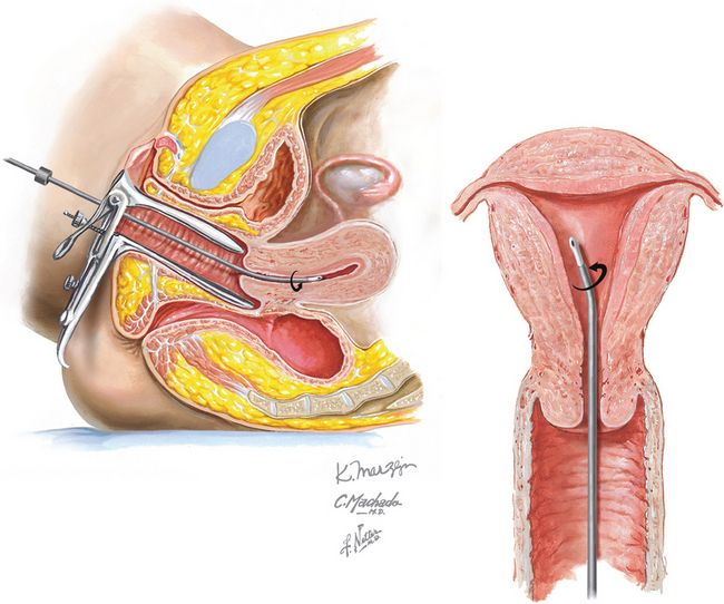
Chapter 243 Endometrial Biopsy
REQUIRED EQUIPMENT
CPT CODE(S)
Lipscomb GH, Lopatine SM, Stovall TG, Ling FW. A randomized comparison of the Pipelle, Accurate, and Explora endometrial sampling devices. Am J Obstet Gynecol. 1994;170:591.
Nolan TE, Smith RP, Smith MT, Gallup DC. A prospective evaluation of an endometrial suction curette. J Gynecol Surg. 1992;8:231.
Dijkhuizen FP, Mol BW, Brolmann HA, Heintz AP. The accuracy of endometrial sampling in the diagnosis of patients with endometrial carcinoma and hyperplasia—A meta-analysis. Cancer. 2000;89:1765.
Ferry J, Farnsworth A, Webster M, Wren B. The efficacy of the Pipelle endometrial biopsy in detecting endometrial carcinoma. Aust NZ J Obstet Gynaecol. 1993;33:76.
Goldchmit R, Katz Z, Blickstein I, et al. The accuracy of endometrial Pipelle sampling with and without sonographic measurement of endometrial thickness. Obstet Gynecol. 1993;82:727.
Larson DM, Broste SK. Histopathologic adequacy of office endometrial biopsies taken with the Z-sampler and Novak curette in premenopausal and postmenopausal women. J Reprod Med. 1994;39:300.
Law J. Histological sampling of the endometrium—A comparison between formal curettage and the Pipelle sampler. Br J Obstet Gynaecol. 1993;100:503.
Warwick A, Ferryman S, Musgrove C, Redman C. An evaluation of the gynocheck for endometrial sampling. J Obstet Gynaecol. 1993;13:198.
American College of Obstetricians and Gynecologists. Management of anovulatory bleeding. ACOG Practice Bulletin 14. Washington, DC: ACOG, 2000.
Chambers JT, Chambers SK. Endometrial sampling: When? Where? Why? With what? Clin Obstet Gynecol. 1992;35:28.
Good AE. Diagnostic options for assessment of postmenopausal bleeding. Mayo Clin Proc. 1997;72:345.
Katz VL. Diagnostic procedures. In: Katz VL, Lentz GM, Lobo RA, Gershenson DM, editors. Comprehensive Gynecology. 5th ed. Philadelphia: Mosby/Elsevier; 2007:227.