Endocrinology

Published on 03/04/2015 by admin

Filed under Endocrinology, Diabetes and Metabolism

Last modified 22/04/2025

Print this page

rate 1 star rate 2 star rate 3 star rate 4 star rate 5 star
Your rating: none, Average: 2.9 (38 votes)

This article have been viewed 3606 times

Endocrinology

PITUITARY DISORDERS

The pituitary is the ‘master controller’ of hormone function in the body, converting signals from the brain and hypothalamus to actions via hormones.

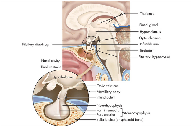
The pituitary normally sits in the sella turcica at the base of the middle cranial fossa (Fig 29.1). It is covered by a dural layer known as the diaphragma sella. The pituitary is joined to the hypothalamus by the infundibulum or pituitary stalk, in front of which sits the optic chiasm. The sella turcica is bounded laterally by the cavernous sinuses and their contents, and the sphenoid sinus antero-inferiorly. The anterior pituitary is embryologically derived from the posterior pharynx and secretes prolactin follicle-stimulating hormone (FSH), luteinising hormone (LH), growth hormone (GH), thyrotropin-stimulating hormone (TSH) and adrenocorticotrophic hormone (ACTH) in response to trophic-releasing hormones from the hypothalamus via a portal blood flow system. The posterior pituitary secretes oxytocin and vasopressin under neural control from the hypothalamus.

PITUITARY TUMOUR

Tumours in the pituitary are common and have been found to occur in around 10% of people in autopsy studies.1 Various series have reported rates of up to 24%. With the high background rate of pituitary masses seen on MRI, clinical questions about how to proceed are likely to occur. Unless the brain is imaged for another reason, it is more prudent to have a clinical diagnosis and confirmatory tests of a pituitary disorder before requesting a CT or MRI of the pituitary.

Tumours of the pituitary may be developmental cysts, blood or infarcted tissue, physiological hyperplasia or adenomas.

Adenomas may be functioning or non-functioning. Physiological effects may be excess autonomous secretion of hormones or trophic hormones, loss of normal pituitary function or mass effects that include compression of nearby structures such as the optic chiasm.

Tumours smaller than 10 mm in size are called microadenomas, and those bigger than 10 mm, macroadenomas. Compressive effects of a pituitary tumour normally occur when the adenoma grows to > 10 mm and enlarges beyond the pituitary fossa. Common symptoms of a mass effect are headaches and loss of peripheral vision that correlates with a bitemporal hemianopia as the optic chiasm is compressed.

As an adenoma grows, there may be loss of function of the normal anterior pituitary in a typical order. The mnemonic: ‘Go Looking For That Adenoma’ is a good aide memoire for the order of loss of pituitary function:

Diagnostic approach

The most important thing is that the diagnosis of a pituitary hormone-stimulated excess or deficiency is made, or at least suspected, and then the pituitary may be imaged. Tumours are sometimes found when a CT head or MRI brain is performed for another reason. In this case, careful history and examination needs to be done, and then testing for any anterior pituitary hormone deficiencies or excesses may be done.

Tumours are defined by their size and secretion of hormones. Tumours do not tend to invoke a mass-related loss of function of other anterior pituitary hormones until they tend toward macroadenomas. The exception to this is a prolactinoma, which will suppress the gonadotrophs in a normal physiological way even when it is a microprolactinoma.

Prolactinomas tend to produce prolactin in a linear relation to their size. Macroadenomas therefore tend to produce levels of prolactin greater than 10 times the upper limit of the reference range. Microadenomas tend to produce levels of prolactin from 1–10 times the upper limit of normal. Other causes of prolactin in this range include medications with a dopamine antagonistic effect, such as antipsychotics and antiemetics. Stress may cause a transient increase in prolactin, as will physical causes such as nipple stimulation and lesions that affect the T4 dermatome, including Varicella zoster. Masses that result in compression or loss of function of the pituitary stalk also limit the inhibitory signals from the hypothalamus and result in microadenoma-level hyperprolactinaemia.

Examination

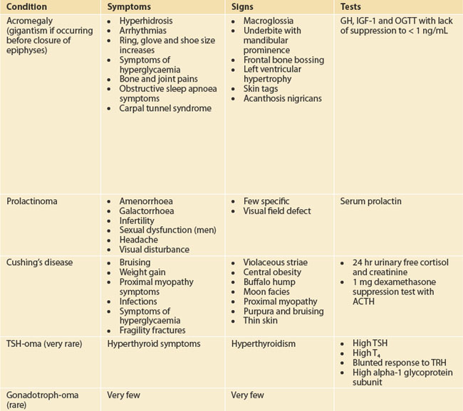
Target the examination to the symptoms and manifestations of hormone excesses or deficiencies as outlined in Table 29.1. Check for galactorrhoea as well as back and skin lesions in patients suspected with hyperprolactinaemia. Galactorrhoea from one breast in the absence of hyperprolactinaemia requires careful examination to ensure that there is no local breast pathology. Always check visual fields to confrontation.

Investigations

Pathology

In a pituitary mass, checking paired trophic hormones and their target hormones is essential for proper interpretation. Check ACTH and cortisol at around 8–9 am, together with FSH, LH and testosterone (oestrogen in females), which also has a diurnal variation, with higher levels in the morning, TSH and thyroxine (T4), prolactin (with dilution in macroadenomas), GH and insulin-like growth factor 1 (IGF-1). Non-functioning adenomas tend to produce higher levels of alpha-1 glycoprotein subunit, which may be requested on a sample of serum. Be aware that ACTH in particular degrades quickly, and so informing the laboratory beforehand of an impending test can help ensure it is put on ice and sent to the central laboratory quickly.

The above are static tests. If both results are normal, beware of the inappropriate levels such as a T4 at the lower limit of normal, with a lower limit of normal TSH (inappropriately normal).

If there are any concerns, dynamic tests may be ordered, although these are often ordered by a specialist and/or in a hospital environment. Of the dynamic test, the oral glucose tolerance test (OGTT) to suppress GH to < 1.0 ng/mL is the only one that is reliable and may safely be done as an outpatient. Twenty-four hour urinary free cortisol and 1 mg overnight dexamethasone suppression tests may also be performed as an outpatient, to help with the investigation of cortisol excess. If an excess is confirmed, then a high-dose dexamethasone suppression test will help delineate whether the problem is Cushing’s disease or ectopic ACTH production.

Integrated management

ACROMEGALY

Acromegaly is a condition of monoclonal growth of pituitary somatotrophs that produces excess growth hormone in a non-regulated way. It has a prevalence of around three per million.2
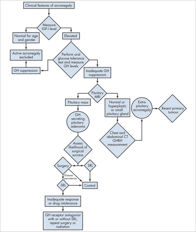
GH is normally secreted in a diurnal and metabolic fashion, with pulsatile release that is highest during sleep. Amino acids and ghrelin from the gut are also stimuli to its release via the hypothalamus.

GH stimulates the production of IGF-1 and IGFBP-3 (insulin-like growth factor binding protein-3) on binding to the receptors in the liver. There are some direct effects on the cartilage but, other than that, the majority of the physiological effects of GH are mediated via IGF-1. The liver’s ability to produce IGF-1 in response to GH is blunted in liver disease, hypothyroidism and poorly controlled diabetes mellitus. Interestingly, malnutrition reduces IGF-1 production, and obesity inhibits GH pulses from the pituitary.

Integrated management

See Fig 29.2 for an overview of management.

HYPOPITUITARISM

Hypopituitarism may be complete (pan-) or partial. It may be congenital, acquired or iatrogenic.

Congenital problems are many and rare, and such things as PIT-1 gene mutations cause a loss of lactotrophs (prolactin), thyrotrophs (TSH) and somatotrophs (GH). Others, such as Kallmann’s syndrome, result in loss of the gonadotrophs and a normal male phenotype but hypogonadotrophic hypogonadism and a degree of olfactory deficit.

Acquired hypopituitarism may occur due to problems such as trauma affecting the pituitary stalk (infundibulum), apoplexy or infarction in Sheehan’s syndrome. Other issues such as lymphocytic hypophysitis are being increasingly recognised with higher-teslar MRI machines. Infections and inflammatory lesions such as sarcoid and histiocytosis may affect the pituitary, as well as intra- and extrasellar masses.

Iatrogenic causes include radiation-associated pituitary damage or surgical complication after attempted removal of a macroadenoma.

Lastly, hypopituitarism can be secondary to hypothalamic disease.

GH

ACTH TSH   ADH/vasopressin

Integrated management

PARATHYROID DISORDERS

Located generally behind the thyroid gland are the four parathyroid glands, which have the role of producing parathyroid hormone (PTH). PTH is the main controller of calcium levels within the blood and bones, and this of course has important implications for neuromuscular function.

HYPERCALCAEMIA

Extracellular calcium is a tightly controlled electrolyte. Significant symptoms, including life-threatening conditions, occur when control is lost and the levels of calcium in the body go outside the tightly controlled range. The largest store of calcium within the body is in the bones, but calcium is stored within skeletal and cardiac muscle, and within the neuronal signalling and neuromuscular junctions.

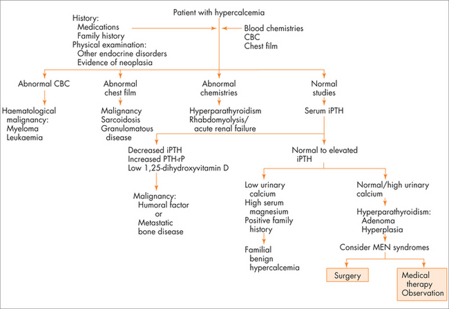
The most common symptoms of hypercalcaemia are the classic ‘stones, bones, moans and groans’ of kidney stones, bone pains, mood changes including depressive symptoms and abdominal pains including constipation. Polyuria and dehydration can occur secondary to high serum calcium. The most common cause for congenital hypercalcaemia is familial hypocalciuric hypercalcaemia. The most common acquired causes of hypercalcaemia are hyperparathyroidism and malignancy. A useful thought map to think about acquired hypercalcaemia is to think about PTH-dependent and PTH-independent causes of hypercalcaemia (see Fig 29.3).

Aetiology

Extracellular calcium is normally controlled very tightly by two interrelated hormones, namely parathyroid hormone (PTH) and vitamin D. There is a sigmoidal inverse relationship between serum calcium levels and PTH levels, such that a decrease in serum calcium results in increased PTH and vice versa.

Familial hypocalciuric hypercalcaemia is a congenital variation in the sensitivity of the calcium-sensing receptor, with a reduced sensitivity to serum calcium and so a higher set point of serum calcium at which the PTH is turned down. Serum calcium is high, as is PTH, but urine 24-hour calcium excretion is low.

Milk–alkali syndrome is less common now that histamine receptor antagonists and proton pump inhibitors are the mainstay treatment of hyperacidity syndromes of the stomach. The use of antacids together with the ingestion of milk products results in increased absorption and mild hypercalcaemia. While there has been a reduction in the presentation of milk–alkali syndrome from these less-used drugs, there have been a number of case reports of similar presentations in those using large doses of calcium carbonate, which provides both calcium and alkali. High calcium level together with high bicarbonate and perhaps some renal impairment should prompt the GP to ask about calcium carbonate intake.

PTH-dependent hypercalcaemia can be due to primary hyperparathyroidism, less commonly secondary hyperparathyroidism, or tertiary hyperparathyroidism. Secondary hyperparathyroidism often does not result in hypercalcaemia alone, as it is an appropriate response to maintain calcium levels. This occurs in the situation of vitamin D deficiency, where high parathyroid levels maintain serum calcium when vitamin D levels are insufficient to provide enough gut and renal absorption to do so. Primary hyperparathyroidism may be due to a parathyroid adenoma, or adenomas on their own or in the setting of MEN1 syndrome of pituitary tumours, hyperparathyroidism and pancreatic tumours, or MEN2 with medullary thyroid cancer and phaeochromocytoma. Parathyroid adenomas are most commonly sporadic and not part of another syndrome, and are mostly single-gland adenomas. Less commonly, PTH-secreting thymus tumours can produce the parathyroid hormone excess. Tertiary hyperparathyroidism is thought to occur through long-standing secondary hyperparathyroidism, which then causes irreversible parathyroid gland hyperplasia and autonomous function.

PTH-independent causes of hypercalcaemia are usually due to excesses in active vitamin D, or lytic lesions of the bone. The far less common production of PTHrp (PTH-related protein) is usually associated with squamous cell carcinomas, or renal, bladder, breast or ovarian cancers. The conditions that result in excess active vitamin D (1,25-dihydroxy vitamin D) are excess intake of calcitriol, or granulomatous diseases such as sarcoid, tuberculosis and lymphoma, both non-Hodgkins and Hodgkins. In these conditions the macrophages in the granulomata convert the inactive vitamin D to active vitamin D without the need for PTH, which normally carries this out by stimulating the 1-alpha-hydroxylase enzyme in the kidneys.

Other causes such as multiple myeloma and breast cancer are the two more common malignancies that can lead to lytic bone lesions and uncontrolled release of calcium into the extracellular fluid, independently of a suppressed PTH level.

Integrative management

Surgery is indicated for primary hyperparathyroidism in symptomatic disease. In the case of asymptomatic primary hyperparathyroidism, surgery can be considered if:

If the patient is not a surgical candidate, due to frailty, then Cinacalcet, a calcium sensing receptor agonist, may be an option, but at the time of publication is only indicated for secondary hyperparathyroidism in renal failure and parathyroid cancer.

HYPERPARATHYROIDISM

Most of the investigation and treatment of hyperparathyroidism is covered above, in the hypercalcaemia section. Brief further information is given below.

Hyperparathyroidism can be primary, secondary or tertiary.

Primary hyperparathyroidism (PHPTH) is due to between one and four functioning adenomas of the parathyroid glands. In 85% of cases there is a single parathyroid gland adenoma, which secretes PTH independent of the serum calcium. It is most common in women over the age of 55 years. The remainder of PHPTH is due to multiple adenomas, hyperplasia and, very rarely, parathyroid carcinoma. The incidence of PHPTH is around 4 per 100,000.

Secondary hyperparathyroidism is usually found in the setting of vitamin D deficiency. With the strength of the sun and concerns about skin cancer, in some countries such as Australia and New Zealand, and the increasingly indoor lifestyle of the population in developed countries, vitamin D deficiency can be very common. Those at highest risk are the elderly, such as those in institutional care, dark-skinned races and societies where it is culturally appropriate to wear head-to-toe clothing.

In vitamin D deficiency, the control of serum calcium falls back onto PTH, which stimulates renal absorption of calcium, liberation of calcium from the mineralised bone at the expense of bone mineral density, and increased hydroxylation of the low levels of 25-OH Vit D to 1,25-OH Vit D. These patients are normally eucalcaemic. Secondary hyperparathyroidism can also occur in the setting of renal failure, with loss of kidney parenchyma to convert 25-OH Vit D to 1,25-OH Vit D under the control of PTH. As such, the patient has reduced active vitamin D, and PTH predominates. 1,25-OH Vit D actively inhibits the growth of the PTH glands. The renal physician may elect to give the patient active vitamin D in the form of calcitriol, but it depends on the levels of calcium and phosphate. Elective parathyroidectomy may be indicated at very high levels of secondary hyperparathyroidism, depending on the stage of their chronic kidney disease.

Tertiary hyperparathyroidism usually occurs in renal patients who have had long-standing secondary hyperparathyroidism. With low levels of active vitamin D, there is loss of inhibition of parathyroid gland hyperplasia, and autonomous PH secretion results from the hyperplastic glands, even when the cause of the secondary hyperparathyroidism is corrected.

Integrative management

HYPOPARATHYROIDISM

Hypoparathyroidism is most commonly acquired, with transient or permanent hypoparathyroidism post surgery for thyroid disorders. Aside from surgery, there are several causes of hypoparathyroidism that arise due to aplasia of the glands, autoimmune destruction or inhibition of the release of PTH by activating antibodies to the calcium-sensing receptor. Many of the causes of hypoparathyroidism that appear in childhood are parts of rare but important syndromes, which should be managed by a paediatric endocrinologist. The more common cause for childhood hypoparathyroidism, as part of the autoimmune polyglandular syndrome type 1, is discussed in this section.

Pathology

Congenital and genetic:

PTH release problems:

Surgery:

Autoimmune:

Pseudohypoparathyroidism:

Integrative management

PARATHYROID TUMOUR

Parathyroid adenoma and carcinoma are difficult to differentiate clinically. Parathyroid carcinoma is an uncommon but important condition occurring in around 2% of investigated hyperparathyroidism. With a different prognosis and natural history, it is important to identify. Typically, parathyroid carcinomas tend to cause higher levels of hyperparathyroidism and hypercalcaemia than primary hyperparathyroidism.

Integrative management

THYROID DISORDERS

The thyroid gland is a butterfly-shaped gland that sits in the neck around the thyroid and cricoid cartilages of the larynx and trachea. It is primarily responsible for producing the hormone thyroxine, which accelerates or brakes the body’s metabolic processes. The symptoms of an over- or underactive thyroid vary, and can be quite different between patients.

HYPERTHYROID DISORDERS

Symptoms of hyperthyroidism are those of a metabolism that is sped up, and include symptoms resembling anxiety and agitation, tremors, weight loss despite an increased appetite, palpitations, a syncopated heartbeat, spread out or absent periods, frequent opening of bowels, heat intolerance and lethargy. There is often a family history of thyroid problems, either hyperthyroidism or hypothyroidism. There are a few common causes for an overactive thyroid, described below.

Graves’ disease

Normally the thyroid gland’s production is controlled by the pituitary gland. The control is finely tuned. Graves’ disease is an autoimmune condition and is the most common cause of hyperthyroidism, affecting up to 2% of women—it is approximately four times more common in women than in men. In Graves’ disease, the immune response, rather than destroying the thyroid gland, stimulates it, making it grow in both size and production of thyroxine. This creates a goitre, which is typically painless, and the symptoms of thyrotoxicosis mentioned above.

The antibodies or proteins that stimulate the thyroid gland may also stimulate the tissues behind the eyes, and the anterior tibial region. If these antibodies stimulate the tissues behind the eyes it leads to exophthalmos, creating an impression of staring, and may affect vision if extreme. Referral to an ophthalmologist is appropriate. Eye problems in Graves’ disease seem to be more common in smokers.

HYPOTHYROID DISORDERS

The symptoms associated with an underactive thyroid gland and low levels of thyroxine include tiredness, weight gain and reduced appetite, dyspnoea on exertion, peripheral oedema, dry skin and hair, cold intolerance, menorrhagia and easy bruising. The most common cause is autoimmune destruction of the thyroid gland, Hashimoto’s thyroiditis.

THYROID DISEASE: SUMMARY OF THERAPEUTICS

Hypothyroidism:

Supplements:

Thyroiditis:

Hyperthyroidism:

ADRENAL GLAND DISORDERS

The adrenal glands sit atop the kidneys and are primarily responsible for releasing a range of chemical mediators of the stress response, such as glucocorticoids (e.g. cortisol) from the outer cortex. The cortex is also responsible for releasing mineralocorticoids such as aldosterone, which has an important role in regulating blood pressure, and androgens such as dehydroepiandrosterone (DHEA). Catecholamines such as adrenaline and noradrenaline are released from the central area of the gland, called the medulla.

ADDISON’S SYNDROME

Adrenal insufficiency is an important treatable condition, and can be life-threatening if not identified and treated properly. The incidence varies between developed and developing countries, due to the higher incidence of infective diseases affecting the adrenal gland, in the latter—it is estimated at 1 in 100,000 in developed countries and up to 11 per 100,000 in developing countries, and is likely to increase in the latter in line with the incidence of Mycobacterium tuberculosis disease and HIV/AIDS.

Aetiology

Adrenal insufficiency may be secondary, due to pituitary disease. It usually occurs in panhypopituitary patients after surgery, trauma affecting the stalk, hypothalamic disease or radiotherapy. ACTH chromophobe cells of the pituitary are usually the last cells to lose their function in the setting of a compressing macroadenoma. ACTH may be selectively lost in isolation, as in lymphocytic hypophysitis. These patients differ clinically in that they do not have typical hyperkalaemia, due to the fact that they have an intact renin-angiotensin-aldosterone system.

More commonly, adrenal insufficiency is primary, due to diseases that directly affect the adrenal gland itself. These patients are more likely to be hyperkalaemic, with loss of the mineralocorticoid as well as the glucocorticoid production. Primary adrenal diseases may be congenital anatomical problems such as adrenal hypoplasia, or congenital enzyme problems, as in congenital adrenal hyperplasia where the genes encoding enzymes involved in the production of cortisol are defective.

Infections such as Mycobacterium tuberculosis may produce a granulomatous destruction of the adrenal cortex and medulla, usually in the setting of disseminated tuberculosis. Syphilis and HIV have been documented to result in adrenal insufficiency. Neisseria meningitidis septicaemia may result in adrenal infarction and the Waterhouse-Friderichsen syndrome of associated adrenal insufficiency. Infarction may occur in any form of septicaemia, as can haemorrhage due to disseminated intravascular coagulation (DIC) and coagulopathy. Supratherapeutic anticoagulation may also result in adrenal haemorrhage and insufficiency.

Fungal infections with Histoplasmosis and Cryptococcus may result in adrenal insufficiency, which may or may not recover with appropriate treatment.

Ketaconazole, which may be used for its antifungal effects or for metastatic prostate cancer, may also block enzymes in the steroidogenesis pathway of cortisol and result in reversible loss of adrenal cortisol production. It and other drugs such as metyrapone etomidate and mitotane may be used with the indication of medically and surgically resistant Cushing’s syndrome, due to their effects on decreasing cortisol production from the adrenal cortex.

The very common but often forgotten cause of adrenal insufficiency is withdrawal of corticosteroids too quickly after a course long enough and at high enough dose to suppress ACTH stimulation of the adrenal cortices, and thus atrophy, and poor ACTH response when exogenous steroids are withdrawn. The static laboratory test of adrenal function with an early-morning cortisol and ACTH or the dynamic short synacthen test are identical, and history should point to the cause. Steroids must then be reintroduced and tapered at a slower rate. It is variable but a prednisone equivalent dose to around 10 mg per day for approximately 3 weeks should be assumed to suppress the adrenal cortex, and will require a slower taper. Adrenocortical suppression is more likely in elderly patients and those with dexamethasone or nocturnal doses of steroids. The nocturnal doses more effectively suppress the early morning peak of ACTH and thus lead more quickly to adrenal cortex atrophy and blunted ACTH response.

Adrenal cortical dysfunction is most commonly autoimmune in the developed world. It may occur in isolation or in the setting of APS types 1 and 2. Type 1 APS is characterised by mucocutaneous candidiasis, hypoparathyroidism and autoimmune adrenalitis that usually occurs in the first years of life and almost invariably occurs before the age of 20 years. APS Type 2 is more common than type 1 and occurs later in life than type 1, with autoimmune adrenalitis, autoimmune thyroid disease (hypo- more than hyperthyroidism) and type 1 diabetes mellitus. Adrenalitis usually occurs first but may follow the other manifestation in a good proportion of patients.

Other congenital causes of adrenal insufficiency such as adrenoleukodystrophy are more rare.

Infiltration from amyloid or metastatic breast or lung cancer is also rare.

Diagnostic approach

History and examination will often lead to a feeling for whether there is a problem with adrenocortical function, and may point to a possible cause. (See the section on history, below, for common symptoms and signs.)

Hyponatraemia is common in adrenal insufficiency, but hyperkalaemia is less common and does not occur in secondary adrenal insufficiency due to intact aldosterone production. An early-morning paired cortisol and ACTH will indicate whether there is a problem with primary or secondary hypoadrenalism. A cortisol peak of 400 nmol/L has a high specificity for adequate adrenal function. Regardless of ACTH levels, there is no need to further investigate adrenocortical insufficiency.

ACTH is released in a peak and is relatively unstable if not collected properly, but, those issues aside, a low cortisol with a low or inappropriately normal ACTH would tend to point to secondary adrenal insufficiency. A high ACTH at any time of the day with a less than adequate cortisol is very indicative of primary adrenal problems. In the indeterminate range, a short synacthen test of 250 μg of synacthen given as an intramuscular injection with cortisol measured at 0, 30 and 60 minutes can be performed. If done at 0800 with a morning ACTH and cortisol, a basal result of > 400 nmol/L or a stimulated rise of about 550 nmol/L indicates adequate adrenal function. Check with your local laboratory as to the normal cut-offs for its particular assay. The short synacthen test does not differentiate between primary and secondary adrenal insufficiency in the chronic setting, where there is inadequate ACTH drive with resulting adrenal atrophy and blunting of adrenal response, as one would see in primary disease.

Some have argued that this is a supraphysiological test, which may not properly assess for partial adrenal insufficiency. It has been suggested that a 1 μg synacthen test may be more of a physiological test dose.

Integrative management

Pharmacological

If the patient has presented unwell, hypotensive and shocked, and you suspect adrenal insufficiency, establish large-bore IV access and take blood for FBC, eLFT, ACTH and cortisol, and then commence treatment for shock, as well as giving 100 mg of IV hydrocortisone. The patient should be transferred to care in hospital. If the cortisol result returns as adequate in 1–2 days, then the steroids treatment may be stopped.

All steroids except dexamethasone have mineralocorticoid effects. Patients with secondary adrenal insufficiency will have an intact mineralocorticoid output, so addition of any extra mineralocorticoid in the form of fludrocortisone is not necessary.

In chronic adrenal insufficiency, replacement regimens are many but the aim is to try to provide enough glucocorticoid replacement in a normal physiological pattern that follows a diurnal variation, with a peak early in the morning that then tapers through the day. This may be achieved with hydrocortisone 20–30 mg per day in 2–3 divided doses in a regimen such as 12 mg on waking, 8 mg at lunch and 4 mg at afternoon tea. A simpler strategy is hydrocortisone 20 mg mane and 10 mg in the early afternoon, or cortisone acetate 25 mg mane and 12.5 mg in the early afternoon. The afternoon dose may be brought closer to midday if the patient reports difficulty sleeping. If the patient reports feeling unwell and quite fatigued in the early evening, then using a three-times-a-day split dose with a smaller proportion in the late afternoon can be trialled.

Be aware that fatigue as a symptom of inadequate replacement may lead to excessive replacement. There are many causes of fatigue. If hydrocortisone is used, checking a 24-hour urine collection for free cortisol may be used to assess whether the replacement is excessive.

In the setting of adrenal suppression from long-term steroids, treatment for other conditions in someone who should otherwise have an intact hypothalamic–pituitary–adrenal (HPA) axis, then a slow taper of the steroids, should allow for recovery of their normal axis function. The longer the patient has been on steroids, and the older the patient, the slower the taper. This may require a taper over several months to a year. Adequacy of their axis may be checked with a morning ACTH and cortisol prior to their normal dose, noting that the dexamethasone with its longer duration of action is likely to interfere with this technique.

If patients with primary adrenal insufficiency have documented postural hypotension or symptoms, and are on adequate glucocorticoid replacement, fludrocortisones 0.05–0.1 mg/day may be added.

CUSHING’S SYNDROME

Cushing’s disease is the disorder described by Harvey Cushing, with pituitary disease with ACTH-dependent hypercortisolism. Cushing’s syndrome describes the other forms of cortisol excess. (See Table 28.3 for a list of other causes of hypercortisolaemia.) The incidence of Cushing’s disease or syndrome is difficult to quantify. There is significant overlap between the manifestation of Cushing’s disease, and that of obesity and its complications. European series have estimated the incidence at 1–3 per million patients.

TABLE 28.3 Causes of hypercortisolaemia

Name Cause
Cushing’s disease ACTH-dependent pituitary disease
Pseudocushing’s disease

Factitious Cushing’s Iatrogenic or surreptitious glucocorticoid excess Ectopic ACTH Bronchogenic tumours producing ACTH Primary hypercortisolism Cortisol-secreting adrenal tumour

Establishing the diagnosis of hypercortisolaemia involves establishing a clinical likelihood or pre-test probability, establishing the biochemical excess of glucocorticoids, and then establishing the cause, in order to plan effective treatment.

The diagnosis may be perplexing, given that there is much overlap between normal levels in biochemical testing and levels in those with true glucocorticoid excess.

Aside from iatrogenic Cushing’s syndrome, Cushing’s disease accounts for the majority of cases of Cushing’s syndrome, at 60–70%. Adrenal disease is also not uncommon, accounting for approximately 20% of cases. Ectopic ACTH secretion and resultant Cushing’s syndrome accounts for approximately 10–15% of cases, with the other causes responsible for the few remaining per cent.

Diagnostic approach

Question 2: Is there glucocorticoid excess?

The Endocrine Society has released guidelines for investigation of Cushing’s Syndrome and Cushing’s disease.3

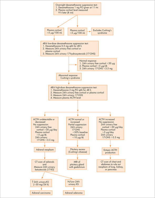
See Figure 29.4 for an overview.

Note: Conditions that may lead to abnormal results with few features of Cushing’s syndrome clinically include:

Integrative management