Endocrine disease

Published on 02/03/2015 by admin

Filed under Internal Medicine

Last modified 22/04/2025

Print this page

rate 1 star rate 2 star rate 3 star rate 4 star rate 5 star
Your rating: none, Average: 0 (0 votes)

This article have been viewed 4392 times

15 Endocrine disease

Pituitary disorders

The hypothalamus regulates many vital functions, including appetite, thirst, thermal regulation and sleep. It controls pituitary function by stimulatory and inhibitory factors and by hormones released via the pituitary stalk.

Many hormones are released in either a pulsatile or a circadian pattern and are regulated by feedback systems. Endocrine tests on serum/plasma take advantage of natural peaks and troughs in secretion, while stimulatory or suppression tests are used to investigate hormone deficiency or excess further.

Pituitary tumours

Pituitary tumours are a relatively common incidental finding in the general population; only a minority cause clinical problems, either due to uncontrolled secretion of pituitary hormones, or due to local compression effects. Tumours may be small (micro-adenomas, < 10 mm diameter) or large (macro-adenomas, > 10 mm) and the vast majority are benign.

Treatment

The aim of therapy is to:

Treatment (Table 15.1) options include surgery, radiotherapy and medical therapy, depending on the aetiology of the pituitary mass. Post-operative radiotherapy is used if significant tumour bulk remains after surgery or the underlying disease is still active. Radiotherapy results in a gradual decline of residual pituitary function over many years and must be monitored.

Table 15.1 Comparison of primary treatment for pituitary tumours

Treatment method Advantages Disadvantages
Surgical
Trans-sphenoidal adenomectomy or hypophysectomy Relatively minor procedure
Potentially curative for micro- and smaller macro-adenomas
Some extrasellar extensions may not be accessible
Risk of CSF leakage and meningitis
Trans-frontal Good access to suprasellar region Major procedure; danger of frontal lobe damage
High chance of subsequent hypopituitarism
Radiotherapy
External (40–50 Gy) Non-invasive
Reduces recurrence rate after surgery
Slow action, often over many years
Not always effective
Possible late risk of tumour induction
Stereotactic Precise administration of dose to lesion Long-term follow-up data limited
Yttrium implantation High local dose Only used in few centres
Medical
Dopamine agonist (e.g. bromocriptine) Non-invasive; reversible Usually not curative; significant side-effects in minority
Somatostatin analogue (octreotide) Non-invasive; reversible Usually not curative; expensive
Growth hormone receptor antagonist (pegvisomant) Highly selective Usually not curative; very expensive

Hypopituitarism

Compression of the pituitary gland by a mass lesion results in progressive loss of function. Growth hormone and gonadotrophins are usually the first hormones to be affected, followed by TSH and finally ACTH (panhypopituitarism). Prolactin levels may rise due to compression of the pituitary stalk and loss of dopaminergic inhibitory control.

Investigation of pituitary function

Basal serum investigations

Dynamic tests

Management

Options for hormone replacement regimes are given in Table 15.2.

Table 15.2 Replacement therapy for hypopituitarism

Axis Usual replacement therapies
Adrenal Hydrocortisone 15–40 mg daily (starting dose 10 mg on rising/5 mg lunchtime/5 mg evening)
(Normally no need for mineralocorticoid replacement)
Thyroid Levothyroxine 100–150 mcg daily
Gonadal  
Male Testosterone IM, orally, transdermally or implant
Female Cyclical oestrogen/progestogen orally or as patch
Fertility HCG plus FSH (purified or recombinant) or pulsatile GnRH to produce testicular development, spermatogenesis or ovulation
Growth Recombinant human growth hormone used routinely to achieve normal growth in children
Also advocated for replacement therapy in adults where growth hormone has effects on muscle mass and well-being
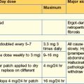
Thirst Desmopressin 10–20 mcg 1–3 times daily by nasal spray or orally 100–200 mcg 3 times daily
Carbamazepine, thiazides and chlorpropamide are very occasionally used in mild diabetes insipidus
Breast (prolactin inhibition) Dopamine agonist (e.g. cabergoline 500 mcg weekly)

FSH, follicle-stimulating hormone; GnRH, gonadotrophin-releasing hormone; HCG, human chorionic gonadotrophin.

Thyroid disorders

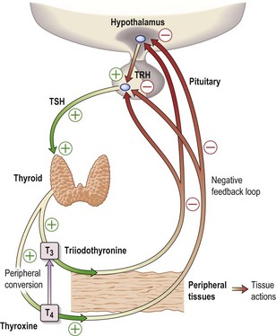
The thyroid hormones, T4 and T3, are produced within the thyroid gland. Synthesis and release are stimulated by TSH, which is released from the pituitary gland in response to the hypothalamic factor, TRH (thyrotrophin-releasing hormone) (Fig. 15.1). Predominantly, T4 is produced, but this is converted in the peripheral tissues (liver, kidney, muscle) to the more active T3. More than 99% of T4 and T3 circulate bound to plasma proteins, mainly thyroid-binding globulin (TBG).

Diagnosis of thyroid disease includes the clinical features, together with measurement of plasma TSH and thyroid hormones T4 and T3.

Hypothyroidism

Hyperthyroidism

Hyperthyroidism is common and is characterized by high levels of T3 and T4 with suppression of plasma TSH. Nearly all cases are due to intrinsic thyroid disease.

Clinical features

Weight loss, increased appetite, irritability, tremor and heat intolerance are classic symptoms of hyperthyroidism. A tachycardia with or without atrial fibrillation is a frequent presentation, particularly in the elderly. A goitre with a bruit occurs. Apathetic thyrotoxicosis occurs in elderly patients, in whom the clinical picture is more like hypothyroidism with few clinical signs of thyrotoxicosis.

Treatment (Table 15.3)

Propranolol is used to gain rapid control of thyrotoxic symptoms. High doses (up to 160 mg 3 times daily) may be required due to increased metabolism of the drug.

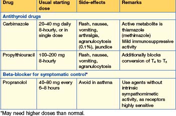
There are three main options and preference varies widely.

Antithyroid medications. Carbimazole initially 20 mg 3 times daily is the drug most commonly used in the UK. Its active metabolite, methimazole, is used preferentially in the USA. These drugs inhibit the formation of thyroid hormone and the clinical benefits can take up to 2 weeks to become apparent. Propylthiouracil 300–600 mg daily is occasionally used as an alternative, particularly during pregnancy and breast feeding. It has additional theoretical benefits in that it prevents peripheral conversion of T4 to T3. The major side-effect of these medications is agranulocytosis, and prior to commencing treatment patients should be told to seek medical help should severe mouth ulcers, sore throat or febrile illness occur. A white blood cell count is then urgently required. Antithyroid medication also reduces the immune processes and thus a trial of therapy for up to 18–24 months may result in remission of autoimmune thyrotoxicosis. Initially doses of antithyroid medications are high. There is then the option of dose reduction as the thyroid tests normalize, or the option of adding thyroxine to high-dose anti-thyroid medication (‘block and replace regimen’). A fall of T4 and T3 in response to treatment guides initial dose reductions (gradually reduce dose to 5 mg over 6 to 24 months if hyperthyroidism is controlled), as the TSH may remain suppressed for some time. Antithyroid medications will never lead to remission in toxic nodular disease, and while long-term antithyroid medications are an option, more definitive treatment is required.

Goitre

Assessment is made of its pathological nature and the patient’s thyroid status. Acute pain within a goitre suggests an acute viral thyroiditis (de Quervain’s), which may be associated with transient hyperthyroidism. Pain may also herald bleeding into a cyst and is rarely associated with thyroid cancer.

Reproduction and sexual disorders

Normal puberty in the male begins between 10 and 14 years with acceleration of growth velocity and development of secondary sexual characteristics. In girls sexual development starts a year earlier and normal menses begin between the ages of 11 and 15.

Female disorders

Polycystic ovarian syndrome (PCOS)

PCOS consists of oligomenorrhoea, hirsutism, acne and weight gain. Hyperandrogenism, insulin resistance and features of the metabolic syndrome (p. 429) are frequently seen. The syndrome probably arises from a combination of environmental and genetic interactions. Increased numbers of ovarian follicles may be found on US, but this is also seen in women without the syndrome.

Investigations

• Treatment. Weight loss, exercise and non-medical management of hirsutism, e.g. shaving, waxing, laser and electrolysis, are used. A topical cream, eflornithine (an antiprotozoal), has become available and reduces hair growth.

Menopause

Menopause is diagnosed with loss of menses for 12 months, occurring at an average age of 50 years.

Premature menopause occurs with loss of menses before the age of 40. Chromosomal abnormalities and autoimmune ovarian failure account for the majority of cases, while chemotherapy and radiotherapy can also result in early gonadal failure.

Hyperprolactinaemia

Prolactin stimulates breast milk production and inhibits the gonadotrophin axis, resulting in oligomenorrhoea and reduced libido.

Assay interference by ‘macroprolactin’, a prolactin–IgG complex that lacks biological activity, should be excluded.

Acromegaly

Growth hormone acts in the liver to synthesize and secrete insulin-like growth factor (IGF)-1, which promotes skeletal and soft tissue growth. Growth hormone excess will result in gigantism prior to epiphyseal fusion, and acromegaly following puberty.

Acromegaly is due to excess growth hormone release, usually from a benign pituitary adenoma.

Somatotroph tumours are frequently large (> 1 cm diameter) and are associated with local compression effects, including chiasmal compression causing visual field defects and ophthalmoplegia. Compromise of normal pituitary function can result initially, with loss of libido or oligomenorrhoea.

Excess growth hormone results in a change of bodily appearance with coarsening of the features and an increase in shoe, glove and in particular ring size. Increased sweating is a common symptom, as is generalized lethargy and weakness — which may result from loss of ACTH but may also reflect loss of intrinsic muscle power despite increasing muscle bulk.

Acromegaly is associated with increased mortality from cardiovascular, respiratory and metabolic complications, e.g. diabetes. Obstructive sleep apnoea is commonly recognized. The incidence of colorectal carcinoma is also higher.

Treatment

Trans-sphenoidal surgery remains the treatment of choice, with medical therapy used to optimize the patient for anaesthesia and to achieve a reduction in tumour bulk to facilitate removal. Normal growth hormone dynamics are rarely achieved with surgery and therefore disease control rather than cure is sought. Following surgery, medical therapy may once more be required to achieve satisfactory growth hormone control — patients with uncontrolled levels are offered radiotherapy, which should improve growth hormone levels over subsequent months and years. A nadir of growth hormone levels following a GTT of < 5 mU/L, and normalization of serum IGF-1 level are predictive of good outcome in the long term.

Cushing’s syndrome

Cortisol, secreted from the adrenal cortex under the influence of pituitary ACTH, is the principle endogenous glucocorticoid. It enhances gluconeogenesis, fat deposition, protein catabolism, sodium retention and potassium loss, and attenuates the immune response.

Cushing’s syndrome is due to glucocorticoid excess, which is seen most frequently following the therapeutic administration of exogenous synthetic steroids.

Clinical features consist of weight gain, change of appearance, depression, lack of libido, excess hair growth, acne and psychosis. Signs include plethora, thin skin, bruising, hypertension, pathological fractures (particularly involving the vertebrae and ribs), striae (purple or red) and a proximal myopathy. Moon face is characteristic.

Diagnosis

Exogenous steroid use should be excluded. Medications such as oestrogen will increase cortisol-binding globulin and result in misleadingly high total cortisol measurements. Such medications should be stopped 6 weeks prior to investigations.

Adrenal insufficiency

Adrenal insufficiency may result from primary adrenal disease, pituitary disease with failure of ACTH production or, very rarely, hypothalamic disease (failure of corticotrophin-releasing hormone (CRH)). In practice, adrenal insufficiency is most commonly seen in the context of stopping exogenous glucocorticoid therapy for inflammatory diseases such as rheumatoid arthritis or asthma.

Clinical features are often vague and non-specific, but include weight loss, anorexia, weakness, fever, depression, diarrhoea, abdominal pain and constipation. Signs include pigmentation (especially of new scars and palmar creases) and postural hypotension. Loss of body hair and vitiligo also occur.

Secondary adrenal failure

Insufficient ACTH production from pituitary disease, or HPA axis suppression following long-term exogenous steroid therapy, leads to failure of adrenal glucocorticoid production. Adrenal mineralocorticoid function is mainly preserved, being stimulated predominantly by angiotensin II rather than ACTH.

Assessment of the HPA axis

Short tetracosactide test: see p. 568. Variations exist, which extend the test to last up to 2 days. These tests help differentiate between primary, secondary and tertiary (hypothalamic) failure. Primary adrenal failure will fail to respond to ACTH, while secondary and tertiary adrenal failure will exhibit a delayed response to exogenous ACTH.

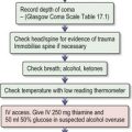
Emergency management of adrenal failure (Box 15.1) (also see Emergencies in Medicine p. 720)

Long-term steroid therapy

Long-term therapy with supra-physiological doses of exogenous steroids results in adrenal failure and compromise of the adrenal response to stress if the glucocorticoid therapy is abruptly withdrawn.

Side-effects. While steroid replacement therapy at physiological doses is not associated with side-effects (Box 15.2), high-dose treatment is frequently associated with unwanted effects. In the short term, high-dose steroid therapy may be associated with mood swings and sleep disturbance. Longer-term therapy may lead to adrenal suppression, osteoporosis and cushingoid appearance. Glucose intolerance can also be seen with high-dose steroid therapy, as can hypertension and hypokalaemia.

Diabetes insipidus

Fluid homeostasis, i.e. thirst and water regulation, is largely controlled by antidiuretic hormone (ADH, also called vasopressin); this is synthesized in the hypothalamus and released from the posterior pituitary in response to rising serum osmolality or perceived hypovolaemia. Other factors influencing ADH release include nausea, hypothyroidism and hypercortisolaemia.

Polyuria and loss of free water (as much as 10–15 L/24 hours) can result from loss of ADH action, either through failure of secretion secondary to hypothalamic damage, or renal insensitivity to ADH. Any cause of polyuria, including psychogenic polydipsia, will give mild renal resistance to ADH. Relative protection from overwhelming dehydration in diabetes insipidus is maintained by an intact thirst axis, which will ensure an adequate fluid intake. Cranial diabetes insipidus may be masked by cortisol deficiency resulting from pituitary damage, since glucocorticoids are necessary for renal handling of free water.

Syndrome of inappropriate ADH secretion (SIADH)

Inappropriate secretion of ADH results in retention of water and subsequent hyponatraemia. Mild symptoms of confusion, irritability and nausea occur as sodium levels fall below 125 mmol/L (125 meq/L); fitting and coma occur as the sodium falls below 115 mmol/L. A diagnosis of SIADH can only be made in a patient who is clinically normovolaemic with normal thyroid and adrenal function.

Hypercalcaemia

The major causes of hypercalcaemia are primary hyperparathyroidism and malignancies (> 90% of cases).

Treatment of primary hyperparathyroidism

Primary hyperaldosteronism

Angiotensin is generated in response to increased renin levels, which occur due to reduced renal perfusion. It causes vasoconstriction and aldosterone release. Aldosterone leads to renal sodium and water retention with potassium loss.

Adrenal adenomas (Conn’s syndrome) and bilateral adrenal hyperplasia account for the majority of cases of primary hyperaldosteronism.

Phaeochromocytoma

Phaeochromocytomas are tumours arising from the adrenal medulla, although 10% occur in extramedullary chromaffin tissue and are referred to as paragangliomas. Around 90% of cases are sporadic and these are typically solitary lesions. The majority are benign, although significant morbidity and mortality can be associated with them. Most phaeochromocytomas release noradrenaline (norepinephrine), some release noradrenaline and adrenaline (epinephrine), and occasionally they release adrenaline alone or dopamine. They are associated with sustained or paroxysmal hypertension; symptoms include headaches, palpitations and sweating.