29. Encephalocele
Definition
Encephalocele is a herniation of a portion of the brain and/or meninges through a defect of the bony skull table. The condition may be congenital or result from trauma or surgery.
Incidence
In the United States the incidence is 1 to 4:10,000 live births; internationally there is no estimate available. Females are affected more often than males.
Etiology
Congenital encephalocele is produced by a defect in the closure of the embryonic neural tube. The closure defect results in an abnormality of the skull and meninges. Encephalocele may also occur as the result of failed basilar ossification. This defect can potentially occur at several skull locations.
Anomalies Associated with Encephalocele
• Arnold-Chiari II malformation
• Brain migrational anomalies
• Chemke syndrome∗
∗See Appendix G: Rare Syndromes.
• Corpus callosum agenesis
• Cryptophthalmos syndrome
• Dandy-Walker malformation∗
• Knobloch syndrome
• Meckel-Gruber syndrome∗
• Roberts syndrome∗
• Spina bifida
• Trisomy 18
• von Voss syndrome∗
| Bony Defect | Occurrence Percentage |
|---|---|
| Occipital | 75% |
| Frontoethmoidal | 13% to 15% |
| Parietal | 10% to 12% |
| Sphenoidal | ∼2% |
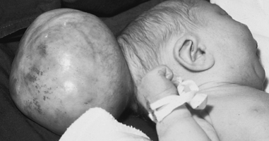 |
| Encephalocele. |
Signs and Symptoms
Frontoethmoidal
• Corpus callosum agenesis
• Hypertelorism
• Interhemispheric lipoma or heterotopias
• Mass almost always contains blood
• Mass near the dorsum of the nose, orbits, or forehead
• Midline craniofacial dysraphism
Parietal
• Aqueduct stenosis
• Arnold-Chiari II malformation (see p. 26)
• Dandy-Walker cysts
• Hydrocephalus
• Microcephaly
Sphenoidal
• Occult and frequently not detected unless computed tomography (CT) and/or magnetic resonance imaging (MRI) of the head is indicated/undertaken
Medical Management
Treatment depends heavily on the size, content, and location of the defect. Surgical correction is the definitive intervention, the ease of which is determined by the size and location of the defect. The best prognosis is found with an anteriorly located encephalocele that is devoid of brain tissue and without any associated anomaly. The poorest prognosis is found in cases with large and/or posterior defects with one or more associated anomalies. Anterior encephaloceles have an almost 100% survival rate, whereas survival rates decline to about 55% in cases of posterior encephaloceles.
Only about 21% of diagnosed cases of encephaloceles are born alive; only about 50% of those neonates live, with only approximately 10% of encephalocele patients surviving the perinatal/neonatal period. Some form of mental deficit is diagnosed in three out of four encephalocele patients who survive birth and surgical correction.
Complications
• Cerebrospinal fluid leak
• Hemorrhage
• Infection
• Meningitis
Anesthesia Implications
Surgical correction of the defect is engaged as soon as feasible after birth. As such, age-appropriate concerns for separation are important, in this case directed more toward the child’s parents. The youngest child (days old) will experience less separation anxiety than one who is several months old. The anxiety of the parents will be high no matter the child’s age. These parents will benefit most by seeing an anesthetist who is comfortable in dealing with and caring for a very young child. Calm confidence and comfort with caring for this child will be of utmost importance in gaining the trust of this child’s parents. The anesthetist should take time to sit with the child’s parents and answer any questions and address any concerns the parents may bring up. Talking about their understanding of the proposed surgery will also help alleviate some measure of their anxiety. At the appropriate time, the anesthetist should confidently assume the custody of the child and proceed to the operating room.
Obtaining/maintaining a secure airway, particularly mask fit, may be difficult owing to the size and location of the encephalocele. For example, a true frontal encephalocele may render mask fit almost impossible and inhalation induction may not be possible. The anesthetist may find it necessary to be “creative” with placement of the mask to accomplish the inhalation induction of this infant. Once somnolence is achieved, intravenous access can be obtained and the induction/intubation process completed in a timely fashion.
Occipital encephalocele may cause the child’s head to be awkwardly positioned if the child is supine. The anesthetist may find it necessary to turn the child’s head to one side or the other so as not to place undue pressure on the defect; it may, in fact, be necessary to place the child’s head on a soft foam or gel “doughnut” to remove pressure from the defect. It may also be advisable to attempt intubation with the child in a lateral decubitus position.
Nasal intubation should not be attempted in the child suspected or known to have a sphenoidal encephalocele, which may not be immediately obvious but is normally detected within the first 10 years of life.
Fluid requirements and replacements must be strictly determined and adhered to because of the patient’s age.
Blood loss can be relatively large, depending on the size and location of the defect. The age and overall health of the patient, including the presence of any associated systemic anomalies, will have a significant impact on the patient’s ability to tolerate blood loss. Large occipital or sphenoidal encephaloceles are of particular importance because they are associated with greater volumes of blood loss. The anesthetist may be called upon to induce deliberate hypotension, with or without hyperventilation, to “relax” the brain and reduce the volume of the encephalocele sac.
A large occipital encephalocele may impede any head/neck extension during the intubation process, making direct laryngoscopy more difficult, and may thus contribute to the inability to intubate the patient with encephalocele. It is prudent to have the pediatric difficult airway cart at hand. In addition, it may be prudent to have a surgeon present to surgically secure the patient’s airway if direct laryngoscopy and other measures are unsuccessful.