Embryology of the Reproductive Tract

Published on 10/03/2015 by admin

Filed under Obstetrics & Gynecology

Last modified 22/04/2025

Print this page

rate 1 star rate 2 star rate 3 star rate 4 star rate 5 star
Your rating: none, Average: 1 (1 votes)

This article have been viewed 2374 times

Chapter 1 Embryology of the Reproductive Tract

Development of the ovary

The germ cells, which will eventually inhabit the gonads, originate from the primitive hind gut. They appear around the 25th day.

By 30 days, the gut, complete with its mesentery, is formed. The germ cells now migrate from the gut to the root of the mesentery. Of the original 6 or 7 million, only 1–2 million are present at birth and this number is reduced to 300,000 at puberty. A smaller number of these cells may be a factor that leads to premature menopause.

At the same time, the coelomic epithelium proliferates and forms thickenings, the genital ridges, together with the underlying mesenchyme on either side of the mesenteric root, near the developing kidney.

At this stage, the primitive gonad (genital ridge) consists of a mesoderm (coelomic epithelium plus mesenchyme) covered by the coelomic epithelium. The germ cells now migrate from the root of the mesentery to the genital ridge.

The coelomic epithelium, growing into the genital ridge, forms the so-called sex cords, which enclose each germ cell.

Until this time, that is, around the 7th week, the gonad is of an indifferent type, the male being indistinguishable from the female.

The germ cells and most of the sex cord cells remain in the superficial part, which is the future cortex of the ovary. The cords then lose contact with the surface epithelium and form small groups of cells, each with its germ cell, a primitive follicle. Some of the sex cord cells grow into the medulla. These tend to regress and form rudimentary tubules, the rete.

As the ovary grows, it projects increasingly into the peritoneal (coelomic) cavity, thus forming a mesentery.

Simultaneously, the ovary descends extraperitoneally within the abdominal cavity. Two ligaments develop and these may help to control its descent, guiding it to its final position and preventing its complete descent through the inguinal ring, in contrast to the testes. The first structure is the suspensory ligament attached to the anterior (cephalic) pole of the ovary which connects it with its site of origin, the genital ridge.

Another ligament, or gubernaculum, develops at the posterior or caudal end of the ovary. At first attached to the genital ridge, it later becomes attached to the developing uterus and becomes the ovarian ligament.

Development of uterus and fallopian tubes

When the embryo reaches a size of 10 mm at 35–36 days, a longitudinal groove appears on the dorsal aspect of the coelomic cavity, lateral to the Wolffian (mesonephric) ridge.

This groove or fold is then sealed off to form a tube, the paramesonephric or Müllerian duct. The tube is open at its upper end, enabling communication with the future peritoneal cavity. The lower end forms a solid tip (Müllerian tubercle), which develops burrowing properties.

The Müllerian ducts from either side grow in a caudal direction, extraperitoneally. They also bend medially and anteriorly and ultimately fuse in front of the hind gut. The mesonephric duct becomes involved in the walls of the paramesonephric ducts.

At first, there is a septum that separates the lumina of the two ducts. Later the septum disappears and a single cavity is formed, the uterus. The upper parts of both the ducts remain separate and form the fallopian tubes.

While this is happening, the ovary is also affected. Its gubernaculum is ultimately attached to the Müllerian duct at the cornu of the developing uterus. This pulls the ovary medially such that its long axis becomes horizontal.

The lower end of the fused Müllerian ducts, beyond the uterine lumen, remains solid, proliferates and forms a solid cord. This cord will canalise to form the vagina, which opens into the urogenital sinus.

At the point of entry into the urogenital sinus, a part of the Müllerian tubercle persists and forms the hymen.

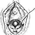
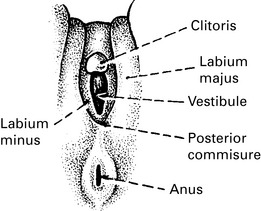
Development of external genitalia

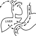
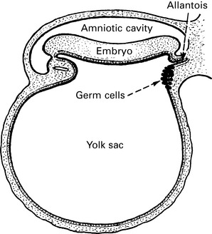
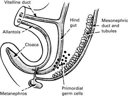
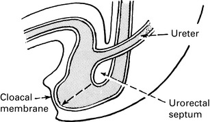
At an early stage, the hind gut and the various urogenital ducts open into a common cloaca.

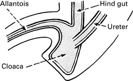
A septum (urorectal) grows down between the allantois and the hind gut during the 5th week.

Eventually, this septum fuses with the cloacal membrane, dividing the cloaca into two compartments – the rectum dorsally and the urogenital sinus ventrally. At the same time, the developing uterus grows down and makes contact with the urogenital sinus.

At the end of the 7th week, the urogenital membrane breaks down and the urogenital sinus opens on to the surface.

The developing uterus and vagina push downwards and cause an elongation and narrowing of the upper part of the urogenital sinus. This will form the urethra.

Meanwhile, on the surface of the embryo, around the urogenital sinus, five swellings appear. At the cephalic end, a midline swelling grows, the genital tubercle, which will become the clitoris. Posterior to the genital tubercle and on either side of the urogenital membrane a fold is formed – urethral folds. Lateral to each of these another swelling appears – the genital or labial swelling. These swellings approach each other at their posterior ends, fuse and form the posterior commissure. The remaining swellings become the labia minora.

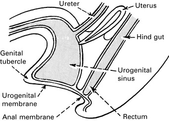
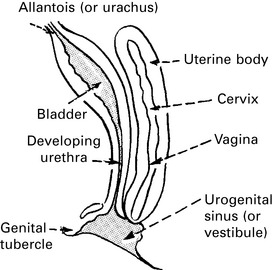
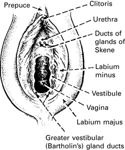
Certain small, but clinically important, glands are formed in and around the urogenital sinus.

In the embryo, the epithelial buds arise from the urethra and also from the epithelium of the urogenital sinus. In the male, these two sets of buds grow together and give rise to the glands of the prostate. They remain separate in the female, with the urethral buds forming the urethral glands and the urogenital buds giving rise to the paraurethral glands of Skene. The ducts of the latter open into the vestibule, on either side of the urethra.

Two other small glands arise by budding from the epithelium of the posterior part of the vestibule, one on either side of the vaginal opening. These are the greater vestibular or Bartholin’s glands. Similar smaller glands also arise in the anterior portion of the vestibule.