Embryo and fetus

Published on 10/03/2015 by admin

Filed under Obstetrics & Gynecology

Last modified 22/04/2025

Print this page

rate 1 star rate 2 star rate 3 star rate 4 star rate 5 star
Your rating: none, Average: 0 (0 votes)

This article have been viewed 2577 times

Chapter 4 Embryo and fetus

Once implantation has occurred the embryo develops rapidly. A neural primitive streak develops in the second week after fertilization; during the third week the fetal heart develops and links up with the primitive vascular system; during the fourth week the gut has formed; and by the sixth week the urogenital sinus has formed.

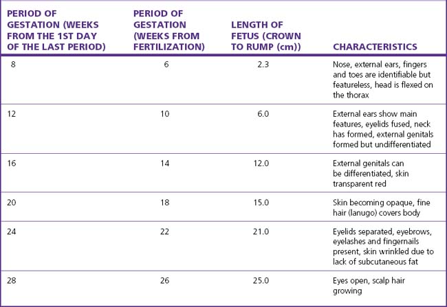
By the seventh week after fertilization most of the organs have formed and the embryo becomes a fetus. The early growth of the fetus is shown in Table 4.1.

NUTRITION

Fetal growth is determined by many factors, both genetic and environmental. Of the latter, adequate placental perfusion and placental function are crucial. Maternal nutrition is not a limiting factor except in cases of extreme starvation, although chronic undernutrition may be associated with anaemia and may lead to a low-birthweight baby.

The fetus, insulated in its protective amniotic sac and relatively weightless, directs most of the energy supplied to it to growth. The energy is derived mainly from glucose. Only small amounts of lipids, as free fatty acids, cross the placenta until the fourth quarter of pregnancy. Any excess carbohydrate, after the growth and metabolic energy needs of the fetus have been met, is converted into lipids, and this conversion increases as term approaches.

From the 30th gestational week the fetal liver becomes increasingly efficient and converts glucose into glycogen, which is stored in the fetal heart muscle, the skeletal muscle and the placenta. Should fetal hypoxia occur the fetus is able to obtain energy from the heart muscle and placenta for anaerobic glycolysis (see Ch. 20).

Free fatty acids are formed and stored in brown and white adipose tissue. Brown fat is deposited around the fetal neck and behind the scapulae and the sternum and around the kidneys. It is metabolized to provide energy to maintain the infant’s body temperature after birth. White adipose tissue forms the subcutaneous cover of the body of a term fetus, but in preterm babies the layer may be thin. It acts as an insulator and as a lipid store.

The fat stores of an 800 g fetus (24–26 weeks’ gestation) constitute 1% of its body weight; by the 35th week fat constitutes 15% of fetal body weight.

As the placenta clears the blood of bilirubin and other metabolic products that require a transferase activity, the fetal (and neonatal) liver is deficient in certain transferases. The result is that unless the deficiencies are corrected in the early neonatal period, bilirubin may accumulate in the neonate’s blood, which is of some consequence in haemolytic disease of the newborn (see p. 129–130).

Amino acids cross the placenta by active transfer and are converted into protein. Protein synthesis exceeds protein breakdown, and the fetus uses some of the breakdown amino acids for resynthesis.

The fetus also synthesizes a specific protein, α-fetoprotein (AFP) in its liver. The peak of AFP is reached between the 12th and 16th gestational weeks, after which a decline occurs until term. The protein is secreted in the fetal urine and swallowed by the fetus, to be degraded in its gut. If the fetus is unable to swallow, as in cases of anencephaly, the level of AFP in the amniotic fluid rises.

LUNGS

In the early embryo the lungs are made up of epithelial tubes surrounded by mesoderm. With further development the epithelium becomes folded and glandular to form primitive alveoli. By the 22nd gestational week a capillary system has developed and the lungs are capable of gas exchange. By term, three or four generations of alveoli have developed and been replaced. Their epithelium, which has a cuboidal appearance, becomes flattened with the first breath. By the 24th week, fluid fills the alveoli and the passages. There are two principal alveolar cell types, the flatter type I pneumocytes, which facilitate gas exchange, and the cuboidal type II pneumocytes that by the 24th week begin to secrete a surface-active lipoprotein surfactant. Surfactant facilitates lung expansion at birth and helps the air-containing lung to maintain its normal volume. However, until the 35th week the amount of surfactant may be insufficient for some babies to expand their lungs after birth, and hyaline membrane disease may develop.

The fetus makes respiratory movements (breathing) from early in pregnancy. At first they are sporadic, but by midpregnancy the movements become regular and increase in frequency as the pregnancy advances. Respiratory activity results in the inspiration of amniotic fluid into the bronchioles but no further, as the fluid secreted into the alveoli is under higher pressure. The reduction in fetal breathing movements when the fetus is subjected to chronic hypoxia can be observed during ultrasound examination. Acute episodes of hypoxia in late pregnancy or during the birth may stimulate gasping. This fetal gasping draws the amniotic fluid, which often contains meconium, deeper into the lungs.

ENDOCRINE ACTIVITY

The fetal hypothalamus secretes corticotrophin-releasing hormone (CRH) by the 13th week, thyrotrophin-releasing hormone (TRH), gonadotrophin-releasing hormone (GnRH) and somatostatin by the 15th week, and growth hormone-releasing hormone (GhRH) by the 18th week after fertilization.

Growth hormone (GH) can be released by the fetal pituitary by the 7th week and luteinizing hormone (LH) and follicle-stimulating hormone (FSH) by 9 weeks. Although adrenocorticotrophic hormone (ACTH) is detectable by the 10th week the pituitary–adrenal axis remains immature and the adrenal gland only becomes sensitive to ACTH late in pregnancy. This is possibly because the main source of ACTH is the placenta.

Thyroid-stimulating hormone (TSH) is released from the 14th week, but T3 and T4 levels remain low throughout pregnancy. Immediately after birth there is a surge of TSH, which leads to a rapid transient rise in T3 and T4 and a fall in reverse T3.

The posterior lobe of the fetal pituitary gland secretes oxytocin from the second trimester and levels rise during labour. Arginine vasotocin is detectable from the 9th week but its function is not understood. Arginine vasopressin is secreted from the 12th week and plays a key role in cardiovascular function under stress conditions.

Antinatriuretic factor (ANF) is released from the atria (predominantly from the right) in response to pressure changes; this helps regulate blood volume by increasing glomerular filtration. Another key component for fetal cardiovascular homeostasis is the renin–angiotensin–aldosterone system. Fetal renin levels are 20 times greater than adult levels, and renin is released in response to a fall in blood volume. Aldosterone is detectable from the second trimester. Aldosterone increases renal sodium reabsorption and has a negative feedback effect on renin release.

Insulin is present in the fetal pancreas by the 10th week, but pancreatic release of insulin is relatively insensitive until 28 weeks. The two growth factors IGF-1 and IGF-2 increase with gestation, especially from 33 weeks, and are related to fetal placental lactogen levels.