28 Eisenmenger syndrome
Salient features
History
• Symptoms may not appear until early late childhood or early adulthood
• Cyanosis (appears as right-to-left shunting develop)
• Dyspnoea on exercise and impaired exercise tolerance
• Palpitations (common and usually caused by atrial fibrillation or flutter)
• Haemoptysis (may occur as a result of pulmonary infarction, or rupture of dilated pulmonary arteries or aorticopulmonary vessels)
• Syncope (owing to inadequate cardiac output or, less commonly, an arrhythmia)
• Symptoms of hyperviscosity including visual disturbances, fatigue, headache, dizziness and paraesthesia
• Symptoms of heart failure are uncommon until the disease is in advanced stages.
Examination
• Clubbing of fingers and central cyanosis
• An ‘a’ waves in the JVP, ‘v’ wave if tricuspid regurgitation is also present
• Left parasternal heave and palpable P2
• Loud P2, pulmonary ejection click, early diastolic murmur of pulmonary regurgitation (Graham Steell murmur)
• Loud pansystolic murmur of tricuspid regurgitation
• Listen carefully to the second sound. The clinical findings from the underlying defect are as follows:
Advanced-level questions
What factors worsen deteriorate pulmonary hypertension in these patients?
What are the complications of Eisenmenger syndrome?
How would you investigate this patient?
• Electrocardiogram shows right ventricular hypertrophy; atrial arrhythmias particularly in those with underlying ASD.
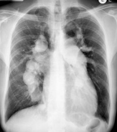
• Chest radiograph shows conspicuous dilatation of the pulmonary artery (Fig. 28.1) with narrowed ‘pruned’ peripheral vessels (caused by pulmonary hypertension); slight to moderate enlargement of the heart (predominantly RV) may be seen in ASD whereas the size of the heart is normal in VSD or PDA.
• Echocardiography provides evidence of right ventricular overload and pulmonary hypertension; the underlying cardiac defect can be visualized, although shunting may be difficult to demonstrate by colour Doppler imaging because of low velocity jet; contrast echocardiography permits localization of shunt.
• Cardiac catheterization determine the extent and severity of pulmonary vascular disease and accurately quantify the magnitude of the intracardiac shunting; assessment of reversibility of shunting is done using pulmonary vasodilators (e.g. oxygen, inhaled nitrous oxide, intravenous adenosine or epoprostenol).
What is the prognosis in these patients?
• Survival is 80% 10 years after diagnosis, 77% at 15 years, and 42% at 25 years.
• Death is usually sudden; other causes include heart failure, haemoptysis, brain abscess or stroke.
• Poor prognostic factors include syncope, clinically evident right ventricular systolic dysfunction, low cardiac output and severe hypoxaemia.
What treatment is available for Eisenmenger syndrome?
• Phlebotomy done cautiously in symptomatic hyperviscosity due to secondary erythrocytosis when the haematocrit >0.65 (but not due to dehydration). Always replace fluid intravenously following phlebotomy
• Long-term intravenous epoprostenol (Circulation 1999;99:1858–65; Ann Intern Med 1999;130:740–3)
• Uncontrolled studies suggest that prostacyclin analogues and phosphodiesterase 5 inhibitors (e.g. sildenafil) may have benefits in advanced pulmonary vascular disease
• In a randomized, controlled trial of bosentan versus placebo, bosentan showed a significant improvement in exercise capacity based on 6-minute walk distance, with serious safety concerns (Circulation 2006;114:48–54)
• Combined heart–lung transplantation (limited success and hence patients should be carefully selected).