27. Ehlers-Danlos Syndrome
Definition
Ehlers-Danlos syndrome (EDS) is a group of inherited connective tissue disorders that vary according to clinical and biochemical evidence, inheritance mode, and disorder severity (mild to lethal). The disorder is characterized by joint hyperextension, skin hyperextensibility, easy bruising, tissue friability, bleeding, poor wound healing, subcutaneous nodules, as well as cardiovascular, orthopedic, intestinal, and ocular defects.
Incidence
There are six types of Ehlers-Danlos syndrome (see table). The frequency of all recognized types of EDS is not exactly known but is estimated to be 1:5000 to 10,000. There is no discernable racial predilection.
Etiology
Ehlers-Danlos syndrome is an autosomal inherited disorder. The type of EDS the patient has differs according to the chromosome locus/loci and specific gene(s) abnormality.
Signs and Symptoms
The following signs and symptoms are common to all EDS types:
• Alveolar bone loss
• Bladder diverticula or rupture
• Bony dysplasias
• Easy bruising
• Excessive joint dislocations
• Joint hypermobility
• Microcornea
• Micrognathia
• Mitral valve prolapse
• Myopia
• Occipital exostoses
• Periodontitis
• Poor wound healing
• Prominent venous markings
• Retinal detachment
• Scoliosis
• Short stature
• Skin hyperextensibility
• Spontaneous medium-sized artery rupture
• Spontaneous viscus rupture(s)
• Tissue fragility
• Wide/thin scars
Medical Management
The first, most critical step is correctly diagnosing the syndrome. Because this disorder family affects connective tissues, careful, thorough cardiac evaluation is essential. Mitral valve prolapse is particularly common in EDS patients and should be carefully described and confirmed using an echocardiogram. Frequent, serial cardiovascular evaluations are particularly important for vascular EDS patients because of the higher risk of aneurysm formation and rupture.
Patients should be closely monitored for development and progression of scoliosis. Patients with scoliosis should be cautioned to minimize activities that produce excessive stress and strain to their back because of the hypermobility of their joints.
Lacerations and similar injuries should be carefully tended. The patient’s tissue friability and fragility, as well as the wide scarring associated with the disorder, are a contraindication for use of sutures for wound closure. Better alternatives for wound closure in the EDS patient include wound/tissue glue and/or adhesive strips.
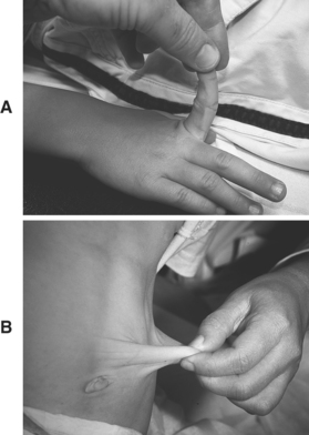 |
| Ehlers-Danlos Syndrome. A, Hyperextensible joint seen in Ehlers-Danlos syndrome. B, Skin laxity in Ehlers-Danlos syndrome. |
Complications
• Bowel perforation
• Death
• Detached retina
• Hollow viscus perforation or rupture
• Pneumothorax
• Spontaneous arterial rupture
• Uncontrollable hemorrhage
• Wound dehiscence
Anesthesia Implications
Correctly positioning a patient with EDS is critical. The hypermobility of the joints predisposes the patient to joint injuries, particularly during emergence from anesthesia. The anesthetist must also take great care to secure the patient’s extremities to armboards or tuck them at the patient’s side while using copious padding to minimize or prevent pressure/compression injury, such as bruising.
The tissue fragility, poor wound healing, scarring, bruising, and propensity for bleeding present something of a quandary to the anesthetist. Intramuscular injections should be avoided, if possible, making adequate anxiolysis difficult. Obtaining intravenous access on the first attempt (ideal for any patient) takes on added importance for the EDS patient. A central venous catheter must be placed with great caution because of the increased risk of pneumothorax, as well as the greater potential for extensive hematoma formation. The increased potential for extensive hematoma formation must be factored into the decision for placement of an intra-arterial catheter. Once any of these invasive lines has been inserted, methods to secure those lines must be carefully considered. Tape applied to the skin should be minimized, and gauze wrap should be used whenever possible. In addition, the intravenous and intra-arterial lines must be inspected frequently to prevent or at least minimize extravasation of fluids, which can be extensive because of the hyperextensibility of the patient’s skin. This measure is of increased importance to the anesthetist because of the tissue damage that can occur with extravasation of any number of anesthesia-related drugs.
The anesthetist must be even more cautious than usual during direct laryngoscopy to minimize trauma to the tissues because of increased potential for bleeding and bruising. Because of the increased potential for pneumothorax, assisted and controlled ventilation can be injurious to the patient with EDS. The anesthetist should choose the pressure-controlled ventilation option that is available on most newer anesthesia machines and opt for the lowest appropriate inspiratory pressure possible to maintain adequate ventilation and gas exchange. The expiratory phase for the patient with EDS should also be prolonged, to as much as 1:2.5 or 1:3, to reduce the possibility of air trapping and barotrauma. Surgical wound dehiscence is another possible complication for the patient with EDS. The anesthetist can help reduce the potential for dehiscence by extubating the patient while relatively deeply anesthetized or by continuing mechanical ventilation and muscle paralysis into the post-anesthesia care unit.
Regional anesthesia for the patient with EDS is relatively contraindicated because of the higher potential for bleeding and extensive hematoma formation. For this same cause, the anesthetist should anticipate greater-than-normal blood volume losses for a given surgical intervention. As a result, it may be prudent to obtain blood type and antibody screening preoperatively, in the event transfusion becomes necessary during the course of surgery.