Chapter 647 Eczematous Disorders
Eczematous skin disorders are characterized by exudation, lichenification, and pruritus. Acute eczematous lesions demonstrate erythema, weeping, oozing, and the formation of microvesicles within the epidermis. Chronic lesions are generally thickened, dry, and scaly, with coarse skin markings (lichenification) and altered pigmentation. Many types of eczema occur in children; the most common is atopic dermatitis (Chapter 139), although seborrheic dermatitis, allergic and irritant contact dermatitis, nummular eczema, and vesicular hand and foot dermatitis (dyshidrosis) are also relatively common in childhood.
647.1 Contact Dermatitis
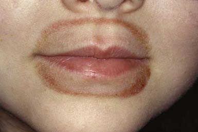
Irritant contact dermatitis can result from prolonged or repetitive contact with various substances that include saliva, citrus juices, bubble bath, detergents, abrasive materials, strong soaps, and proprietary medications. Saliva is probably one of the most common offenders; it may cause dermatitis on the face and in the neck folds of a drooling infant or a retarded child. In older children who habitually lick their lips because of dryness, frequently without being aware, a striking, sharply demarcated perioral rash may develop (Fig. 647-1). Among the exogenous irritants, citrus juices, proprietary medications, and bubble bath preparations are relatively common. Excessive accumulation of sweat and moisture as a result of wearing occlusive shoes may also cause irritant dermatitis.
Irritant contact dermatitis may be indistinguishable from atopic dermatitis or allergic contact dermatitis. A detailed history and consideration of the sites of involvement, the age of the child, and contactants usually provide clues to the etiologic agent. The propensity for development of irritant dermatitis varies considerably among children; some may respond to minimal injury, making it difficult to identify the offending agent through history. Irritant contact dermatitis usually clears after removal of the stimulus and temporary treatment with a topical corticosteroid preparation (Chapter 638). Education of patients and parents about the causes of contact dermatitis is crucial to successful therapy.
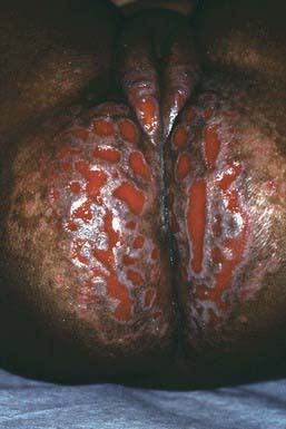
Diaper dermatitis can be regarded as the prototype of irritant contact dermatitis. As a reaction to overhydration of the skin, friction, maceration, and prolonged contact with urine and feces, retained diaper soaps, and topical preparations, the skin of the diaper area may become erythematous and scaly, often with papulovesicular or bullous lesions, fissures, and erosions (Fig. 647-2). The eruption can be patchy or confluent, but the genitocrural folds are often spared. Chronic hypertrophic, flat-topped papules and infiltrative nodules may occur. Secondary infection with yeast is common. Discomfort may be marked because of intense inflammation. Allergic contact dermatitis, seborrheic dermatitis, psoriasis, candidosis, atopic dermatitis, and rare disorders such as Langerhans cell histiocytosis (histiocytosis X) and acrodermatitis enteropathica should be considered when the eruption is persistent or is recalcitrant to simple therapeutic measures.
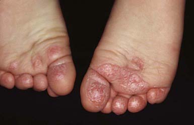
Juvenile plantar dermatosis is a common form of irritant contact dermatitis occurring mainly in prepubertal children. The dermatitis characteristically involves the weight-bearing surfaces, may be pruritic or painful, and causes a glazed appearance of the plantar skin (Fig. 647-3). Fissuring may become extensive, producing considerable discomfort. The dermatitis results from alternating excessive hydration and rapid moisture loss, which cause chapping of the skin and cracking of the stratum corneum. Affected children often have hyperhidrosis, wear occlusive synthetic footwear, and subject their feet to rapid drying without moisturization. Immediate application of a thick emollient when socks and shoes are removed or immediately after swimming usually minimizes this condition. Severe inflammatory cases may require short-term (1-2 wk) application of a medium- to high-potency topical steroid.
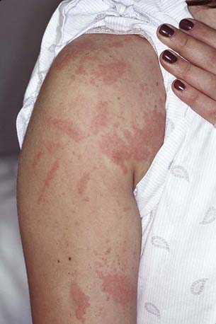
Rhus dermatitis (poison ivy, poison sumac, poison oak) is often vesiculobullous and may be distinguished by linear streaks of vesicles where the plant leaves have brushed against the skin (Fig. 647-4). Fluid from ruptured cutaneous vesicles does not spread the eruption; antigen retained on the skin, under the fingernails, and on clothing initiates new plaques of dermatitis if not removed by washing with soap and water. Antigen may also be carried by animals on their fur. Black spot poison ivy is a rare variant that manifests as small discrete black lacquer-like glossy papules with surrounding erythema and edema. The saplike allergen (oleoresin) is present on live and dead leaves, and sensitization to one plant produces cross reactions with the others.
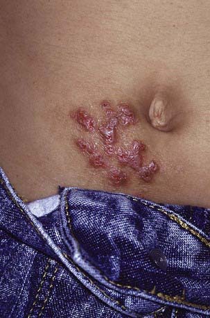
Nickel dermatitis usually develops from contact with jewelry or metal closures on clothing and is seen most frequently on the earlobes, such as when earring posts of nickel-containing metal rather than nonmetallic materials or stainless steel are used to keep a pierced tract open. Metal closures on pants frequently cause periumbilical dermatitis (Fig. 647-5). Some children are exquisitely sensitive to nickel, with even the trace amounts found in gold jewelry provoking eruptions.
Beattie PE, Green LC, Lowe G, et al. Which children should we patch test? Clin Exp Dermatol. 2007;32:6-11.
Humphrey S, Bergman JN, Au S. Practical management strategies for diaper dermatitis. Skin Ther Lett. 2006;11:1-6.
Jacob SE, Zapolanski T, Chayavichitsilp P, et al. p-Phenylenediamine in black henna tattoos. Arch Pediatr Adolesc Med. 2008;162:790-792.
Krakowski AC, Eichenfield LF, Dohil MA. Management of atopic dermatitis in the pediatric population. Pediatrics. 2008;122:812-824.
Schram SE, Willey A, Lee PK, et al. Black-spot poison ivy. Dermatitis. 2008;19:48-51.
647.2 Nummular Eczema
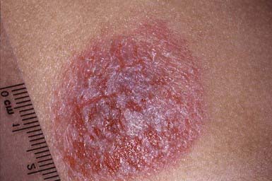
Nummular eczema is unrelated to other types of eczema and is characterized by more or less coin-shaped eczematous plaques. Common sites are the extensor surfaces of the extremities (Fig. 647-6), buttocks, and shoulders. The plaques are relatively discrete, boggy, vesicular, severely pruritic, and exudative; when chronic, they often become thickened and lichenified. The cause is unknown. Most frequently, these lesions are mistaken for tinea corporis, but plaques of nummular eczema are distinguished by the lack of a raised, sharply circumscribed border; the lack of fungal organisms on a potassium hydroxide (KOH) preparation; and frequent weeping or bleeding when scraped. Control of pruritus and inflammation is usually achieved with a potent topical corticosteroid preparation. Steroid-impregnated tapes may simultaneously treat and provide barrier protection to these circumscribed eczematous plaques. An antihistamine may be helpful, particularly at night. Antibiotics are indicated for secondary infection.
Fergusson DM, Horwood J, Shannon FT. Early solid feeding and recurrent childhood eczema: a 10-year longitudinal study. Pediatrics. 1990;86:541-546.
Krafchik BR. Eczematous disorders. In: Eichenfield LF, Frieden IJ, Esterly NB, editors. Textbook of neonatal dermatology. Philadelphia: WB Saunders; 2001:241.
647.3 Pityriasis Alba
Pityriasis alba occurs mainly in children; the lesions are hypopigmented, round or oval, macular or slightly elevated patches with fine adherent scale (Fig. 647-7). They may be mildly erythematous and relatively well defined but lack a sharply marginated border. Lesions occur on the face, neck, upper trunk, and proximal portions of the arms. Itching is minimal or absent. The cause is unknown, but the eruption appears to be exacerbated by dryness and is often regarded as a mild form of eczema. Pityriasis alba is frequently misdiagnosed as vitiligo, tinea versicolor, or tinea corporis. The lesions wax and wane but eventually disappear. Application of a lubricant may ameliorate the condition; if pruritus is troublesome, a low-potency topical steroid may used. Normal pigmentation often takes months to return.
647.4 Lichen Simplex Chronicus
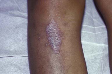
Lichen simplex chronicus is characterized by a chronic pruritic, eczematous, circumscribed, solitary plaque that is usually lichenified and hyperpigmented (Fig. 647-8). The most common sites are the posterior aspect of the neck, the dorsum of the feet, the wrists, and the ankles. Although the initiating event may be a transient lesion such as an insect bite, trauma from rubbing and scratching accounts for persistence of the plaque. Pruritus must be controlled to permit healing. A topical fluorinated corticosteroid preparation is often helpful, but constant irritation of the skin must be avoided. A covering to prevent scratching may be necessary.
647.5 Vesicular Hand and Foot Dermatitis (Dyshidrotic Eczema, Dyshidrosis, Pompholyx)
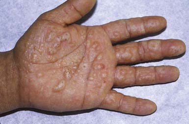
A recurrent, sometimes seasonal, blistering disorder of the hands and feet, vesicular hand and foot dermatitis occurs in all age groups but is uncommon in infancy. The pathogenesis is unknown; no genetic factor has been identified, although an increased incidence of atopy has been recorded in affected patients and their relatives. The disease is characterized by recurrent crops of small, intensely pruritic vesicles on the hands and feet. Sites of predilection are the palms, soles, and lateral aspects of the fingers and toes. Primary lesions are noninflammatory and are filled with clear fluid, which, unlike sweat, has a physiologic pH and contains protein. Larger vesicles and bullae may occur (Fig. 647-9), and maceration and secondary infection are frequent because of scratching. The chronic phase is characterized by thickened, fissured plaques that may cause considerable discomfort. Hyperhidrosis is common in many patients, but the association may be fortuitous. The diagnosis is made clinically. The disorder may be confused with allergic contact dermatitis, which usually affects the dorsal rather than the volar surfaces, and with dermatophytosis, which can be distinguished by a KOH preparation of the roof of a vesicle and by appropriate cultures.
647.6 Seborrheic Dermatitis
Clinical Manifestations
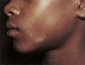
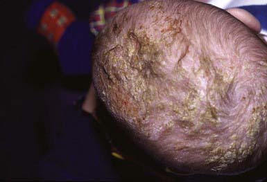
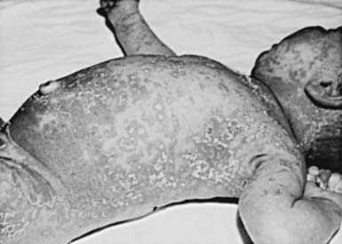
The disorder may begin in the 1st mo of life and may be most troublesome in the 1st yr. Diffuse or focal scaling and crusting of the scalp, sometimes called cradle cap (Fig. 647-10), may be the initial and at times the only manifestation. A greasy, scaly, erythematous papular dermatitis, which is usually nonpruritic, may involve the face, neck, retroauricular areas, axillae, and diaper area. The dermatitis may be patchy and focal or may spread to involve almost the entire body (Fig. 647-11). Postinflammatory pigmentary changes are common, particularly in black infants. When the scaling becomes pronounced, the condition may resemble psoriasis and, at times, can be distinguished only with difficulty. The possibility of coexistent atopic dermatitis must be considered when there is an acute weeping dermatitis with pruritus, and the two are often clinically inseparable at an early age. An intractable seborrhea-like dermatitis with chronic diarrhea and failure to thrive may reflect systemic dysfunction of the immune system. A chronic seborrhea-like pattern, which responds poorly to treatment, may also result from cutaneous histiocytic infiltrates in infants with Langerhans cell histiocytosis. Seborrheic dermatitis is a common cutaneous manifestation of AIDS in young adults and is characterized by thick, greasy scales on the scalp and large hyperkeratotic erythematous plaques on the face, chest, and genitals.
Treatment
Scalp lesions should be controlled with an antiseborrheic shampoo (selenium sulfide, sulfur, salicylic acid, zinc pyrithione, tar), used daily if necessary. Inflamed lesions usually respond promptly to low- to medium-potency topical corticosteroid therapy. Topical immunomodulatory agents (tacrolimus, pimecrolimus) approved for the treatment of atopic dermatitis in children ≥2 yr of age (Chapter 139) may have a role in the treatment of other eczematous disorders such as seborrheic dermatitis. Concerns for systemic absorption and potential immunosuppression are higher in the younger patient population typically afflicted with seborrheic dermatitis. Topical antifungal agents (ketoconazole, ciclopirox, bifonazole) effective against Malassezia have been advocated. These are available as gels, creams, foams, and shampoos. The efficacy of antifungal agents is well documented in controlled trials in adults. Wet compresses should be applied to the moist or fissured lesions before application of the steroid ointment. Many patients require continued use of an antiseborrheic shampoo. Response to therapy is usually rapid unless there are complicating factors or the diagnosis is in error.


