[level-membership-for-dermatology-category]
Eczematous Disorders
Wynnis L. Tom, lawrence F. Eichenfield
Introduction
Eczematous eruptions represent a significant proportion of the skin diseases affecting neonates and infants. Clinically they are characterized by erythema, edema, scale, and sometimes crusts. The most common disorder is atopic dermatitis (AD) and, in fact, the term ‘eczema’ (which means ‘boiling over’) is often used by laymen to refer to AD. Seborrheic dermatitis and irritant dermatitis also frequently affect infants, while allergic contact dermatitis (ACD) is less common. Some nutritional, metabolic, and immunologic diseases have clinical manifestations that include eczematous eruptions which may be difficult to differentiate from AD, and they require a high index of suspicion for their occurrence.
Atopic dermatitis
Atopic dermatitis is a chronic, pruritic skin condition with a frequently relapsing course. It was first described by Besnier in 1892 as ‘prurigo diasthétique’. In 1933, Wise and Sulzberger coined the term ‘atopic dermatitis’, and Hill and Sulzberger characterized the clinical entity 2 years later.1,2 AD is notable for inflammation, xerosis, crusts, excoriations, and lichenification, and it is the most common chronic inflammatory dermatosis of early childhood. The disease imposes an enormous burden on the personal, social, emotional, and financial resources of patients and their families. In the USA, annual direct costs for AD care are estimated at US$1–4 billion,3 which does not include the effects of lost productivity such as missed work and school days nor the impact on quality of life.
The prevalence of AD has increased markedly over the past 50 years, with current rates of 15–29% in the USA, Europe, Japan, and other industrialized countries and lower but increasing rates in developing nations.4–6 AD affects individuals of all races, with several studies showing a slightly higher prevalence in black children.6 However, different countries with similar ethnic demographics have markedly different recorded prevalences, supporting the concept that environmental factors are important in disease expression.7 AD may be slightly more common in girls (female : male ratio of 1.3 : 1), although some studies note that it affects the sexes approximately equally.8
Risk factors
Multiple epidemiologic studies have examined risk factors for disease development. Only two factors show consistent, strong association: a family history of atopic diseases (i.e., asthma, allergic rhinitis, atopic dermatitis) and loss of function mutations in the filaggrin (FLG) gene. A positive family history of atopy is noted in approximately 70% of affected individuals. The odds of developing AD are 2–3-fold higher in children with one atopic parent, and this increases to as high as 3–5-fold if both parents are atopic.9,10 Some have noted a maternal history of AD itself to be more predictive.11,12 The FLG gene encodes a key protein involved in terminal differentiation of the epidermis, and its breakdown products are important in the formation of the skin barrier, including the stratum corneum, and natural moisturizing factor (see below). Loss of function mutations confer a risk for earlier-onset AD, as well as for more severe disease.13
AD is more frequent in higher social and socioeconomic classes.14 Other factors that may be associated with a greater risk for disease include small family size, migration from rural to urban environments, and increased parental education.7 Some have noted a higher incidence in association with maternal smoking and elevated birthweight, though not all studies corroborate these findings.15,16 The effect of exposure to pets in the home is unclear, due to conflicting data.17,18
Clinical findings
History
The onset of disease is most commonly between 3 and 6 months of age, with approximately 60% of patients developing the eruption within the first year of life and 90% by 5 years of age.19,20 Atopic dermatitis is usually the first manifestation of the ‘atopic march,’ which refers to the subsequent development of food allergies, asthma, and allergic rhinitis, although this progression does not happen in all individuals and/or the sequence may vary.20 More recently, eosinophilic esophagitis and gastroenteritis have been added to the list of associated disorders.21
The predominant symptom is pruritus, which is difficult to control and a major cause of morbidity and poor quality of life for both the child and caregiver.22 It frequently interferes with sleep and can lead to complications such as secondary infection of excoriated lesions. Parents should be questioned about the infant scratching and rubbing against objects such as bedding or carpeting. Unfortunately, these actions further incite inflammation and the itch sensation, leading to a vicious cycle.23
Physical examination
There are three age-related phases to AD: infantile, childhood, and adult disease. During each phase both the sites of involvement and the morphology of the lesions change, although the phases often overlap. The infantile phase generally lasts until 2–3 years of age, the childhood phase from 2 years until puberty, and the adult phase from puberty onward.
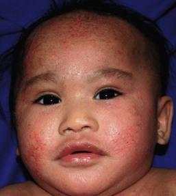
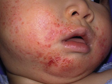
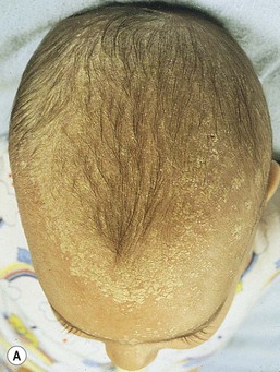
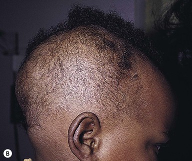
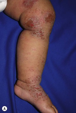
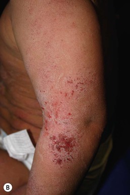
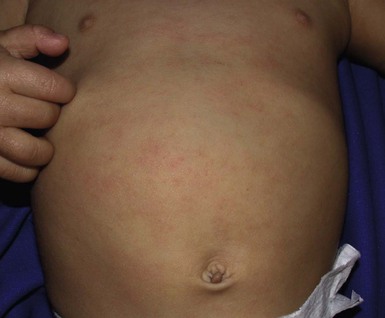
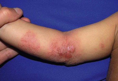
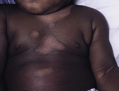
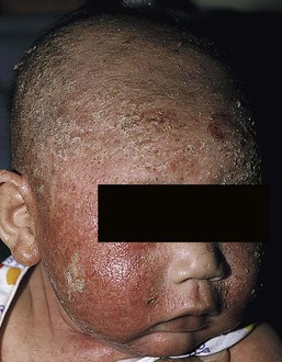
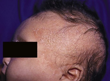
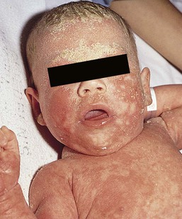
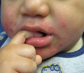
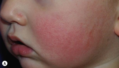
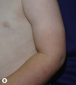
In infancy, AD characteristically begins on the cheeks (Figs 15.1, 15.2) and scalp (Fig. 15.3) and evolves over time to involve the lateral and extensor aspects of the arms and legs (Fig. 15.4). The periauricular areas are often affected and can lead to infra-auricular fissuring.24 While the trunk may have lesions, the diaper area is usually spared due to the retention of moisture and protection from rubbing and external triggers provided by these coverings (Figs 15.5, 15.6). Lesions are symmetric, scaly, erythematous patches and plaques, within which crusting is common. Acute lesions are more exudative. Lymphadenopathy may be noted and reflects local inflammation. During late infancy and the childhood phase, the flexural surfaces of the extremities become the most commonly involved sites, particularly the antecubital and popliteal fossae (Fig. 15.7). Other frequently involved locations include the neck, wrist and ankle flexures, and the creases between the thighs and buttocks. Lesions continue to be erythematous patches and plaques with crusting, but often there is increased excoriation. Chronic findings, such as lichenification (enhanced skin markings) and pigmentary changes start to become prominent (Fig. 15.8). The pigmentary alterations (hypo- or hyperpigmentation) often evoke parental concerns about scarring, although lesions generally do not leave scars unless severely infected or deeply excoriated. From puberty onwards, AD lesions tend to prefer the face, back, wrists, hands, and dorsal feet.

There are a number of additional physical findings that, if present, aid in the diagnosis of atopic dermatitis (Box 15.1). These include other conditions associated with xerotic skin, such as ichthyosis vulgaris, keratosis pilaris, and pityriasis alba. Follicular lichenification with papules may be noted, especially in those with darker skin type. These are not specific to AD, however, and may be seen in otherwise healthy individuals as well. Other findings suggestive of an atopic diathesis that become more prominent with rubbing or scratching include an additional line or groove in the lower eyelid (the Dennie-Morgan fold), infraorbital darkening (‘allergic shiners’), and a prominent horizontal nasal crease (‘allergic salute’).
Diagnosis
The diagnosis of AD is made clinically. This is often more challenging in early infancy, when it can be difficult to distinguish AD from other infantile eczematous disorders. Family history is often helpful and absence of an atopic disorder in the family history makes the diagnosis much less likely, though does not preclude its occurrence. The most recognized diagnostic criteria are those proposed by Hanifin and Rajka in 1980, but the four major and 23 minor criteria are too numerous to be used routinely in clinical practice.25 Several expert groups have proposed more condensed sets of criteria,26–28 including the following,27 which is applicable to both children and adults:
A. Essential features (must be present)
a. Pruritus
b. Eczematous dermatitis (acute, subacute or chronic)
i. Typical morphology and age-specific patterns
ii. Chronic or relapsing history
B. Important features (seen in most cases, adding support to the diagnosis)
b. Atopy
i. Personal and/or family history
ii. IgE reactivity
c. Xerosis
C. Associated features (these clinical associations help to suggest the diagnosis of AD but are too nonspecific to be used to define or detect AD for research or epidemiologic studies)
a. Atypical vascular responses (e.g., facial pallor, white dermatographism, delayed blanch response)
b. Keratosis pilaris/hyperlinear palms/ichthyosis
d. Other regional findings (e.g., perioral changes/periauricular lesions)
e. Perifollicular accentuation/lichenification/prurigo lesions.
To date, there is no specific laboratory test to confirm the diagnosis of AD. Serum immunoglobulin E (IgE) is elevated in approximately 70–80% of patients, although even severe cases may have normal levels.23 The majority of patients also have increased circulating eosinophils. Recent discoveries of novel T-lymphocyte subsets and cytokine proteins have generated additional candidate markers. Thymus and activation-regulated chemokine (TARC, also called CCL17) is one such molecule suggested to increase with disease activity.29 However, to date, none have demonstrated adequate sensitivity and specificity to serve as a biomarker for the disease or its response to therapy. Laboratory testing is thus seldom needed in the routine evaluation of uncomplicated AD.
Skin biopsies may be helpful in distinguishing AD from other inflammatory skin disorders, but the biopsy findings are not pathognomonic. The histology varies from an acute dermatitis with spongiosis and a perivascular lymphocytic infiltrate, to chronic changes in lichenified skin showing acanthosis, hyperkeratosis, and accumulation of Langerhans’ cells, macrophages, and mast cells in addition to lymphocytes around blood vessels. The superficial venular plexus has endothelial cell hypertrophy, basement membrane thickening, and increased numbers of immunoreactive nerve fibers.30,31 Eosinophils are not commonly present in acute AD, but are increased in number in chronic lesions. Mast cells may be normal to increased, and in different stages of degranulation.
Etiology and pathogenesis
The pathogenesis of AD involves a complex interplay of genetic, immunologic, infectious, and other environmental factors. While still not completely understood, it appears to entail two major defective processes: dysfunction of the epidermal barrier and dysregulation of the immune system. Which process occurs first to initiate disease (‘outside-in’ vs ‘inside-out’) is an ongoing debate, although it is clear that they interact and exacerbate one another. This is compounded by a heightened response to environmental allergens, irritants, and microbial antigens.31,32
For many years, it has been known that AD involves genetic susceptibility, given familial clustering, concordance in monozygotic twins but discordance in dizygotic twins, and reports of transfer of AD by bone marrow transplantation.33,34 It has been most highly linked to genes of the epidermal differentiation complex located on chromosome 1q21, particularly the FLG gene, which is also the main cause of ichthyosis vulgaris. The association of FLG mutations with AD is one of the most robust known for complex genetic disorders.13 Large, population-based studies have identified over 35 mutations in affected individuals, with distinct mutations in different ethnic groups.35 As mentioned above, loss of function mutations are associated with early-onset and more severe, persistent disease, and additionally appear to give increased allergic sensitization and total IgE levels, a greater risk for asthma and food and other allergies, and a higher incidence of skin infections with herpes simplex virus (eczema herpeticum). But despite the strong association, FLG null mutations are seen in only approximately 18–48% of individuals with AD (depending on the population) and conversely, about 40% of individuals with such mutations do not develop the disease.13 Genetic screening of affected families has highlighted other chromosome regions that contribute to disease susceptibility, including cytokine and immune-related genes (e.g., those encoding interleukins (IL)-4, -5, and -13 and the regulated on activation of normal T-cell expressed and secreted (RANTES) chemotactic cytokine), genes encoding innate immunity receptors (e.g., the genes for toll-like receptors (TLR)-2 and -9), and genes implicated in other inflammatory diseases (e.g., those encoding nucleotide-binding oligomerization domain protein (NOD)-1 and NOD2).36,37 There are likely additional genes that remain to be discovered, and of those already identified to have high disease association, a number still require determination of exact function.
Defects in both the structure and function of the epidermal barrier are present in AD. FLG mutations lead to disorganized keratin filaments and abnormal architecture of the lamellar bilayer and assembly of the cornified envelope. Decreased filaggrin breakdown products affect levels of natural moisturizing factor and the skin pH, resulting in increased transepidermal water loss and skin permeability. In addition, barrier function is markedly reduced due to a decline in ceramide levels, which are major water-retaining molecules, and from an imbalance of proteolytic enzymes that affect epidermal adhesion and their anti-protease counterparts.35,38 Abnormalities in tight junction expression that form a second barrier in the stratum granulosum further add to the pathology.34 These factors all lead to dry, xerotic skin, while facilitating entry of antigens through the damaged epidermis that stimulate cutaneous inflammation.
Immunologic abnormalities are another major pathogenic mechanism. Antigens bind to and activate epidermal Langerhans’ cells and dermal dendritic cells. Circulating T cells that have increased expression of the cutaneous lymphocyte-associated antigen (CLA) receptor home to the skin, where they are primed by these antigen-presenting cells. In the acute phase of disease, these activated T cells overexpress cytokines of the Th2 class, notably IL-4 and IL-13, which mediate switching of immunoglobulin isotypes to IgE synthesis and upregulated expression of adhesion molecules on endothelial cells. IL-4 also stimulates mast cells to produce mediators such as histamine. IL-5 predominates in subsequent stages and is involved in eosinophil development and survival.31,39 While AD begins acutely as a Th2-mediated disorder, in its chronic phase it is characterized by Th0 cells (cells that share some activities of both Th1 and Th2 cells) and Th1 cells; associated cytokines include interferon-γ. The switch to Th0/1 cells involves the infiltration of the epidermis by inflammatory dendritic epidermal cells (IDEC) and production of IL-12 and IL-18, as well as several remodeling-associated cytokines. Also of significance is an increase in Th22 cells in chronic AD skin, which produce IL-22 that inhibits terminal differentiation and induces epidermal hyperplasia.40 Additional T cell subsets have been identified, but their roles in AD remain to be fully defined. T regulatory cells with normal immunosuppressive activity appear to be expanded in the peripheral blood of patients, but not in lesional skin or atopy patch test sites. Th17 cells have been found to be elevated in patients with acute AD, but not as prominently as in psoriasis.31
Enhanced allergen penetration through the damaged epidermis gives increased production of thymic stromal lymphopoietin (TSLP) by keratinocytes, which may be a critical link between the barrier and immune defects. TSLP has been shown to drive both the initial Th2 cytokine response and the switch to the Th0/1 phenotype.39 Mechanical injury such as scratching or rubbing, microbes, and proinflammatory cytokines themselves further stimulate release of TSLP, thus perpetuating the inflammation. The immunologic response contributes to additional barrier abnormalities, since IL-4 and IL-13 are known to suppress filaggrin expression and downregulate proper processing of profilaggrin.
Antigens that gain entry include microbes. The antimicrobial barrier is compromised in AD from a combination of the defective physical barrier, an increased pH which may allow greater adherence and multiplication of bacteria, and toll-like receptor defects which diminish recognition of microbial agents. In addition, the increase in Th2 cytokines suppresses the synthesis of antimicrobial peptides by keratinocytes, specifically LL-37 (cathelicidin) and β-defensins 2 and 3.41 This predisposes atopic individuals to widespread skin infections due to bacteria and viruses (including herpes, molluscum, and vaccinia), and possibly to dermatophytes. Staphylococcus aureus, in particular, can trigger multiple inflammatory cascades. Staphylococcal toxins activate T cells in a superantigen-driven fashion and induce IgE-specific responses. These further skew the immune response toward Th2 cytokine production and explain the association of S. aureus infection with exacerbations of AD.42 The superantigens also cause a profound reduction in steroid responsiveness of T cells, giving another possible mechanism for disease flares. Exogenous protease inhibitors produced by S. aureus may also damage the epidermal barrier, thereby potentiating the absorption of allergens into the skin and leading to increased bacterial colonization.
The impact of food exposure on the development of atopic dermatitis is controversial. Although it is known that food allergy and sensitization more commonly develop in children with atopic dermatitis, it is unclear how important food exposures are to the onset and course of atopic dermatitis, nor how common food-induced eczematous dermatitis is during infancy.43 Aeroallergens may also trigger immune responses, but sensitization is usually after infancy. The house-dust mite, Dermatophagoides pteronyssinus, has been cited in the pathogenesis of AD, but measures to decrease dust mite exposure are not necessarily protective.44 Malassezia yeasts have also been suggested but not confirmed to cause exacerbations of disease.
The mechanisms underlying pruritus in AD are the subject of much investigation. In addition to histamine, several other mediators, such as neuropeptides, kinins, and cytokines, can induce pruritus. Studies have shown that the number of sensory and neuropeptide-containing nerve fibers is increased in lesional AD skin.30,45 Mast cells store large amounts of proteases, including tryptase, which may induce pruritoceptive itch when released in the proximity of these nerve fibers. These release stress-induced neuropeptides in parallel, which might activate mast cells through neurokinin receptors to perpetuate pruritic signals. The Th2 response contributes via increased levels of IL-31, which has recently been found to enhance pruritus. Staphylococcal superantigens also markedly enhance IL-31 production.45,46
Differential diagnosis
The common disorders in the differential diagnosis of AD include seborrheic dermatitis, contact dermatitis, psoriasis, scabies, ichthyosis, tinea corporis, and keratosis pilaris (Box 15.2). In early infancy, seborrheic dermatitis may be virtually indistinguishable from AD due to the similarity in sites of involvement and morphology (Fig. 15.9). Irritant dermatitis is not uncommon, but allergic contact dermatitis is rare in the newborn period, although it may affect infants. A configuration suggesting an external source of exposure may be evident and helpful in determining the presence of a contact dermatitis.
Psoriasis is not as common in infancy, but psoriasis vulgaris and pustular psoriasis may occur (see Chapter 16). In the case of psoriasis vulgaris, the diaper area is most commonly affected and consists of erythematous, well-demarcated plaques surmounted by some to little scale, which is less thick and silvery relative to other sites due to moisture and occlusion. About 5% of children appear to an overlap of psoriasis and atopic dermatitis lesions, and often have a family history of both disorders.47
Scabies may sometimes be difficult to distinguish from AD because both can cause severe pruritus, but it is rarely considered in the newborn, and is more relevant to infants. Here the face is not usually involved, whereas eczematous patches on the cheeks and xerosis are typical of AD. Moreover, the eruption of scabies has polymorphous lesions, including burrows, papules, nodules, eczematous and urticarial lesions, as well as pustules on the palms and soles. A recent onset of itching in family members is also helpful in differentiating the two conditions.
When the skin lesions are associated with failure to thrive, diarrhea, infection, and/or signs in other organ systems, it is important to consider systemic disorders, such as nutritional and metabolic abnormalities, and genetic conditions including immunodeficiency (Box 15.3). Also in the differential diagnosis are proliferative disorders, most notably Langerhans’ cell histiocytosis, which often includes hemorrhagic lesions. Occasionally, AD may be so severe that the whole body becomes erythrodermic, causing confusion with other causes of erythroderma, particularly Netherton syndrome and nutritional deficiencies (see Chapter 18).
Prognosis
Data from follow-up surveys show enormous variation in the long-term course and prognosis of AD, owing to differences in patient sampling techniques. Vickers48 reported that AD cleared by age 20 in 90% of patients, but the inclusion of infants with seborrheic dermatitis, which resolves within weeks to months, biased these results. Wüthrich49 found that of 121 infants followed to a mean of 23.5 years, 11% had resolution in childhood, an additional 25% cleared in adolescence, but 32% had a chronic continuous course from infancy into adulthood and 20% had reappearance of disease in adolescence. Despite these differences, there is a steady decline in AD prevalence over the childhood years. Up to 60–70% resolution of disease by adulthood appears to be a reasonable estimate, with an overall prevalence rate of 2–10% in adults.49,50
Early onset within the first 6 months of life, greater disease severity, and persistence into adolescence are predictors that the disease is more likely to continue into adult life.51 Other reported risk factors for adult disease include the presence of other atopic conditions such as asthma and allergic rhinitis, a family history of AD in parents or siblings, high serum IgE levels, and FLG null mutations in early-onset cases.49,51 It is, however, difficult to precisely predict if and when a particular child will have resolution or remission and management should continue until there is notable quiescence of lesions. But even after what appears to be resolution, easily irritated or dry skin may persist or hand eczema may affect the individual later in life.52
Figures on the incidence of asthma and allergic rhinitis following or in association with AD vary from 10–70%.51,53 In one survey, 36% suffered from allergic rhinitis, 28% from asthma, and 15% from both.54 There is an increased risk of developing asthma or AD if there is a family history of either condition. Higher rates have also been reported with more severe AD, with 60% having asthma and 62% with allergic rhinitis reported in one study.55 Mechanisms to link their occurrence are under study. Filaggrin mutations appear to increase the risk of development of asthma and other atopic conditions.13,53 It has been suggested that early or severe AD and epicutaneous sensitization to environmental allergens may lead to inflammation and development of allergic disease on re-exposure at other epithelial barrier sites (e.g., the respiratory or gastrointestinal tract). Systemic circulation of keratinocyte-derived TSLP may play an important role in this process.56 Although supported by animal models, this remains to be confirmed, along with whether prevention or early treatment of AD mitigates subsequent development of other atopic conditions.
Treatment
The management of AD should be directed at reducing signs and symptoms of disease and decreasing the frequency and severity of flares. It requires a multimodal approach that includes meticulous general skin care, hydration of the skin, avoidance of potential irritants and allergens, and reduction of pruritus and inflammation. Secondary infections should be treated and prevented. It is extremely important to educate the parents on the chronic nature of the disease, as many seek an immediate cure which is not possible. The goals of therapy, including the benefits, risks, and side effects of treatments, should be detailed. Because parents are inundated with much information, careful attention to detail, repetition of advice, and written handouts and action plans can improve treatment outcomes. Educational programs in the form of structured, multidisciplinary classes and nursing-led care are themselves effective primary interventions that can give some decrease in disease severity.57
Xerosis/dry skin
Xerosis is a significant component of the disease, and hydrating the skin is a foundation of atopic dermatitis therapy. This is accomplished through frequent application of emollients or moisturizers and care in bathing techniques. The appropriate frequency of bathing is controversial, but limiting to a short duration using warm rather than hot water is recommended. Bathing alone leads to a decrease in hydration status of the skin once the water evaporates.58 On the other hand, there is evidence that bathing followed by the immediate application of an emollient while the skin is wet (the ‘soak and seal’ method) has an excellent hydrating effect, while removing scale and crust, and can be a tremendously useful treatment measure for patients during flares.59,60
The application of emollients and moisturizers independent of bathing can also improve hydration status, enhance barrier function, and be steroid sparing.59,61 They should be applied all over the body and after topical corticosteroids have been applied to lesional skin. There is no one preferred emollient or moisturizing agent. Ideally, it should be safe, effective, inexpensive, and free of additives, fragrances, and other potential sensitizing agents. Ointments are the thickest agents but creams and oils may also be used, while lotions should generally be avoided as they are less effective in decreasing xerosis. Products containing urea, lactic acid, and α-hydroxy acids may sting and their absorption in infants is unknown.
Topical therapies have been developed to target skin barrier dysfunction and replace abnormal epidermal lipids. These include preparations having distinct compositions of lipids and ceramides to mimic endogenous lipids, and creams containing palmitoylethanolamide, hydrolipids, and/or filaggrin breakdown products.62 While they have shown some benefit and may be helpful as adjuvant agents, the few studies performed to date have not demonstrated superiority relative to traditional, less expensive options such as petrolatum ointment.62,63 But addressing the defective barrier is an important therapeutic concept, given the current understanding of AD pathogenesis.
Anti-inflammatory agents
Topical corticosteroids.
Topical corticosteroids were first introduced by Sulzberger and Witten in 1952 and remain a mainstay of AD treatment because of their excellent anti-inflammatory effects. They are grouped according to potency into seven classes, from very weak (VII) to superpotent (I). There is often fear and reluctance on the part of parents to use them, despite the extremely small number of reported side-effects in those treated, the majority of which have occurred in patients receiving inappropriate treatment.64,65 Physicians should encourage the correct use of topical corticosteroid preparations on affected inflamed areas. Both amount and potency are important and close monitoring during treatment is appropriate for safety and compliance. Precise information should be given to the parents regarding the manner of application, and written instructions are very useful.
There are many methods of prescribing topical corticosteroids and other anti-inflammatory therapies. Published algorithms are available and may be used.66 Mild cases and/or facial or diaper areas are generally treated with low-potency steroids. For more significant flares, or if low-potency steroids are inadequate to control the disease, more potent topical corticosteroids are used for truncal and body surfaces. For severe flares, wet-wrap therapy (a damp layer of gauze or clothing with an outer dry layer) may be utilized with low- to mid-strength topical steroids for several days to increase their effect.67 Prescriptions should be given for sufficient treatment of the area involved, but with more caution and set limits with higher potency agents. In general, one finger-tip unit (the amount from the distal interphalangeal joint to the finger tip, ~0.5 g) should be applied over an area of two adult palms.64,68 For acute flares, areas should be treated until the inflammation disappears, with frequent and liberal use of emollients. Most should be used twice a day, although some agents are designated for once-daily use. Emollients should be continued after corticosteroids are discontinued and may help lessen disease recurrence and the need for anti-inflammatory agents.
Systemic corticosteroids, particularly long-term use, are not indicated for the treatment of AD in neonates or infants. In addition to hypothalamic-pituitary-adrenal axis suppression, they can affect linear growth and have known rebound and other negative effects.
Topical calcineurin inhibitors.
Topical calcineurin inhibitors are another class of anti-inflammatory therapy for atopic dermatitis. In the USA, topical tacrolimus and pimecrolimus are approved as second-line agents for short-term and non-continuous chronic use in children aged 2 years and older, but they are sometimes used off-label. Unlike topical steroids, topical calcineurin inhibitors do not carry a risk of skin atrophy, and therefore may be helpful for sites of thinner skin (i.e., eyelids, face, and diaper area where steroid atrophy are a bigger concern) or to decrease the use of topical steroids in those with moderate or severe disease. There is histologic evidence that while both classes improve epidermal differentiation and decrease transepidermal water loss, topical steroids do not restore the epidermal barrier structures (e.g., lamellar body extrusion and lipid bilayer formation in the lower stratum corneum) to the same extent as calcineurin inhibitors.69
The most common side-effects with topical calcineurin inhibitors are local reactions such as stinging and burning, although this may improve after several applications or when first preceded by a short period of topical corticosteroid use. Patients should be monitored for signs and symptoms of cutaneous viral infections, although ongoing surveillance has not shown a consistent increase in frequency.70,71 These agents do carry a boxed warning stating that their long-term safety has not been established because of rare post-marketing cases of malignancy, specifically, of skin cancer and lymphoma. Interim analyses of ongoing, 10-year surveillance studies to address these concerns have not found evidence of increased malignancy rates relative to that expected in the general pediatric population.72,73 Several studies, including a large case–control study of 293 253 patients, have found an increased risk of lymphoma that correlates with AD severity, but not with topical calcineurin inhibitor use.74,75
Maintenance therapy.
After obtaining clearance/control of a flare, the goal is to prolong the period until the next flare with a long-term maintenance regimen, as well as to establish a rescue strategy in the event of disease relapse. The maintenance regimen may use emollients, newer barrier cream therapies, intermittent topical corticosteroids, and/or topical calcineurin inhibitors, depending on disease severity and frequency. For sites with frequent recurrence, proactive therapy can be beneficial, where a topical anti-inflammatory agent is used on a scheduled intermittent basis 2–3 times weekly on clear or almost clear skin, rather than waiting until the disease recurs. Randomized controlled trials using topical steroids and topical calcineurin inhibitors in this manner have noted a reduction in relapse risk, fewer exacerbations, and increased time to first recurrence, without significant adverse effects.76
Pruritus
One of the major challenges of treatment is controlling the intense pruritus, which also causes sleep deprivation in both patients and their family members. Though commonly prescribed, there is little supportive evidence for effectiveness of antihistamines in decreasing actual itch or disease.77 Sedating H1 antihistamines likely help by causing drowsiness from central nervous system (CNS) depression. Nonsedating antihistamines show variable results in controlling AD-associated pruritus and are more helpful for urticaria, dermatographism, and rhinitis. Effective decrease of skin inflammation and increased measures for hydration are the most effective interventions at this time to reduce pruritus.45 But with ongoing study of the mechanisms underlying pruritus, additional targets for therapy are being identified (e.g., IL-31 and the H4 histamine receptor on Th2 cells that mediates its release, prostaglandins, and neuropeptides such as substance P).
Environmental control
Dry skin, excessive sweating, changes in temperature and humidity, and exogenous irritants and allergens are all triggers of eczema that are influenced by the environment.78 Skin hyperreactivity in infants with atopic dermatitis is common, allowing factors that are not often discernible to induce pruritus and the ‘itch–scratch’ cycle that may cause eczema flares. Avoidance of known triggers and irritants is an important part of therapy, although based more on expert opinion than robust supportive evidence. Neonates with AD are irritated by coarse fibers, such as wool; 100% cotton clothing is traditionally recommended, although similar smooth-fibered clothing, such as commercially available silk fabrics, may also be used.79 As reviewed, bathing followed by emollients can minimize skin dryness, and also lessen vulnerability to irritants, allergens and microbes. Mattress covers, low-pile carpet in sleeping areas, and non-dander-producing pets may be beneficial for patients in whom aeroallergens are suspected of playing a causative role, particularly for children with concomitant asthma and/or rhinitis.78,80 But it should be emphasized that such measures may not result in clearance of AD, though avoidance of known triggers is a reasonable approach in management.
Infections
Staphylococcus aureus frequently colonizes the skin and nares of atopic individuals. Topical corticosteroids alone can reduce bacterial counts.81 Oral antibiotics may be necessary to treat obvious secondary bacterial infection due to S. aureus and/or Streptococcus pyogenes, presenting as pustules or honey-colored crusts. The use of antibiotics for AD flares without clear infection is more controversial.82 Although prolonged oral antibiotic therapy is alleged to reduce the S. aureus superantigens, resistant bacteria may develop. The usual first-line systemic treatment is a first- or second-generation cephalosporin. Macrolides are of limited utility, with significant antibiotic resistance reported in most geographic areas. Methicillin-resistant S. aureus (MRSA) is a growing concern, although there are data for both increased and decreased rates of MRSA in atopic patients relative to the general population.83,84 If MRSA infection is suspected, oral clindamycin or trimethoprim-sulfamethoxazole are reasonable options. Bacterial culture and sensitivities may be needed to guide antibiotic therapy.
Mupirocin (pseudomonic acid) decreases the carrier rate of S. aureus on the skin, but application in large areas is impractical. Irritation and potential toxicity limit the use of antibacterial scrubs in neonates and young infants. Dilute bleach baths (0.005% is recommended and equals 3 cc/gallon of water) have been used to reduce the staphylococcal colony count, and one study showed decreased disease severity when used with intranasal mupirocin in children with frequent secondary infections.85 Recolonization does occur, but there may be less concern with resistance with use of dilute bleach relative to other topical and systemic antibiotics that may be needed long term for other indications.75 Topical hypochlorite products are also available as an alternative to dilute bleach baths, but at higher cost.
Atopic individuals are also more prone to secondary infection with the herpes simplex virus (eczema herpeticum) and this should be suspected when there are vesiculopustular lesions or ‘punched-out’ erosions. Children with FLG mutations, more severe AD disease, and/or with other atopic conditions are at greatest risk.86 Systemic antivirals are needed, with hospitalization and intravenous treatment warranted with systemic symptoms or widespread lesions.
It may also be more difficult to rid extensive common or flat warts or molluscum contagiosum in atopic individuals. Vaccination against smallpox is contraindicated in persons with AD, even when the dermatitis is in remission. In infants and young children, the greater concern is secondary contraction by contact with a recently vaccinated individual. Widespread and even fatal vaccinia can occur in patients with an atopic diathesis and counseling is important for family members who may be considered for vaccination (now selectively reinstituted due to threats of bioterrorism), particularly military personnel. A new smallpox vaccine using a replication-incompetent modified vaccinia virus is being studied, with preliminary safety and efficacy in patients with AD.87
Addressing diet and potential allergens
As discussed, food-induced eczematous dermatitis is controversial, and the more common scenario is that of food allergy occurring concomitantly with AD. Food exposures in allergic children tend to induce type I hypersensitivity reactions such as urticaria, contact urticaria, and non-cutaneous allergy symptoms such as gastrointestinal effects, wheezing, and nasal congestion. Egg, milk, wheat, soy, and peanuts account for more than 90% of the foods producing allergies in children with AD.80 It is important to note that while food allergies are usually associated with positive immediate skin tests or elevated serum IgE specific to various foods, positive tests do not necessarily correlate with a clinically important allergy.
In 2010, a National Institute of Allergy and Infectious Diseases (NIAID) expert panel published guidelines stating that a reproducible adverse health effect (i.e., skin, gastrointestinal, or respiratory symptoms) must occur with exposure to constitute a food allergy.88 Testing for the five foods above is recommended for children less than 5 years of age with moderate to severe AD only if they have either: persistent disease despite appropriate therapy or a reliable history of an immediate reaction after ingestion of a specific food. In the absence of documented or proven food allergy, it is not recommended to avoid potentially allergenic foods as a means of managing AD. Extensive elimination diets are rarely, if ever, necessary because even in patients with multiple positive skin tests or moderate to severe disease, the majority do not show a reaction on controlled challenge.78,89 Cases of malnutrition and kwashiorkor have been reported due to unnecessary, pre-emptive dietary avoidance.90 Thus, if food allergies are suspected, testing and dietary modification should be performed carefully under the supervision of an allergy specialist. Positive in vitro tests should be confirmed with controlled food challenges and re-testing performed with older age, as children will often develop tolerance to milk, egg, soy, and wheat, over time.
Adjunctive and alternative therapies
Families of patients with atopic dermatitis often use alternative therapies. In one study, the second most common reason for patients to seek alternative medicine was for treatment of allergic conditions such as atopic dermatitis.91 In many cases, the safety and efficacy of these treatments have not been established, although the number of published reports evaluating such therapies is increasing. Larger, long-term randomized trials are needed in order to determine safety, efficacy, optimal dosing, and treatment duration before such therapies can be recommended, particularly in infants. Chinese herbal therapy is the modality most extensively studied for the treatment of AD. A number of controlled trials have been conducted, but results have been mixed and inconclusive.92,93 There are also significant safety concerns: a number of cases of serious hepatotoxicity have been reported with their use for AD and some topical forms of these herbal medications have been found to be contaminated with corticosteroids.
Essential fatty acid supplementation has also been studied for use in AD. It is theorized that the disturbed barrier function in the skin of these patients may be caused by altered metabolism of fatty acids and that this disturbance could potentially be treated with agents high in certain fatty acids. These include, among others, evening primrose oil, fish oil, and borage oil. Results to date have been mixed, and there is not sufficient evidence to support their use.93 Other alternative therapies include acupuncture, massage therapy, hypnotherapy, transepidermal nerve stimulation, biofeedback, and other oral herbal supplements, but supportive data for their use in infantile AD are scant.
Primary prevention
Despite attempts to mitigate risk, there are no known methods that effectively prevent the development of AD. Prebiotic and probiotic use in pregnant mothers and infants has given conflicting results, with dosing, timing, and the type of probiotic given varying greatly among studies.94 Prolonged or exclusive breast-feeding beyond the recommended first 4–6 months of infancy does not convincingly impact the development of disease and there is even some suggestion of a higher risk of AD with longer duration of breast-feeding. Maternal avoidance diets during pregnancy and the lactation period and delayed introduction of solid foods (including possible allergenic foods) also have not decreased the incidence of disease. There has been a modest but inconsistent benefit with the use of hydrolyzed protein formulas compared with cow’s milk formulas.95,96
More recently, research has turned to skin barrier protection in the newborn period to prevent AD. One open-label, prospective pilot study of at least daily use of a common over-the-counter emollient and petrolatum in high-risk newborns found that 3 of 20 (15%) developed disease during the follow-up period.97 While similar high-risk cohorts have a reported 30–50% rate of AD by age 2, the time of follow-up of this study was not adequate for comparison and the small sample size was an additional limiting factor. Nevertheless, early barrier protection for the primary prevention of AD is an intriguing concept under further investigation.
Seborrheic dermatitis
First described by Unna in 1887, seborrheic dermatitis is a distinct inflammatory eruption primarily involving the head and intertriginous areas. One survey of 1116 children reported a prevalence of approximately 10% in early childhood, with the majority having minimal to mild disease.98 Seborrheic dermatitis affects both sexes and is most common within the first 4–6 weeks of life, but may occur up to 1 year of age, followed by a rapid decline in frequency.
Cutaneous findings
The scalp is the first area to be involved, with greasy, yellow scale on the vertex (often called ‘cradle-cap’; Fig. 15.10) and variable erythema. Hair loss is uncommon. Lesions often affect the face, primarily the central and lower forehead, eyebrows, malar eminences, nasolabial folds, external ears, and retroauricular creases (Fig. 15.11). Other commonly affected sites include the diaper area, the axillary area and other creases (Fig. 15.12), and the presternal and interscapular areas. Well-demarcated, erythematous plaques with a variable degree of scale are noted at the diaper area and folds, whereas eczematous patches are common on the trunk.

Candida albicans can enter the affected areas, causing maceration, crusting, and at times, papules or pustules (see Chapter 14). Secondary bacterial infection may rarely occur, with crusting and pustules on the existing lesions. Post-inflammatory hypopigmentation is common in dark-skinned infants, but improves after the dermatitis has resolved.
Etiology and pathogenesis
Despite how common it is, the etiology and pathogenesis of seborrheic dermatitis remain unknown. Given the ages of occurrence, theories of increased sebaceous gland activity as a result of maternal hormones and nutritional factors have been proposed, but not validated.99
Studies have implicated the yeast-like organism Malassezia (previously Pityrosporum ovale) in the etiology of seborrheic dermatitis, as the organism was detected in the majority of affected infants, but in only a few of the controls.100 In treatment studies, topical ketoconazole 2% and bifonazole shampoos resulted in favorable response in most of those treated. But as Malassezia yeast is known to be present on all human skin, the condition may reflect an abnormal reaction in predisposed individuals.
Diagnosis
The diagnosis is usually made clinically. The histology of the lesions is not diagnostic and consists of a subacute dermatitis with elongation of the epidermal rete ridges. Lymphocytes and histiocytes are found in association with mild to moderate spongiosis and psoriasiform hyperplasia associated with parakeratosis around follicular ostia. The presence of neutrophils is more suggestive of seborrheic dermatitis than other forms of dermatitis. There are no specific laboratory markers for diagnosis of this condition.
Differential diagnosis
Seborrheic dermatitis may be difficult to distinguish from psoriasis. Psoriasis in infants often starts in the diaper area with persistent, well-demarcated, erythematous plaques surmounted by scale. But the greasy scale in the scalp and creases that are typical of seborrheic dermatitis are not seen in psoriasis. Similarly, AD in newborns is easy to confuse with seborrheic dermatitis, particularly during the first few weeks of life, although the diagnosis usually becomes clear with time. The morphology of the lesions and the presence or absence of pruritus and xerosis are helpful in differentiating between the two. Seborrheic dermatitis is more likely when the axillae are affected, and AD is more likely to be the diagnosis when the shins and forearms are involved. Scalp involvement in AD has a more dry scale.
Persistent hemorrhagic, atrophic, or ulcerative lesions should alert physicians to consider the diagnosis of Langerhans’ cell histiocytosis (LCH; see Chapter 28). LCH in neonates and young infants can resemble seborrheic dermatitis, given a similar distribution in the scalp and the retroauricular and diaper areas. Well-demarcated, erythematous patches and crusts are evident, but the presence of petechiae and purpura is typical of LCH. Lesions of LCH may also involve other areas of the body and with varying morphologies, including erythematous papules, vesicles, and nodules. Skin biopsy easily differentiates between the two conditions and should be performed when LCH is in the differential diagnosis, given the potential systemic implications.
Other eczematous disorders, including immunodeficiency diseases, nutritional and metabolic disorders, may appear similar to seborrheic dermatitis but will have associated infections or systemic manifestations such as diarrhea and failure to thrive.
Course, management, treatment, and prognosis
Follow-up studies of patients with infantile seborrheic dermatitis have produced a variety of findings. Vickers48 classified both AD and seborrheic dermatitis as one disease. Others concluded that many of the cases diagnosed as infantile seborrheic dermatitis evolved into AD. These reports may have based their diagnosis on the finding of cradle cap, which was thought to be diagnostic of seborrheic dermatitis but is now known to also frequently occur in AD.101 Reports of seborrheic dermatitis evolving into psoriasis may reflect the previous lack of recognition of psoriasis in infancy. Based on current understanding of the entity, infantile seborrheic dermatitis is thought to resolve over weeks to months.
Treatment of seborrheic dermatitis, when needed, consists of using a mild shampoo, such as selenium or zinc, or anti-yeast shampoos such as ketoconazole. A low-potency topical corticosteroid may be needed if more inflammatory lesions are noted. Scale can be softened before removal with an oil preparation or a weak keratolytic agent. Such measures are usually effective in controlling the disease within several weeks. Salicylic acid preparations should not be used because absorption can cause salicylism and irritation.
Recurrence is rare in infancy. Although this is a condition known to affect adolescents and adults, there is no evidence that infantile disease predisposes to the development of the eruption later in life.
Contact dermatitis
Contact dermatitis is an inflammatory process caused by exposure either to an irritating substance or to one causing an allergic reaction of the skin. Irritant contact dermatitis occurs in many individuals who contact the offending agent, while allergic contact dermatitis occurs only in a small number of exposed individuals who become sensitized and have subsequent re-exposure. The classic picture of contact dermatitis is a scaly or vesicular pink plaque with well-defined margins corresponding to the area of contact.
Irritant contact dermatitis
The severity of an irritant contact dermatitis (ICD) depends on the chemical and physical properties of the irritant, the location and condition of the skin at the time of exposure, and the concentration and duration of contact. Symptoms usually consist of burning and pain rather than itch and their onset ranges from minutes to 1–2 days post-exposure.102
ICD of the diaper region is one of the most common eruptions of infancy. It consists of erythema with maceration and may include papules, erosions, and even ulcerations, when severe. The convex surfaces of the buttocks, genitalia, lower abdomen and upper thighs tend to be involved, while the skin folds are spared, given less exposure to sources of irritation (see Chapter 17). The constant moisture, occlusion, and friction in the diaper area make it prone to damage. This is compounded by contact with fecal proteolytic enzymes and lipases that are activated in a damp, alkaline environment, along with irritation from urine and chemicals such as soaps, detergents, and topical preparations.103 Secondary infection with bacteria or Candida albicans can occur and should be addressed in management. Premature neonates, as well as infants between 9 and 12 months of age, have a higher rate of irritant diaper dermatitis.103,104 Underlying medical conditions or medications that cause diarrhea can increase the risk. Cases of severe erosive dermatitis with desquamation and blistering have been reported with accidental and occasional therapeutic ingestion of senna-containing laxatives by toddlers, with overnight diaper occlusion likely augmenting the reaction.105
Another common site for ICD in infants is the face and particularly, the perioral area. This age group is more susceptible due to frequent drooling, the putting of hands and objects into the mouth (Fig. 15.13), and contact with wet foods during feeding.
Pathogenesis
Although inflammatory cells have a role in the development of ICD, allergen-specific immune lymphocytes are not involved in pathogenesis and prior sensitization is not necessary. Irritants all tend to lead to skin barrier disruption, epidermal cellular damage, and release of pro-inflammatory mediators.102 Keratinocytes play a major role with release of cytokines such as IL-1α, IL-1β, and tumor necrosis factor (TNF)-α, and by upregulation of major histocompatability class II antigens and cell adhesion molecules. The chemokine CCL21 is produced by dermal lymphatic endothelial cells and facilitates the migration of naive T-lymphocytes, resulting in a cutaneous inflammatory response.106
Diagnosis and differential diagnosis
The diagnosis of ICD depends on the patient history and clinical presentation. Histology is generally thought to be unhelpful, as findings vary by irritant mode of action and concentration, along with the severity and duration of the eruption. Parakeratosis and some degree of intercellular edema or spongiosis in the epidermis are often present. There may be mild dermal edema and a superficial, perivascular infiltrate of lymphocytes. Depending of the concentration of irritant, apoptotic keratinocytes may range from occasional to marked ballooning with confluent areas of necrosis. More chronic ICD has hyperkeratosis and parakeratosis, moderate to marked acanthosis, and elongation of the rete ridges. It can sometimes be difficult to clinically differentiate irritant from allergic contact dermatitis. There do not appear to be histologic findings or specific cytokines that clearly distinguish the two reactions and patch testing may be needed to exclude the latter.102
Candidal infection of the diaper area usually gives sharply marginated, beefy-red plaques that include the skin folds. White scale may be at the border and a hallmark is the presence of satellite papules and pustules near the plaques. Psoriasis at this site gives well-demarcated, erythematous plaques that also involve the folds, but it may be difficult to diagnose except by long-term observation, especially when there is little scale. The development of more classic psoriatic lesions at other body sites or the presence of nail pitting is helpful. Diaper psoriasis also tends to be more resistant to treatment with low-potency topical anti-inflammatory agents.
Management
Efforts to avoid the causative irritant and contributory factors are key to the improvement of ICD. Gentle cleansers should be used and the area kept free from excessive moisture. Thick barrier creams or ointments can help to protect the skin from exogenous substances and accelerate barrier recovery. Petrolatum jelly is one example, and at the diaper area, zinc oxide paste can also be helpful. Frequent diaper changes help to minimize contact with urine and stool, with superabsorbent diapers being better than cloth diapers.107 Low-potency topical corticosteroids or topical calcineurin inhibitors may be needed for short courses if there is significant inflammation.
Allergic contact dermatitis
Allergic contact dermatitis (ACD) was previously believed to be rare in childhood, but is now recognized to occur even in very young children, though the data in neonates and infants are limited. This may be due to increased contact sensitization as a result of exposure at younger ages to cosmetic products and piercings. One study of 321 children under 3 years of age with suspected ACD showed that 200 children (62.3%) had at least one positive reaction to 30 allergens considered as the standard pediatric series.108 Nickel was the most frequent allergen, with over one-quarter of cases testing positive. The next most frequent allergens were potassium dichromate, cocamidopropyl betaine, cobalt chloride, neomycin sulfate, and methylchloroisothiazolinone/methylisothiazolinone. Rates were highest in those with truncal or widespread dermatitis.
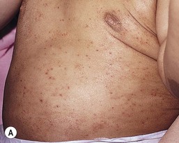
Nickel sensitivity in neonates and infants is predominantly caused by contact with nickel snaps on clothing. The most commonly involved areas are on the center of the chest and upper abdomen, where there may be a pruritic, well-defined dermatitis corresponding to the area of contact. Id reactions may occur, particularly in the antecubital fossae. Another possible source of nickel allergy is via ear-piercing, which in some cultures is performed in very young infants.108 FLG null mutations appear to be associated with nickel contact allergy.109 Allergic contact dermatitis may also occur in the diaper area, although at much lower frequency than irritant dermatitis. It also often spares the folds. Allergens include fragrances, disperse dye, and sorbitan sesquioleate, an emulsifier in topical preparations. Iodopropyl carbonate, methylchloroisothiazolinone/methylisothiazolinone, and bronopol in baby wipes, as well as mercapto compounds in the elastic borders of diapers, may also cause ACD.107 ACD has been reported from contact exposure to certain car seats composed of a shiny, nylon-like material, although the exact allergen has yet to be determined.110
Patients with AD may be predisposed to both forms of contact dermatitis because of their impaired barrier function. However, in some studies, the rate of positive patch tests was similar in children with and without AD.111
Sensitization may occur after only one or a few exposures to the offending agent, or it may be after years of contact. When sensitization does occur, it typically takes 10–14 days from exposure. On repeat exposure, the eruption develops within 24–48 h.102 It is pruritic, with acute lesions having more intense erythema, papulation, or vesiculation, and oozing. The lesions then become more crusted and with scale. Chronic ACD lesions tend to have lichenification and scale with little vesiculation.
Pathogenesis
ACD is produced by a T-lymphocyte-mediated, type IV, delayed hypersensitivity reaction. There are two distinct phases in its development: the induction (sensitization) phase and the elicitation phase.102 In the first phase, the allergen, usually a small molecular weight chemical (hapten or prehapten), penetrates the epidermis and binds with self-proteins to generate immunogenic complexes. The hapten and/or the complexes bind toll-like receptors on keratinocytes and activate the innate immune response. This leads to elaboration of proinflammatory mediators, such as IL-1β, that cause skin-resident dendritic cells to take up the hapten or haptenated proteins, further process them, and then migrate to regional lymph nodes where they present the processed peptides to naive antigen-specific T cells. These naive T cells become primed, activated, and generate a clone of differentiated effector T cells, which have skin-specific homing antigens. On repeat contact of skin-resident dendritic cells with the same hapten, the activated effector T cells are recruited back to the initial site of antigen encounter in the skin (the elicitation phase). They release proinflammatory cytokines, such as interferon-γ, and lead to the development of eczematous skin inflammation.
Diagnosis and differential diagnosis
A pruritic eruption with linearity or sharp edges, especially if on localized or exposed areas only, should raise suspicion for ACD. A careful medical and environmental exposure history are needed to try to confirm this and to elicit potential culprits. Patch testing is the gold standard to identify the external chemicals to which a person is allergic, but this is not usually performed in neonates or infants, unless the eruption is persistent or refractory to treatment. The thin-layer rapid use epicutaneous (TRUE) Test® is a pre-prepared, commercially available patch testing kit that tests for reactivity against 36 of the most common contact allergens. More comprehensive patch testing is indicated to identify allergies to other chemicals not found in the TRUE Test®, or if specific allergens are of interest based on the history and clinical distribution of the dermatitis. While available patch tests are approved only for the adult population at this time, they have been used in children in studies and in the clinical setting. Patch tests are usually placed on unaffected areas on the patient’s back and sometimes the arms, with removal between 24 and 48 h and evaluation at 48 h and at 72 or 96 h. However, there is some debate as to whether tests should be removed earlier or if different allergen concentrations are needed in children.112 Positive tests do need correlation for clinical relevance and sometimes, further confirmed by repeat open application testing of products containing the allergen that have been in contact with the patient.
The histology of allergic contact dermatitis is similar to that of other forms of eczematous dermatitis and thus, biopsy is more helpful to rule out other non-eczematous eruptions. Early lesions have spongiosis in the epidermis that then forms vesicles. The dermis has a superficial perivascular infiltrate consisting predominately of lymphocytes and other mononuclear cells. Eosinophils are usually present, but sometimes only in small numbers. In lesions that persist, scale crust and epidermal hyperplasia develop and the dermal inflammatory cell infiltrate becomes denser. Chronic lesions may show little spongiosis but have a prominent psoriasiform hyperplasia of the epidermis.
As mentioned, ACD needs to be differentiated from ICD and atopic dermatitis, although it may also occur as a consequence of the two conditions. AD usually does not involve the diaper area.
Management
First and foremost, contact with the offending allergen should be avoided. For instance, metals containing nickel should be eliminated if allergic, including zippers and underwear with nickel snaps. Information is available online on common products containing particular allergens (http://truetest.com). The American Contact Dermatitis Society also has a database that allows member physicians to create a list of products free of the allergens to which the patient is allergic. Topical corticosteroids are helpful until the inflammation has subsided, with moderate to potent agents sometimes needed. Topical calcineurin inhibitors may be useful for more sensitive sites.
Keratosis pilaris
This very common condition is seen in patients with atopic dermatitis as well as in otherwise healthy children. It is caused by perifollicular retention of scale, leading to plugging in the orifice of the hair follicle (see also Chapter 25). Affected infants and children develop small, rough, flesh-colored papules on the lateral aspects of the upper arms and thighs; the cheeks may be involved, with or without surrounding erythema (Fig. 15.14). Some have a more extensive distribution to include the trunk. Lesions are generally asymptomatic, but may become red and inflamed. Treatment is aimed at skin hydration with emollients. Mild keratolytics and exfoliants may be used, but are not curative and need careful application to avoid causing irritation. Occasionally, low potency topical steroids or topical calcineurin inhibitors may be utilized for inflamed lesions. The condition usually continues into adulthood, although some sites may improve over time.
Other etiologies of eczematous eruptions (see Boxes 15.2, 15.3)
Many nutritional deficiencies, metabolic disorders, and conditions causing erythroderma may appear eczematous (see Chapter 18).
Zinc deficiency
Zinc is an essential nutrient involved in numerous biologic functions necessary for growth and development.113 Deficiency can result from inherited or acquired conditions that cause inadequate dietary intake, malabsorption, excessive loss, or a combination of these. In particular, acrodermatitis enteropathica is a rare, autosomal recessive disorder caused by an inborn error of metabolism that results in zinc malabsorption. Mutations in the SLC39A4 gene are thought to be the underlying cause. It typically presents when the child is weaned from breast to cow’s milk or in the case of formula-fed infants, after 4–10 weeks, when stores of zinc have been depleted. While both cow’s and breast-milk contain adequate amounts of zinc, in acrodermatitis enteropathica the transport mechanism for intestinal absorption of zinc is defective. Absence of a binding ligand necessary for the transfer of zinc across the Paneth cells of the small intestine further contributes to deficiency.114
In the past, zinc deficiency resulting from inadequate intake was particularly seen in infants receiving parenteral alimentation without adequate zinc supplementation, but its routine addition to parenteral feeds has eliminated this problem. Zinc deficiency is still seen in premature infants who are exclusively breast-fed. The nutritional needs of the premature infant increase in the first few months of life, while at the same time the mother’s breast-milk zinc content decreases. This may be exacerbated by increased zinc secretion into the gut and poor absorption. Zinc deficiency may also be seen in term breast-fed infants, resulting from low or completely absent zinc in the mother’s breast-milk, despite normal maternal plasma zinc levels. In this instance, the transfer of zinc from plasma to breast-milk is defective. The condition may be inherited, as there are reports of absence of breast-milk zinc in multiple members of the same family.115
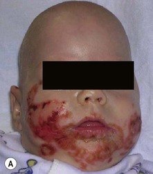
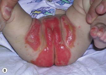
The skin eruption is usually the first clinical sign of zinc deficiency. Lesions are present on the cheeks and chin in a horseshoe distribution with erythematous, dermatitic erosions (Fig. 15.15A). Initially bullae may be present, but these rapidly erode. Over time, a more psoriasiform eruption develops. Periorbital involvement is common. A typical eruption occurs in the diaper area, consisting of a sharply demarcated area of erythema with accentuation of scale at the margin (Fig. 15.15B). The fingers and toes may have similar erosions and be accompanied by nail dystrophy. Skin lesions often become secondarily infected with Staphylococcus aureus and Candida. Overall, the eruption has a periorificial and acral pattern, though occasionally it may occur without any facial involvement. Irritability, refractory diarrhea, hair loss, and failure to thrive are additional notable clinical features.
A low plasma zinc level (<50 µg/dL) is the characteristic finding, and the level of the zinc-dependent enzyme alkaline phosphatase may also be low. Care should be taken to use a plastic syringe when drawing zinc levels and to prevent zinc contamination from rubber stoppers on glass tubes. High measured levels of zinc are due to laboratory error, as the body excretes zinc and limits absorption when the levels get too high.116
Treatment with zinc sulfate 1–3 mg/kg per day leads to rapid improvement, with irritability being the first symptom to respond.113 The inherited condition, acrodermatitis enteropathica, typically needs the higher dose of supplementation and may require long-term therapy. Occasionally, zinc sulfate causes gastrointestinal symptoms and zinc gluconate may be substituted.
Biotin deficiency
Biotin deficiency (multiple carboxylase deficiency) is an autosomal recessive disorder that becomes apparent in the neonatal period or late infancy. In affected neonates, the defect is a deficiency of holocarboxylase synthetase. In the later juvenile form of the disease, biotinidase is absent and leads to impairment of biotin recycling.117 Systemic signs and symptoms of biotin deficiency include vomiting, seizures, developmental delay, hypotonia, and eventual ataxia. Cutaneous lesions resemble those of zinc deficiency, affecting the area around the eyes, face, and perianal area. The lesions are eczematous or psoriasiform and are unresponsive to topical treatment with steroids. Secondary candidal infection is common and remains unresponsive to treatment until the biotin deficiency is corrected.
The pathology can be attributed to the decreased activity of any or all of four critical carboxylase enzymes: 3-methylcrotonyl CoA carboxylase, propionyl CoA carboxylase, pyruvate carboxylase, and acetyl CoA carboxylase. All are biotin-dependent, where biotin acts as a cofactor. The absence of carboxylase enzymes leads to an accumulation of carboxyls in the urine, resulting in lactic acidosis or ketosis. Treatment consists of biotin 5–20 mg/day and gives rapid improvement.118 The biotin is well-absorbed and does not accumulate in tissues.
Cystic fibrosis
Cystic fibrosis is an autosomal recessive disorder due to defects in the CFTR gene, which encodes a protein that functions as a chloride channel and also regulates the flow of other ions across the apical surface of epithelial cells. The condition is characterized by widespread dysfunction of the exocrine glands, resulting in chronic pulmonary disease, pancreatic insufficiency, and elevated sweat electrolytes. It can present in neonates or young infants with an erythematous, scaly rash with periorificial and/or perineal accentuation, also closely resembling that of zinc deficiency. The exact cause of the skin eruption is unknown, although essential fatty acid deficiency, low zinc levels, and protein deficiency may all be involved.119 Pedal edema is common. Failure to thrive, malabsorption, hepatosplenomegaly, chronic sinus inflammation, and frequent pulmonary infections are often noted. Many newborn screens now allow early detection of cystic fibrosis. Treatment is multipronged and includes maintenance of lung function as close to normal as possible via controlling respiratory infections and clearing the airways of mucus; administration of nutritional therapy to allow adequate growth (i.e., enzyme supplements, multivitamins, and mineral supplements), and management of gastrointestinal, urogenital, and other complications. A new agent, ‘ivacaftor’, is a CFTR potentiator that targets the defective protein and is under study.120
Hartnup disease
Hartnup disease is an extremely rare, heterogeneous autosomal recessive disorder characterized by a pellagra-like photosensitive eruption, cerebellar ataxia, emotional instability, encephalopathy, seizures, and aminoaciduria. The defect involves the disordered metabolism of tryptophan associated with the intestinal and renal transport of certain neutral α-amino acids. The disease is usually associated with consanguinity in the parents; it is quite rare in the Western hemisphere.121
Phenylketonuria
Phenylketonuria (PKU) is a rare autosomal recessive disorder involving defective metabolism of phenylalanine (see Chapter 23). Infants may appear normal at birth, although most have blond hair and blue eyes, or are notably fairer than their parents. About 50% of patients develop a dermatitis that is indistinguishable from AD. Intellectual disability becomes evident over time, and some individuals have epilepsy or extrapyramidal manifestation.122 PKU is caused by the absence of phenylalanine hydroxylase, the enzyme required for conversion of phenylalanine to tyrosine. To prevent the accumulation of phenylalanine in the blood, a low-phenylalanine diet is instituted as soon as possible after birth to prevent the resultant mental retardation. Recently, treatment with sapropterin in tetrahydrobiopterin (BH4)-responsive patients with phenylketonuria was noted to increase tolerance to dietary phenylalanine and improve quality of life.123 At birth, all babies in the USA are screened for PKU.
Netherton syndrome
Netherton syndrome is a rare disorder characterized by severe generalized erythroderma, scaling, and alopecia, caused by mutations in the SPINK-5 gene (see Chapters 18 and 19, Figs 18.4 and 19.6). Sparse eyebrows and hair are a hallmark of the syndrome. Light microscopy of affected hair may reveal trichorrhexis invaginata or ‘bamboo hairs’ (see Chapters 18 and 31).
Ichthyosis linearis circumflexa
Ichthyosis linearis circumflexa is a characteristic serpiginous, migratory rash with double-edged scale that is pathognomonic and usually occurs after 2 years of age. Affected patients tend to have dramatic increases in metabolic demands due to their skin disease, often resulting in failure to thrive and/or electrolyte imbalances, including dehydration from increased insensible losses. Cutaneous and systemic infections are common. The immunologic defects include reduced memory B cells, impaired specific antibody responses, and deficient natural killer cell-cytotoxicity.124 An atopic predisposition is noted and laboratory examination often reveals elevated IgE levels. Definitive diagnosis may be made by genetic testing. Management includes emollients, careful use of keratolytics, topical steroids, and topical calcineurin inhibitors on the skin to avoid excessive systemic absorption, and antibiotics for infections.125
Omenn syndrome
Omenn syndrome is an autosomal recessive form of severe combined immunodeficiency (SCID) that presents in the neonatal period with erythroderma or with an intensely pruritic, generalized eczematous eruption that can be difficult to distinguish from AD. Failure to thrive, alopecia, eosinophilia, diarrhea, and repeated infections (bacterial, viral, and fungal but particularly by Staphylococcus aureus) accompany the rash. Hepatosplenomegaly and lymphadenopathy are common. Bone marrow transplantation or other stem cell reconstitution is the first-line therapy for Omenn syndrome, although there are notable treatment-related complications and mortality.126 Cyclosporine may help with the erythroderma. Interferon gamma has been administered to downregulate IL-4 and IL-5 and modulate the inflammatory reaction. Nutritional supplementation is mandatory to decrease the risk of infection and increase the likelihood of successful stem cell reconstitution. Ancillary treatment with intravenous immunoglobulin (IVIG) further decreases the risk of infection.
Wiskott-aldrich syndrome
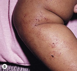
Wiskott–Aldrich syndrome is an X-linked recessive disease, and therefore it affects almost entirely males. A defect in the WASP protein results in defective interaction of T-lymphocytes and antigen-presenting cells such as dendritic cells and macrophages.127 Wiskott–Aldrich syndrome presents in the first few months of life with petechiae and ecchymoses on the skin, along with a bloody diarrhea, caused by both quantitative and qualitative abnormalities of platelets. Nosebleeds are also often seen. The skin eruption, which usually develops after the bleeding diathesis, is characterized by an extremely pruritic, generalized eczematous eruption with bloody crusts (Fig. 15.16). Apart from its hemorrhagic component, the eruption is indistinguishable from AD and fulfils the Hanifin and Rajka criteria.128 IgM deficiency is typical and there may also be cellular and humoral immunodeficiency. Skin abscesses from recurrent bacterial infections with pneumococci, meningococci, and Haemophilus influenzae, as well as lesions of molluscum contagiosum, verrucae, and herpes simplex, are common. Cutaneous vasculitis occurs in ~20% of patients within the first 15 years of life, and in some, presents as Henoch–Schönlein purpura with arthritis and renal involvement. There is a tendency to develop nephropathy and lymphoreticular malignancies later in life.129 Patients with thrombocytopenia may require IVIG and/or corticosteroids, and those with bleeding may need platelet and/or red blood cell transfusions. Bone marrow transplantation can be curative in patients with a histocompatible donor.
Hyper IgE syndrome
Via advances in genetics, hyper-IgE syndrome is now recognized to be a group of primary immunodeficiency diseases characterized by chronic eczematous dermatitis and immune, skeletal, and dental abnormalities. The autosomal dominant form (AD-HIES) is also called Job syndrome and is due to dominant-negative mutations in the STAT3 gene.130 The rash has often been described as similar to AD, but in neonates inflammatory facial papules (Fig. 15.17) and pustules may predominate and initially mimic neonatal acne. The eruption often begins at or soon after birth, becomes generalized, and is usually more extensive and resistant to treatment than typical AD. The disease course usually allows differentiation between the two entities. Also characteristic of hyper-IgE syndrome are recurrent skin infections, both secondary infection of the dermatitis as well as in the form of ‘cold abscesses.’ These abscesses lack the warmth, redness, and tenderness of typical furuncles due to a hampered influx and impaired function of granulocytes that is a part of the syndrome. Staphylococcus aureus is the most common bacteria involved, but Streptococcus, Haemophilus, and enteric Gram-negative bacteria have also been isolated. AD-HIES patients may have intertriginous and retroauricular lesions, external otitis, recurrent folliculitis especially at the upper back and shoulders, and pitted scarring of the face. Chronic mucocutaneous candidiasis affects about 60–80% of patients.130,131 A typical coarse facies emerges by late childhood or adolescence, with asymmetric deepened eyes, a prominent forehead and chin, and bulbous nose. The skeletal abnormalities consist of repeated fractures with minimal trauma and many also have joint hyperextensibility. Cranial synostosis and scoliosis have been reported. Dental abnormalities include retained primary teeth. As the name implies, IgE levels are quite elevated (>2000 IU/mL, or ten times the age-appropriate level), although they may decline and even normalize in adulthood. Eosinophilia (>800/mm3) is also seen. AD-HIES is complicated by recurrent episodes of pneumonia that frequently lead to pneumatoceles and bronchiectasis from aberrant healing. A higher rate of vascular anomalies, including coronary artery tortuosity and dilation, has recently been reported.132 There may be a higher incidence of malignancy, particularly of non-Hodgkin lymphoma.
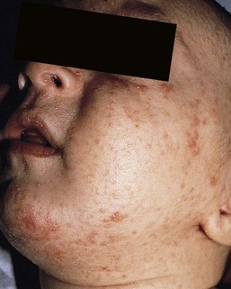
It is now known that DOCK8 (dedicator of cytokinesis 8) gene mutations account for nearly all of the cases categorized as autosomal recessive HIES. Features in common with AD-HIES are elevated serum IgE levels, eosinophilia, chronic dermatitis, recurrent sinopulmonary infections, and cutaneous staphylococcal abscesses. However, the dermatitis in those with DOCK8 deficiency may not be present at birth and is often more severe, while following the classic distribution for AD (Box 15.4).131,133 Affected individuals also suffer from widespread, recalcitrant cutaneous viral infections (by herpes simplex, human papilloma, and molluscipox viruses) and many have asthma and food and environmental allergies. While they also have frequent pneumonia, pneumatoceles are rare. Mucocutaneous candidiasis, coarse facies, retention of primary teeth, joint hyperextensibility, and pathologic fractures are much less common in this form of hyper-IgE syndrome. There is an increased risk of malignancy, particularly of squamous cell carcinoma and lymphoma, which may be due to impaired immune surveillance and chronic viral infections in the case of squamous cell carcinoma. Reduced serum IgM levels and lymphopenia can be noted. Mortality is often by the second to third decade of life, whereas those with AD-HIES often live into their fifth and sixth decades.130 Thus, those with DOCK8 deficiency need close monitoring and more frequent physical examinations.
Management of the hyper IgE syndromes involves aggressive treatment of infections and good skin care. These include topical antimicrobials and dilute bleach baths to decrease S. aureus. colonization and systemic anti-staphylococcal antibiotic therapy to treat and prophylactically reduce skin and lung infections and abscesses. Topical steroids must be used very carefully for the dermatitis, particular in the case of DOCK8 deficiency, where cutaneous viral infections could worsen. These viral infections are a treatment challenge, often refractory to traditional methods and with only occasional response even to systemic agents such as cidofovir and interferon alpha.133 Oral antifungals are needed for those with mucocutaneous candidiasis or fungal infections of the lung, which are also difficult to eradicate. IVIG has been given to some individuals, with a few reports of reduced respiratory tract infections but not viral skin infections, and thus its use is recommended mainly for those with demonstrated impairment of antibody response. Hematopoietic stem cell transplantation has been tried for both major forms of hyper-IgE syndrome, though its exact role and timing still remain to be defined. While helpful for correction of immunodeficiency and some of the severe skin findings, there are risks with additional immunosuppression with myeloablation and prophylaxis of graft-versus-host disease, and the effects on the development of malignancy are unknown at this time.
Conclusion
Eczematous eruptions are important diagnoses to recognize, not only for their common occurrence, but also for potential systemic associations and long-term complications that require careful management in some instances.
Access the full reference list at ExpertConsult.com 
[/level-membership-for-dermatology-category][not-level-membership-for-dermatology-category]
Eczematous Disorders
Wynnis L. Tom, lawrence F. Eichenfield
Introduction
Eczematous eruptions represent a significant proportion of the skin diseases affecting neonates and infants. Clinically they are characterized by erythema, edema, scale, and sometimes crusts. The most common disorder is atopic dermatitis (AD) and, in fact, the term ‘eczema’ (which means ‘boiling over’) is often used by laymen to refer to AD. Seborrheic dermatitis and irritant dermatitis also frequently affect infants, while allergic contact dermatitis (ACD) is less common. Some nutritional, metabolic, and immunologic diseases have clinical manifestations that include eczematous eruptions which may be difficult to differentiate from AD, and they require a high index of suspicion for their occurrence.
Atopic dermatitis
Atopic dermatitis is a chronic, pruritic skin condition with a frequently relapsing course. It was first described by Besnier in 1892 as ‘prurigo diasthétique’. In 1933, Wise and Sulzberger coined the term ‘atopic dermatitis’, and Hill and Sulzberger characterized the clinical entity 2 years later.1,2 AD is notable for inflammation, xerosis, crusts, excoriations, and lichenification, and it is the most common chronic inflammatory dermatosis of early childhood. The disease imposes an enormous burden on the personal, social, emotional, and financial resources of patients and their families. In the USA, annual direct costs for AD care are estimated at US$1–4 billion,3 which does not include the effects of lost productivity such as missed work and school days nor the impact on quality of life.
The prevalence of AD has increased markedly over the past 50 years, with current rates of 15–29% in the USA, Europe, Japan, and other industrialized countries and lower but increasing rates in developing nations.4–6 AD affects individuals of all races, with several studies showing a slightly higher prevalence in black children.6 However, different countries with similar ethnic demographics have markedly different recorded prevalences, supporting the concept that environmental factors are important in disease expression.7 AD may be slightly more common in girls (female : male ratio of 1.3 : 1), although some studies note that it affects the sexes approximately equally.8
Risk factors
Multiple epidemiologic studies have examined risk factors for disease development. Only two factors show consistent, strong association: a family history of atopic diseases (i.e., asthma, allergic rhinitis, atopic dermatitis) and loss of function mutations in the filaggrin (FLG) gene. A positive family history of atopy is noted in approximately 70% of affected individuals. The odds of developing AD are 2–3-fold higher in children with one atopic parent, and this increases to as high as 3–5-fold if both parents are atopic.9,10 Some have noted a maternal history of AD itself to be more predictive.11,12 The FLG gene encodes a key protein involved in terminal differentiation of the epidermis, and its breakdown products are important in the formation of the skin barrier, including the stratum corneum, and natural moisturizing factor (see below). Loss of function mutations confer a risk for earlier-onset AD, as well as for more severe disease.13
AD is more frequent in higher social and socioeconomic classes.14 Other factors that may be associated with a greater risk for disease include small family size, migration from rural to urban environments, and increased parental education.7 Some have noted a higher incidence in association with maternal smoking and elevated birthweight, though not all studies corroborate these findings.15,16 The effect of exposure to pets in the home is unclear, due to conflicting data.17,18
Clinical findings
History
The onset of disease is most commonly between 3 and 6 months of age, with approximately 60% of patients developing the eruption within the first year of life and 90% by 5 years of age.19,20 Atopic dermatitis is usually the first manifestation of the ‘atopic march,’ which refers to the subsequent development of food allergies, asthma, and allergic rhinitis, although this progression does not happen in all individuals and/or the sequence may vary.20 More recently, eosinophilic esophagitis and gastroenteritis have been added to the list of associated disorders.21
The predominant symptom is pruritus, which is difficult to control and a major cause of morbidity and poor quality of life for both the child and caregiver.22 It frequently interferes with sleep and can lead to complications such as secondary infection of excoriated lesions. Parents should be questioned about the infant scratching and rubbing against objects such as bedding or carpeting. Unfortunately, these actions further incite inflammation and the itch sensation, leading to a vicious cycle.23
Physical examination
There are three age-related phases to AD: infantile, childhood, and adult disease. During each phase both the sites of involvement and the morphology of the lesions change, although the phases often overlap. The infantile phase generally lasts until 2–3 years of age, the childhood phase from 2 years until puberty, and the adult phase from puberty onward.
In infancy, AD characteristically begins on the cheeks (Figs 15.1, 15.2) and scalp (Fig. 15.3) and evolves over time to involve the lateral and extensor aspects of the arms and legs (Fig. 15.4). The periauricular areas are often affected and can lead to infra-auricular fissuring.24 While the trunk may have lesions, the diaper area is usually spared due to the retention of moisture and protection from rubbing and external triggers provided by these coverings (Figs 15.5, 15.6). Lesions are symmetric, scaly, erythematous patches and plaques, within which crusting is common. Acute lesions are more exudative. Lymphadenopathy may be noted and reflects local inflammation. During late infancy and the childhood phase, the flexural surfaces of the extremities become the most commonly involved sites, particularly the antecubital and popliteal fossae (Fig. 15.7). Other frequently involved locations include the neck, wrist and ankle flexures, and the creases between the thighs and buttocks. Lesions continue to be erythematous patches and plaques with crusting, but often there is increased excoriation. Chronic findings, such as lichenification (enhanced skin markings) and pigmentary changes start to become prominent (Fig. 15.8). The pigmentary alterations (hypo- or hyperpigmentation) often evoke parental concerns about scarring, although lesions generally do not leave scars unless severely infected or deeply excoriated. From puberty onwards, AD lesions tend to prefer the face, back, wrists, hands, and dorsal feet.
There are a number of additional physical findings that, if present, aid in the diagnosis of atopic dermatitis (Box 15.1). These include other conditions associated with xerotic skin, such as ichthyosis vulgaris, keratosis pilaris, and pityriasis alba. Follicular lichenification with papules may be noted, especially in those with darker skin type. These are not specific to AD, however, and may be seen in otherwise healthy individuals as well. Other findings suggestive of an atopic diathesis that become more prominent with rubbing or scratching include an additional line or groove in the lower eyelid (the Dennie-Morgan fold), infraorbital darkening (‘allergic shiners’), and a prominent horizontal nasal crease (‘allergic salute’).
Diagnosis
The diagnosis of AD is made clinically. This is often more challenging in early infancy, when it can be difficult to distinguish AD from other infantile eczematous disorders. Family history is often helpful and absence of an atopic disorder in the family history makes the diagnosis much less likely, though does not preclude its occurrence. The most recognized diagnostic criteria are those proposed by Hanifin and Rajka in 1980, but the four major and 23 minor criteria are too numerous to be used routinely in clinical practice.25 Several expert groups have proposed more condensed sets of criteria,26–28 including the following,27 which is applicable to both children and adults:
A. Essential features (must be present)
a. Pruritus
b. Eczematous dermatitis (acute, subacute or chronic)
i. Typical morphology and age-specific patterns
ii. Chronic or relapsing history
B. Important features (seen in most cases, adding support to the diagnosis)
b. Atopy
i. Personal and/or family history
ii. IgE reactivity
c. Xerosis
C. Associated features (these clinical associations help to suggest the diagnosis of AD but are too nonspecific to be used to define or detect AD for research or epidemiologic studies)
a. Atypical vascular responses (e.g., facial pallor, white dermatographism, delayed blanch response)
b. Keratosis pilaris/hyperlinear palms/ichthyosis
d. Other regional findings (e.g., perioral changes/periauricular lesions)
e. Perifollicular accentuation/lichenification/prurigo lesions.
To date, there is no specific laboratory test to confirm the diagnosis of AD. Serum immunoglobulin E (IgE) is elevated in approximately 70–80% of patients, although even severe cases may have normal levels.23 The majority of patients also have increased circulating eosinophils. Recent discoveries of novel T-lymphocyte subsets and cytokine proteins have generated additional candidate markers. Thymus and activation-regulated chemokine (TARC, also called CCL17) is one such molecule suggested to increase with disease activity.29 However, to date, none have demonstrated adequate sensitivity and specificity to serve as a biomarker for the disease or its response to therapy. Laboratory testing is thus seldom needed in the routine evaluation of uncomplicated AD.
Skin biopsies may be helpful in distinguishing AD from other inflammatory skin disorders, but the biopsy findings are not pathognomonic. The histology varies from an acute dermatitis with spongiosis and a perivascular lymphocytic infiltrate, to chronic changes in lichenified skin showing acanthosis, hyperkeratosis, and accumulation of Langerhans’ cells, macrophages, and mast cells in addition to lymphocytes around blood vessels. The superficial venular plexus has endothelial cell hypertrophy, basement membrane thickening, and increased numbers of immunoreactive nerve fibers.30,31 Eosinophils are not commonly present in acute AD, but are increased in number in chronic lesions. Mast cells may be normal to increased, and in different stages of degranulation.
Etiology and pathogenesis
The pathogenesis of AD involves a complex interplay of genetic, immunologic, infectious, and other environmental factors. While still not completely understood, it appears to entail two major defective processes: dysfunction of the epidermal barrier and dysregulation of the immune system. Which process occurs first to initiate disease (‘outside-in’ vs ‘inside-out’) is an ongoing debate, although it is clear that they interact and exacerbate one another. This is compounded by a heightened response to environmental allergens, irritants, and microbial antigens.31,32
For many years, it has been known that AD involves genetic susceptibility, given familial clustering, concordance in monozygotic twins but discordance in dizygotic twins, and reports of transfer of AD by bone marrow transplantation.33,34 It has been most highly linked to genes of the epidermal differentiation complex located on chromosome 1q21, particularly the FLG gene, which is also the main cause of ichthyosis vulgaris. The association of FLG mutations with AD is one of the most robust known for complex genetic disorders.13 Large, population-based studies have identified over 35 mutations in affected individuals, with distinct mutations in different ethnic groups.35 As mentioned above, loss of function mutations are associated with early-onset and more severe, persistent disease, and additionally appear to give increased allergic sensitization and total IgE levels, a greater risk for asthma and food and other allergies, and a higher incidence of skin infections with herpes simplex virus (eczema herpeticum). But despite the strong association, FLG null mutations are seen in only approximately 18–48% of individuals with AD (depending on the population) and conversely, about 40% of individuals with such mutations do not develop the disease.13 Genetic screening of affected families has highlighted other chromosome regions that contribute to disease susceptibility, including cytokine and immune-related genes (e.g., those encoding interleukins (IL)-4, -5, and -13 and the regulated on activation of normal T-cell expressed and secreted (RANTES) chemotactic cytokine), genes encoding innate immunity receptors (e.g., the genes for toll-like receptors (TLR)-2 and -9), and genes implicated in other inflammatory diseases (e.g., those encoding nucleotide-binding oligomerization domain protein (NOD)-1 and NOD2).36,37 There are likely additional genes that remain to be discovered, and of those already identified to have high disease association, a number still require determination of exact function.
Defects in both the structure and function of the epidermal barrier are present in AD. FLG mutations lead to disorganized keratin filaments and abnormal architecture of the lamellar bilayer and assembly of the cornified envelope. Decreased filaggrin breakdown products affect levels of natural moisturizing factor and the skin pH, resulting in increased transepidermal water loss and skin permeability. In addition, barrier function is markedly reduced due to a decline in ceramide levels, which are major water-retaining molecules, and from an imbalance of proteolytic enzymes that affect epidermal adhesion and their anti-protease counterparts.35,38 Abnormalities in tight junction expression that form a second barrier in the stratum granulosum further add to the pathology.34 These factors all lead to dry, xerotic skin, while facilitating entry of antigens through the damaged epidermis that stimulate cutaneous inflammation.
Immunologic abnormalities are another major pathogenic mechanism. Antigens bind to and activate epidermal Langerhans’ cells and dermal dendritic cells. Circulating T cells that have increased expression of the cutaneous lymphocyte-associated antigen (CLA) receptor home to the skin, where they are primed by these antigen-presenting cells. In the acute phase of disease, these activated T cells overexpress cytokines of the Th2 class, notably IL-4 and IL-13, which mediate switching of immunoglobulin isotypes to IgE synthesis and upregulated expression of adhesion molecules on endothelial cells. IL-4 also stimulates mast cells to produce mediators such as histamine. IL-5 predominates in subsequent stages and is involved in eosinophil development and survival.31,39 While AD begins acutely as a Th2-mediated disorder, in its chronic phase it is characterized by Th0 cells (cells that share some activities of both Th1 and Th2 cells) and Th1 cells; associated cytokines include interferon-γ. The switch to Th0/1 cells involves the infiltration of the epidermis by inflammatory dendritic epidermal cells (IDEC) and production of IL-12 and IL-18, as well as several remodeling-associated cytokines. Also of significance is an increase in Th22 cells in chronic AD skin, which produce IL-22 that inhibits terminal differentiation and induces epidermal hyperplasia.40 Additional T cell subsets have been identified, but their roles in AD remain to be fully defined. T regulatory cells with normal immunosuppressive activity appear to be expanded in the peripheral blood of patients, but not in lesional skin or atopy patch test sites. Th17 cells have been found to be elevated in patients with acute AD, but not as prominently as in psoriasis.31
Enhanced allergen penetration through the damaged epidermis gives increased production of thymic stromal lymphopoietin (TSLP) by keratinocytes, which may be a critical link between the barrier and immune defects. TSLP has been shown to drive both the initial Th2 cytokine response and the switch to the Th0/1 phenotype.39 Mechanical injury such as scratching or rubbing, microbes, and proinflammatory cytokines themselves further stimulate release of TSLP, thus perpetuating the inflammation. The immunologic response contributes to additional barrier abnormalities, since IL-4 and IL-13 are known to suppress filaggrin expression and downregulate proper processing of profilaggrin.
Antigens that gain entry include microbes. The antimicrobial barrier is compromised in AD from a combination of the defective physical barrier, an increased pH which may allow greater adherence and multiplication of bacteria, and toll-like receptor defects which diminish recognition of microbial agents. In addition, the increase in Th2 cytokines suppresses the synthesis of antimicrobial peptides by keratinocytes, specifically LL-37 (cathelicidin) and β-defensins 2 and 3.41 This predisposes atopic individuals to widespread skin infections due to bacteria and viruses (including herpes, molluscum, and vaccinia), and possibly to dermatophytes. Staphylococcus aureus, in particular, can trigger multiple inflammatory cascades. Staphylococcal toxins activate T cells in a superantigen-driven fashion and induce IgE-specific responses. These further skew the immune response toward Th2 cytokine production and explain the association of S. aureus infection with exacerbations of AD.42 The superantigens also cause a profound reduction in steroid responsiveness of T cells, giving another possible mechanism for disease flares. Exogenous protease inhibitors produced by S. aureus may also damage the epidermal barrier, thereby potentiating the absorption of allergens into the skin and leading to increased bacterial colonization.
The impact of food exposure on the development of atopic dermatitis is controversial. Although it is known that food allergy and sensitization more commonly develop in children with atopic dermatitis, it is unclear how important food exposures are to the onset and course of atopic dermatitis, nor how common food-induced eczematous dermatitis is during infancy.43 Aeroallergens may also trigger immune responses, but sensitization is usually after infancy. The house-dust mite, Dermatophagoides pteronyssinus, has been cited in the pathogenesis of AD, but measures to decrease dust mite exposure are not necessarily protective.44 Malassezia yeasts have also been suggested but not confirmed to cause exacerbations of disease.
The mechanisms underlying pruritus in AD are the subject of much investigation. In addition to histamine, several other mediators, such as neuropeptides, kinins, and cytokines, can induce pruritus. Studies have shown that the number of sensory and neuropeptide-containing nerve fibers is increased in lesional AD skin.30,45 Mast cells store large amounts of proteases, including tryptase, which may induce pruritoceptive itch when released in the proximity of these nerve fibers. These release stress-induced neuropeptides in parallel, which might activate mast cells through neurokinin receptors to perpetuate pruritic signals. The Th2 response contributes via increased levels of IL-31, which has recently been found to enhance pruritus. Staphylococcal superantigens also markedly enhance IL-31 production.45,46
Differential diagnosis
The common disorders in the differential diagnosis of AD include seborrheic dermatitis, contact dermatitis, psoriasis, scabies, ichthyosis, tinea corporis, and keratosis pilaris (Box 15.2). In early infancy, seborrheic dermatitis may be virtually indistinguishable from AD due to the similarity in sites of involvement and morphology (Fig. 15.9). Irritant dermatitis is not uncommon, but allergic contact dermatitis is rare in the newborn period, although it may affect infants. A configuration suggesting an external source of exposure may be evident and helpful in determining the presence of a contact dermatitis.
Psoriasis is not as common in infancy, but psoriasis vulgaris and pustular psoriasis may occur (see Chapter 16). In the case of psoriasis vulgaris, the diaper area is most commonly affected and consists of erythematous, well-demarcated plaques surmounted by some to little scale, which is less thick and silvery relative to other sites due to moisture and occlusion. About 5% of children appear to an overlap of psoriasis and atopic dermatitis lesions, and often have a family history of both disorders.47
Scabies may sometimes be difficult to distinguish from AD because both can cause severe pruritus, but it is rarely considered in the newborn, and is more relevant to infants. Here the face is not usually involved, whereas eczematous patches on the cheeks and xerosis are typical of AD. Moreover, the eruption of scabies has polymorphous lesions, including burrows, papules, nodules, eczematous and urticarial lesions, as well as pustules on the palms and soles. A recent onset of itching in family members is also helpful in differentiating the two conditions.
When the skin lesions are associated with failure to thrive, diarrhea, infection, and/or signs in other organ systems, it is important to consider systemic disorders, such as nutritional and metabolic abnormalities, and genetic conditions including immunodeficiency (Box 15.3). Also in the differential diagnosis are proliferative disorders, most notably Langerhans’ cell histiocytosis, which often includes hemorrhagic lesions. Occasionally, AD may be so severe that the whole body becomes erythrodermic, causing confusion with other causes of erythroderma, particularly Netherton syndrome and nutritional deficiencies (see Chapter 18).
Prognosis
Data from follow-up surveys show enormous variation in the long-term course and prognosis of AD, owing to differences in patient sampling techniques. Vickers48 reported that AD cleared by age 20 in 90% of patients, but the inclusion of infants with seborrheic dermatitis, which resolves within weeks to months, biased these results. Wüthrich49 found that of 121 infants followed to a mean of 23.5 years, 11% had resolution in childhood, an additional 25% cleared in adolescence, but 32% had a chronic continuous course from infancy into adulthood and 20% had reappearance of disease in adolescence. Despite these differences, there is a steady decline in AD prevalence over the childhood years. Up to 60–70% resolution of disease by adulthood appears to be a reasonable estimate, with an overall prevalence rate of 2–10% in adults.49,50
Early onset within the first 6 months of life, greater disease severity, and persistence into adolescence are predictors that the disease is more likely to continue into adult life.51 Other reported risk factors for adult disease include the presence of other atopic conditions such as asthma and allergic rhinitis, a family history of AD in parents or siblings, high serum IgE levels, and FLG null mutations in early-onset cases.49,51 It is, however, difficult to precisely predict if and when a particular child will have resolution or remission and management should continue until there is notable quiescence of lesions. But even after what appears to be resolution, easily irritated or dry skin may persist or hand eczema may affect the individual later in life.52
Figures on the incidence of asthma and allergic rhinitis following or in association with AD vary from 10–70%.51,53 In one survey, 36% suffered from allergic rhinitis, 28% from asthma, and 15% from both.54 There is an increased risk of developing asthma or AD if there is a family history of either condition. Higher rates have also been reported with more severe AD, with 60% having asthma and 62% with allergic rhinitis reported in one study.55 Mechanisms to link their occurrence are under study. Filaggrin mutations appear to increase the risk of development of asthma and other atopic conditions.13,53 It has been suggested that early or severe AD and epicutaneous sensitization to environmental allergens may lead to inflammation and development of allergic disease on re-exposure at other epithelial barrier sites (e.g., the respiratory or gastrointestinal tract). Systemic circulation of keratinocyte-derived TSLP may play an important role in this process.56 Although supported by animal models, this remains to be confirmed, along with whether prevention or early treatment of AD mitigates subsequent development of other atopic conditions.
Treatment
The management of AD should be directed at reducing signs and symptoms of disease and decreasing the frequency and severity of flares. It requires a multimodal approach that includes meticulous general skin care, hydration of the skin, avoidance of potential irritants and allergens, and reduction of pruritus and inflammation. Secondary infections should be treated and prevented. It is extremely important to educate the parents on the chronic nature of the disease, as many seek an immediate cure which is not possible. The goals of therapy, including the benefits, risks, and side effects of treatments, should be detailed. Because parents are inundated with much information, careful attention to detail, repetition of advice, and written handouts and action plans can improve treatment outcomes. Educational programs in the form of structured, multidisciplinary classes and nursing-led care are themselves effective primary interventions that can give some decrease in disease severity.57
Xerosis/dry skin
Xerosis is a significant component of the disease, and hydrating the skin is a foundation of atopic dermatitis therapy. This is accomplished through frequent application of emollients or moisturizers and care in bathing techniques. The appropriate frequency of bathing is controversial, but limiting to a short duration using warm rather than hot water is recommended. Bathing alone leads to a decrease in hydration status of the skin once the water evaporates.58 On the other hand, there is evidence that bathing followed by the immediate application of an emollient while the skin is wet (the ‘soak and seal’ method) has an excellent hydrating effect, while removing scale and crust, and can be a tremendously useful treatment measure for patients during flares.59,60
The application of emollients and moisturizers independent of bathing can also improve hydration status, enhance barrier function, and be steroid sparing.59,61 They should be applied all over the body and after topical corticosteroids have been applied to lesional skin. There is no one preferred emollient or moisturizing agent. Ideally, it should be safe, effective, inexpensive, and free of additives, fragrances, and other potential sensitizing agents. Ointments are the thickest agents but creams and oils may also be used, while lotions should generally be avoided as they are less effective in decreasing xerosis. Products containing urea, lactic acid, and α-hydroxy acids may sting and their absorption in infants is unknown.
Topical therapies have been developed to target skin barrier dysfunction and replace abnormal epidermal lipids. These include preparations having distinct compositions of lipids and ceramides to mimic endogenous lipids, and creams containing palmitoylethanolamide, hydrolipids, and/or filaggrin breakdown products.62 While they have shown some benefit and may be helpful as adjuvant agents, the few studies performed to date have not demonstrated superiority relative to traditional, less expensive options such as petrolatum ointment.62,63 But addressing the defective barrier is an important therapeutic concept, given the current understanding of AD pathogenesis.
Anti-inflammatory agents
Topical corticosteroids.
Topical corticosteroids were first introduced by Sulzberger and Witten in 1952 and remain a mainstay of AD treatment because of their excellent anti-inflammatory effects. They are grouped according to potency into seven classes, from very weak (VII) to superpotent (I). There is often fear and reluctance on the part of parents to use them, despite the extremely small number of reported side-effects in those treated, the majority of which have occurred in patients receiving inappropriate treatment.64,65 Physicians should encourage the correct use of topical corticosteroid preparations on affected inflamed areas. Both amount and potency are important and close monitoring during treatment is appropriate for safety and compliance. Precise information should be given to the parents regarding the manner of application, and written instructions are very useful.
There are many methods of prescribing topical corticosteroids and other anti-inflammatory therapies. Published algorithms are available and may be used.66 Mild cases and/or facial or diaper areas are generally treated with low-potency steroids. For more significant flares, or if low-potency steroids are inadequate to control the disease, more potent topical corticosteroids are used for truncal and body surfaces. For severe flares, wet-wrap therapy (a damp layer of gauze or clothing with an outer dry layer) may be utilized with low- to mid-strength topical steroids for several days to increase their effect.67 Prescriptions should be given for sufficient treatment of the area involved, but with more caution and set limits with higher potency agents. In general, one finger-tip unit (the amount from the distal interphalangeal joint to the finger tip, ~0.5 g) should be applied over an area of two adult palms.64,68 For acute flares, areas should be treated until the inflammation disappears, with frequent and liberal use of emollients. Most should be used twice a day, although some agents are designated for once-daily use. Emollients should be continued after corticosteroids are discontinued and may help lessen disease recurrence and the need for anti-inflammatory agents.
Systemic corticosteroids, particularly long-term use, are not indicated for the treatment of AD in neonates or infants. In addition to hypothalamic-pituitary-adrenal axis suppression, they can affect linear growth and have known rebound and other negative effects.
Topical calcineurin inhibitors.
Topical calcineurin inhibitors are another class of anti-inflammatory therapy for atopic dermatitis. In the USA, topical tacrolimus and pimecrolimus are approved as second-line agents for short-term and non-continuous chronic use in children aged 2 years and older, but they are sometimes used off-label. Unlike topical steroids, topical calcineurin inhibitors do not carry a risk of skin atrophy, and therefore may be helpful for sites of thinner skin (i.e., eyelids, face, and diaper area where steroid atrophy are a bigger concern) or to decrease the use of topical steroids in those with moderate or severe disease. There is histologic evidence that while both classes improve epidermal differentiation and decrease transepidermal water loss, topical steroids do not restore the epidermal barrier structures (e.g., lamellar body extrusion and lipid bilayer formation in the lower stratum corneum) to the same extent as calcineurin inhibitors.69
The most common side-effects with topical calcineurin inhibitors are local reactions such as stinging and burning, although this may improve after several applications or when first preceded by a short period of topical corticosteroid use. Patients should be monitored for signs and symptoms of cutaneous viral infections, although ongoing surveillance has not shown a consistent increase in frequency.70,71 These agents do carry a boxed warning stating that their long-term safety has not been established because of rare post-marketing cases of malignancy, specifically, of skin cancer and lymphoma. Interim analyses of ongoing, 10-year surveillance studies to address these concerns have not found evidence of increased malignancy rates relative to that expected in the general pediatric population.72,73 Several studies, including a large case–control study of 293 253 patients, have found an increased risk of lymphoma that correlates with AD severity, but not with topical calcineurin inhibitor use.74,75
Maintenance therapy.
[/not-level-membership-for-dermatology-category]
