Echocardiographic Imaging of Single-Ventricle Lesions

Published on 21/06/2015 by admin

Filed under Cardiovascular

Last modified 22/04/2025

Print this page

rate 1 star rate 2 star rate 3 star rate 4 star rate 5 star
Your rating: none, Average: 5 (1 votes)

This article have been viewed 2833 times

10 Echocardiographic Imaging of Single-Ventricle Lesions

SV lesions are complex heart defects that result in one of the ventricles being hypoplastic or absent. The most common of these lesions are those that comprise HLHS. SV lesions are divided into three categories based on the adequacy of pulmonary blood flow (PBF) and systemic blood flow (SBF).

This chapter discusses in detail the imaging of these three lesions:

Hypoplastic Left Heart Syndrome

Definition and Prevalence

The goals of the initial echocardiogram in HLHS are to provide a complete anatomic survey and assess the underlying physiology, which play a critical role in determining the initial management of the patient. In addition to identifying the degree of hypoplasia of the left ventricular (LV) chamber and that of the MV and AV, it is important to assess the function of the RV and tricuspid valve (TV), the size and the direction of flow across the AS, ductal physiology, and associated findings such as ventriculocoronary communications or fistulae and anomalous pulmonary venous return.

Pertinent information is obtained from each of the standard views by two-dimensional (2D) and Doppler.

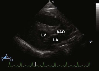
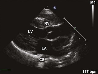
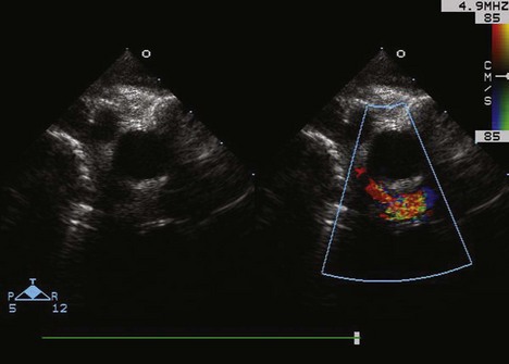
Parasternal Long Axis View (Fig. 10-1)

Parasternal Short Axis View
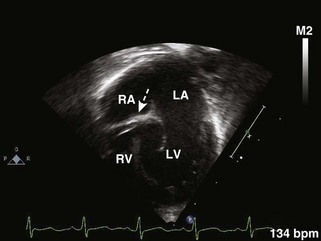
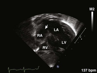
Subcostal Four-Chamber or Coronal View

Double-Inlet Left Ventricle

Definition and Prevalence

The most common form of univentricular AV connection. It is defined as the presence of two AVVs committed to one ventricular chamber of LV morphology. DILV is associated with a rudimentary outlet chamber, the BVF. The inlet septum is absent, and both AVVs lie in fibrous continuity with a semilunar valve. The ventricles are L-looped, most commonly with the LV being the more posteroinferior and rightward chamber. The bulboventricle is thus located anterosuperiorly.

The purpose of the initial echocardiogram is to provide a complete anatomic survey and assess the underlying physiology. The basic anatomy of atrial and visceral situs and location of the cardiac apex must be defined. This information is best obtained from subcostal views. It is important to determine the presence of a straddling, atretic, or stenotic AVV; the relationship of the GVs; the position and degree of restriction of the BVF leading to either systemic or pulmonary outflow tract obstruction; and the physiology of the PDA and other associated anomalies such as abnormal pulmonary or systemic venous connections.

Pertinent information is obtained from the standard views by 2D and Doppler.

Tricuspid Atresia

In postoperative SV management, the echocardiogram is an important diagnostic tool in evaluating the adequacy of PBF and SBF (PA band gradient, aortic arch flow), ventricular function, atrioventricular regurgitation, restriction across the atrial-level communication, Glenn, Fontan pressures, and branch PA stenosis.

Definition and Prevalence

Absence of a direct connection between the RA and the RV. The atretic valve is usually muscular and rarely fibrous. An atrial-level communication, either a secundum ASD or a PFO, is necessary for survival.

The goal of the initial echocardiogram in TA is to provide a comprehensive evaluation of the anatomy and physiology, paying special attention to the absence of a direct communication between the RA and RV, the size of the VSD, relationship of the GVs, the size of the atrial-level communication, and the physiology of the PDA. In addition, associated anomalies should be ruled out such as left superior vena cava (LSVC), left juxtaposition of the RAA, and coarctation of the aorta. Associated findings are more common with transposed great arteries.

Pertinent information is obtained from the standard views.

Postoperative Evaluation

Echocardiographic evaluation following first-stage palliation includes the following:

Norwood and Damus-Kaye-Stansel Procedures

The goals of the postoperative echocardiogram after a Norwood or Damus-Kaye-Stansel (DKS) procedure and before a bidirectional Glenn/Hemi-Fontan procedure are to evaluate for the following.

Echocardiographic Evaluation After Bidirectional Glenn/Hemi-Fontan Procedure

The goals of the echocardiogram after a bidirectional Glenn/Hemi-Fontan procedure and before a Fontan procedure completion are to evaluate for the following.

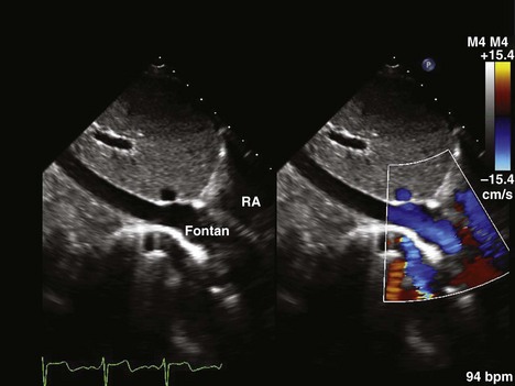
Echocardiographic Evaluation After Fontan Procedure Completion

The goals of the echocardiogram after completion of the Fontan procedure are to evaluate for the following.

Suggested Reading

1 Bevilacqua M, Sanders SP, Van Praagh S, et al. Double-inlet single left ventricle: echocardiographic anatomy with emphasis on the morphology of the atrioventricular valves and ventricular septal defect. J Am Coll Cardiol. 1991;18:559-568.

It discusses the echocardiographic anatomy of DILV with special attention to the morphology, size, and function of the AVVs and VSD and their relationship to PS, aortic stenosis, and AAO.

2 Orie JD, Anderson C, Ettedgui JA, et al. Echocardiographic morphologic correlations in tricuspid atresia. J Am Coll Cardiol. 1995;26:750-758.

It describes how TA in most cases is secondary to the absence of an RA-RV junction along with the associated findings in TA.

3 Fraisse A, Colan SD, Jonas RA, et al. Accuracy of echocardiography for detection of aortic arch obstruction after Stage I Norwood procedure. Am Heart J. 1998;135(2 Pt 1):230-236.

This study evaluates the accuracy of echo in the diagnosis of AAO after a stage I Norwood procedure, identifies echocardiographic predictors of arch obstruction, and examines the time course of its development. It shows that echo is a highly specific but poorly sensitive modality in detecting AAO after a stage I Norwood procedure. Early cardiac catheterization with possible intervention should be considered in patients with moderate or severe RV dysfunction, moderate or severe TR, or an abnormal abdominal Doppler flow pattern during that period.

4 Jacobs ML, Mayer JEJr. Congenital Heart Surgery Nomenclature and Database Project: single ventricle. Ann Thorac Surg. 2000;69(Suppl 4):S197-S204.

The STS-Congenital Heart Surgery Database Committee and the European Association for Cardiothoracic Surgery have proposed a classification that is relevant to surgical therapy.

5 Cook AC, Anderson RH. The anatomy of hearts with double inlet ventricle. Cardiol Young. 2006;16(Suppl 1):22-26.

6 Munoz-Castellanos L, Espinola-Zavaleta N, Keirns C. Anatomoechocardiographic correlation double inlet left ventricle. J Am Soc Echocardiogr. 2005;18(3):237-243.

Anatomoechocardiographic correlation between the morphologic features of equivalent anatomic specimens and the echocardiographic images of patients to provide a means of interpreting the image correctly and making a more precise diagnosis of the cardiac defect.

7 Cardis BM, Fyfe DA, Ketchum D, et al. Echocardiographic features and complications of the modified Norwood operation using the right ventricle to pulmonary artery conduit. J Am Soc Echocardiogr. 2005;18(6):660-665.

8 Galantowicz M, Cheatham JP. Lessons learned from the development of a new hybrid strategy for the management of hypoplastic left heart syndrome. Pediatr Cardiol. 2005;26:90-99.

9 Jacobs ML, Anderson RH. Nomenclature of the functionally univentricular heart. Cardiol Young. 2006;16(Suppl 1):3-8.

It provides an anatomic distinction between the functionally univentricular heart or the functional SV and the hearts with an SV chamber.

10 Khairy P, Poirier N, Mercier LA. Univentricular heart. Circulation. 2007;115(6):800-812.

An overview of the nomenclature and classification of the univentricular heart, epidemiology and pathological subtypes, genetic factors, physiology, clinical features, diagnostic assessment, therapy, and postoperative sequelae. The focus is on information of interest and relevance to the adult cardiologist and cardiac sonographer.