Chapter 24 ECG Differential Diagnoses Instant Reviews
Low-Voltage QRS Complexes
1. Artifactual or spurious, e.g., unrecognized standardization of the ECG at half the usual gain (i.e., 5 mm/mV). Always check this first!
2. Adrenal insufficiency (Addison’s disease)
3. Anasarca (generalized edema)
4. Cardiac infiltration or replacement (e.g., amyloid, tumor)
5. Cardiac transplantation, especially with acute or chronic rejection
6. Cardiomyopathies: dilated, hypertrophic, or restrictive types
7. Chronic obstructive pulmonary disease
9. Hypothyroidism/myxedema (usually with sinus bradycardia)
10. Left pneumothorax (mid-left chest leads)
11. Myocardial infarction, usually extensive
12. Myocarditis, acute or chronic
15. Pericardial effusion/tamponade (latter usually with sinus tachycardia)
Wide QRS Complex (Normal Rate)
I. Intrinsic intraventricular conduction delay (IVCD)∗
II. Extrinsic (“toxic”) intraventricular conduction delay
III. Ventricular beats: premature, escape, or paced
IV. Ventricular preexcitation: Wolff-Parkinson-White pattern and variants
Right Axis Deviation (QRS Axis of +90° or More Positive)
I. Artifact: left-right arm electrode reversal (look for negative P wave and negative QRS complex in lead I)
II. Normal variant, especially in children and young adults
IV. Right ventricular overload
V. Lateral wall myocardial infarction
VI. Left posterior (hemiblock) fascicular block; note: need to exclude all other causes of right axis deviation and rigorously requires marked rightward axis (+110–120° or more)
QT(U) Prolongation (Long QT Syndromes)
∗ For an excellent, updated review of drugs associated with the acquired long QT syndrome and torsades de pointes risk, see the Arizona Cert website: http://www.azcert.org/medical-pros/drug-lists/browse-drug-list.cfm.
∗∗ The Romano-Ward syndrome is the classic, general term used to designate a number of specific, inherited abnormalities in ion channel function (“channelopathies”) that are associated with prolongation of ventricular repolarization (long QT-U) and increased risk of torsades de pointes (see Chapter 16). These hereditary ion channel (potassium, sodium, or calcium) disorders can prolong and increase heterogeneity of ventricular repolarization.
Q Waves
I. Physiologic or positional factors
II. Myocardial injury or infiltration
III. Ventricular hypertrophy or enlargement
ST Segment Elevations
I. Myocardial ischemia/infarction
III. Normal variant (benign “early repolarization” and related patterns)
IV. Left ventricular hypertrophy/left bundle branch block (V1-V2 or V3 and other leads with QS or rS waves, only)
V. Brugada patterns (right bundle branch block patterns with ST elevations in right precordial leads)
VI. Myocardial injury (noncoronary injury or infarction)
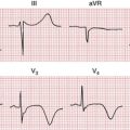
Deep T Wave Inversions
II. Myocardial ischemia/infarction
III. Takotsubo (stress; apical ballooning) cardiomyopathy
IV. Cerebrovascular accident (especially intracranial bleeds) and related neurogenic patterns
V. Left or right ventricular overload
VI. Idiopathic global T wave inversion syndrome
VII. Secondary T wave alterations: bundle branch blocks, Wolff-Parkinson-White patterns
VIII. Intermittent left bundle branch block, preexcitation, or ventricular pacing (“memory T waves”)
Tall, Positive T Waves
Major Bradyarrhythmias
Major Tachyarrhythmias (Basic List, Excluding Artifact)
II. Wide QRS complex tachycardias
Wide QRS Complex Tachycardias (More Comprehensive Classification)
I. Artifact (e.g., tooth-brushing; parkinsonian tremor)
II. Ventricular tachycardia: monomorphic or polymorphic
III. Sinus tachycardia, PSVT, or atrial fibrillation/flutter, with aberrant ventricular conduction caused by:
Atrial Fibrillation: Major Causes and Contributors
1. Alcohol abuse (“holiday heart” syndrome)
4. Cardiomyopathies or myocarditis
9. Idiopathic (“lone” atrial fibrillation)
10. Obstructive sleep apnea (OSA)
11. Paroxysmal supraventricular tachycardias or the Wolff-Parkinson-White preexcitation syndrome
12. Pericardial disease (usually chronic)
13. Pulmonary disease (e.g., chronic obstructive pulmonary disease)
16. Thyrotoxicosis (hyperthyroidism)
17. Valvular heart disease (particularly mitral valve disease)

