E
‘E’ game A technique used to evaluate visual acuity in young children. The letter ‘E’ is shown to the child, who is instructed to either state or point his or her fingers in the direction of the open side of the letter. The ‘E’ is subsequently rotated left, right, up and down randomly as its size is decreased, and acuity is obtained when the smallest letter is just recognizable.
See chart, illiterate E; test, Cardiff acuity.
Eales’ disease See disease, Eales’.
eccentric fixation; viewing See under the nouns.
eccentricity Term referring to the angular distance from the centre of the visual field or from the foveola of the retina. Example: the maximum density of rods in the retina is at a retinal eccentricity of about 20°.
echo See ultrasonography.
echography See ultrasonography.
echothiophate iodide See acetylcholinesterase.
eclipse blindness See blindness, eclipse.
econazole See antifungal agent.
ectasia, corneal A forward bulging and thinning of the cornea. It may result from a disease of the cornea (e.g. keratoconus), trauma, atrophy, raised intraocular pressure or as a complication of photorefractive surgery in which the corneal stroma has been left thinner than about 250 μm. If uveal tissue is included in the protrusion, the condition is called a staphyloma. If the ectasia is limited to a peripheral part of the cornea, it is called Terrien’s disease or Terrien’s marginal degeneration. It is due to degeneration of marginal corneal tissue with superficial vascularization and lipid deposition. It affects adult males more commonly than females and the eye has progressive astigmatism, typically against the rule. Therapy includes rigid contact lenses and occasionally keratoplasty. Syn. keratectasia; keratoectasia; kerectasis.
See degeneration, pellucid marginal; keratoglobus; staphyloma.
ectasia, scleral A bulging and thinning of the sclera, due to disease, trauma, atrophy or raised intraocular pressure. It may be total as in buphthalmos, or partial as in staphyloma. Syn. sclerectasia.
ectoderm The outermost of the three primary germinal layers of an embryo (the other layers being mesoderm and endoderm) from which the eye is derived. It differentiates into outer surface ectoderm and inner neuroectoderm, which gives rise to neural crest cells. The surface ectoderm gives rise to the crystalline lens, the lacrimal gland, the meibomian glands, the corneal and conjunctival epithelium and the epidermis of the eyelids. The neuroectoderm (neural ectoderm) will form the retina, retinal pigment epithelium, the pigmented and non-pigmented layers of the ciliary and iris epithelium, the dilator and sphincter muscles of the iris and the optic nerve fibres. Neural crest cells will form the corneal stroma and endothelium, sclera, iris and choroidal stroma, ciliary muscle and trabecular meshwork.
See cup, optic; mesoderm; vesicles, optic.
ectopia lentis See luxation of the lens.
ectopia of the macula See macula, ectopia of the.
ectopia pupillae See corectopia.
ectropion Outward turning of the eyelid margin. The most common cause is a loss of tonus of the pretarsal orbicularis muscle combined with laxity of the medial and lateral canthal tendons, which occurs in old people and affects only the lower eyelid (involutional ectropion). Tears collect in the lacrimal lake and overflow onto the skin of the face. Other causes of ectropion are scarring, burns, trauma (called cicatricial ectropion), spasm of the orbicularis muscle, which may affect either the upper or lower eyelid, or paralysis of the orbicularis muscle in which only the lower eyelid is affected. Ectropion may lead to exposure keratitis as the lower part of the cornea remains exposed. Management includes instilling an ocular lubricant and patching the eye during sleep as a temporary measure, but, if severe, the treatment is surgical.
cicatricial e.; involutional e. See ectropion.
congenital e . A rare congenital eversion of the eyelid (most often the lower). It may be due to a deficiency of the anterior eyelid lamina. It is usually associated with other disorders, such as blepharophimosis syndrome or Down’s syndrome.
e. uveae A turning of a portion of the posterior pigment epithelium of the iris growing or being drawn around the pupillary margin onto the anterior iris surface. It may be acquired (e.g. following iris neovascularization, neovascular glaucoma, iris melanoma) or congenital (e.g. neurofibromatosis).
eczema An inflammatory disease of the skin characterized by a rash of red spots, rough scaling, dryness and soreness of the skin sometimes leading to the formation of blisters. It often gives rise to itching or to a burning sensation. It may occur on the skin of the face where parts of spectacles rest. Frames should be cleaned regularly to avoid causing skin irritation. Syn. contact dermatitis.
edema See oedema.
edge clearance A small, peripheral gap between the edge of a rigid contact lens and the cornea. It is important as it allows tear exchange and eases lens removal. The absence of edge clearance in rigid contact lenses may lead to superficial corneal damage.
edge detection See contour.
edge lift Deviation of the posterior surface of a contact lens from a sphere at a given diameter. This is produced by either the peripheral curve(s) or the edging process. Edge lift provides peripheral clearance of a rigid contact lens, which is assessed by fluorescein pattern. If the edge lift is specified axially (as an extension of the back central optic zone, measured parallel to the axis of symmetry) it is referred to as axial edge lift. If specified radially as an extension along the back optic zone radius it is referred to as radial edge lift.
See optic zone diameter; optic zone radius, back.
edging Grinding the edge of a lens to the finished shape and size required, at the same time imparting the desired edge form, e.g. flat, bevelled, etc. This is accomplished with a machine called an edger, either by hand with a grinding wheel or automatically operating from a lens pattern or former.
See cribbing; former; glass cutter; glazing.
Edinger–Westphal nucleus See nucleus, Edinger–Westphal.
Edridge–Green lantern An occupational colour vision test that consists of small round and variable sized coloured lights produced by coloured and neutral density filters.
edrophonium chloride See anticholinesterase.
effect The result of an action or condition.
Aubert’s e. See , phenomenon Aubert’s.
Bezold–Brücke e. See phenomenon, Bezold–Brücke.
Broca–Sulzer e. The brightness produced by a flash of a given luminance depends upon its duration. It is maximum for durations around 30–40 ms when the flash luminance is photopic.
Brücke–Bartley e. An increased brightness produced by an intermittent light source (usually around 8–10 Hz) compared to the same light source viewed in steady illumination. Syn. brightness enhancement.
Cheshire cat e. A form of binocular rivalry in which a moving object seen by one eye can cause the entire image, or parts of the image, of a stationary object seen by the other eye to disappear. The effect can be observed by dividing the field of vision with a mirror placed edge-on in front of the nose at a slight angle. One eye looks straight at a stationary object, such as a sleeping cat, while the other eye sees a reflection through the mirror of a white wall or background. If a hand is waved on the mirror side in the region of the field where the cat is seen, the whole cat or part of it may be seen to disappear.
See retinal rivalry.
Craik–O’Brien–Cornsweet e. A phenomenon in which the brightness of an area on one side of a transition strip appears greater than the brightness of the area on the other side of the strip, although both areas outside the transition strip have exactly the same luminance. The transition strip consists of two opposing luminance gradients that meet along a linear edge (called Cornsweet edge); on one side the luminance gradually increases to the edge and on the other side the luminance gradually decreases to the edge. The area adjoining the gradient of increasing luminance appears brighter than the area adjoining the gradient of decreasing luminance. One possible explanation is that the edge information predominates and the visual system and brain ‘fill-in’ the area next to it to construct a higher brightness percept. Note: by covering the transition strip it is easy to confirm that the two areas have the same luminance. Syn. Craik–O’Brien–Cornsweet illusion; Cornsweet illusion.
crowding e. See phenomenon, crowding.
differential prismatic e. The difference in prism power induced by a pair of ophthalmic lenses of different power when the eyes look in various directions of gaze (except through the optical centres). Large amounts of differential prismatic effect can hinder fusion and give rise to diplopia. Example: A patient’s right eye is corrected by +5 D, the left eye by +2 D. When the eye rotates upward so that the visual axes intersect the lenses 1 cm above the optical centres, the induced prism power becomes 5 Δ base down on the right and 2 Δ base down on the left. The differential prismatic effect is 3 Δ base down in front of the right eye, probably too large for fusion to be maintained. Syn. prismatic imbalance; relative prismatic effect.
See anisophoria; law, Prentice’s.
Gelb e. In a faintly illuminated room a piece of black paper (or a rotating black disc) is illuminated by a high intensity projector. The beam of the projector falls exactly on the area of the black surface. The paper or disc will then appear to be white. A reversal of the perception is accomplished by placing a small piece of white paper near the disc in front of the projected light, at which time the paper or disc reappears in its true colour, i.e. black.
kinetic depth e. An impression of a three-dimensional structure of a moving two-dimensional shadow cast by a three-dimensional object. It is most easily demonstrated by casting a shadow onto a translucent screen.
Mandelbaum e. A tendency for the accommodative response to be altered when interposing a conflicting visual stimulus to the one being viewed. If the eyes are viewing a distant object through a dirty window or a wire fence, the actual accommodative response will tend to be raised. If the eyes are viewing a near object in front of a dirty window or wire fence the actual accommodative response will be less than if there were no conflicting stimulus.
McCollough e. A visual after-effect of colour that is seen when viewing, for a minute at least, two differently oriented and differently coloured gratings, such as a vertical grating with blue and black stripes and a horizontal grating with orange and black stripes. After adapting to these the subject looks at a figure containing a grating of vertical black and white stripes and a grating of black and white horizontal stripes of the same size as the original coloured gratings. The white stripes will then appear to be of the complementary colour, that is, the vertical stripes appear pinkish and the horizontal stripes appear bluish.
moiré e. An illusory shimmering movement produced by moving one pattern superimposed on another pattern very similar to it. The phenomenon occurs because parts of the periodic patterns are in phase in some locations, and out of phase in other locations. Examples: passing by a set of railings; if a transilluminated square wave grating is superimposed on an identical grating but cross each other at an angle of less than 45°, moiré fringes will appear at the intersections. Syn. moiré pattern.
See Toposcope.
oblique e. In central vision, contours with oblique orientations are perceived and discriminated less easily than those close to the horizontal or vertical.
Pulfrich e. See stereophenomenon, Pulfrich.
Raman e. In certain substances scattered light may be of a slightly different wavelength from that of the incident light.
Stiles–Crawford e. Variation of the luminosity of a pencil of light stimulating a given receptor with the position of entry of the pencil through the pupil. The maximum luminosity occurs for pencils passing through the centre of the pupil and stimulating the receptor along its axis. This phenomenon is attributed to the particular shape of the cone cells of the retina and occurs only in photopic vision.
Tyndall e. Diffusion of light by the particles present in a liquid or gas. It is because of this effect that heterogeneities (e.g. increased proteins) of the media of the eye can be seen, as occurs in iris and/or ciliary body inflammation. Syn. Tyndall scatter.
effective power See power, effective.
efferent Carrying nervous impulses away from the central nervous system to the periphery.
See afferent.
efficacy, luminous The amount of light emitted by a lamp for each watt of power consumed. It is expressed in lumens/watt.
efficiency scale, Snell–Sterling See visual efficiency scale , Snell–Sterling.
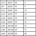
efficiency, spectral luminous (of a monochromatic radiation of wavelength λ) Ratio of the radiant flux at wavelength λm to that at wavelength λ such that both radiations produce equally intense luminous sensations under specified photometric conditions and λm is chosen so that the maximum value of this ratio is equal to one. Symbols: V(λ) for photopic vision. V’(λ) for scotopic vision (Fig. E1). Note: unless otherwise indicated, the values used for the spectral luminous efficiency in photopic vision are the values agreed internationally in 1931 by the CIE and adopted in 1933 by the International Committee on Weights and Measures. For scotopic vision the CIE in 1951 provisionally adopted new values for young observers (CIE). Syn. luminosity curve.
Efron grading scales See grading scales.
Egger’s line See ligament of Wieger.
egocentre A point of reference in the self usually located between the eyes. Absolute judgement of distances and visual directions of objects fixated binocularly are referred to the egocentre.
See localization; oculocentre.
egocentric localization See localization.
Ehlers–Danlos syndrome See syndrome, Ehlers– Danlos.
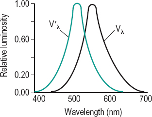
Table E1
Photopic and scotopic relative luminous efficiency factors. The data are based on an average from a large number of individuals agreed by the CIE in 1931 for the photopic factor Vλ and in 1951 for the scotopic factor V’λ
| wavelength (in nm) | Vλ | V’λ |
| 380 | 0.000 0 | 0.000 589 |
| 400 | 0.000 4 | 0.009 292 |
| 420 | 0.004 0 | 0.096 61 |
| 440 | 0.023 0 | 0.328 1 |
| 460 | 0.060 0 | 0.567 2 |
| 480 | 0.139 0 | 0.793 0 |
| 500 | 0.323 0 | 0.981 8 |
| 507 | – | 1.000 0 |
| 520 | 0.710 0 | 0.935 2 |
| 540 | 0.954 0 | 0.649 7 |
| 555 | 1.000 0 | – |
| 580 | 0.870 0 | 0.121 2 |
| 600 | 0.631 0 | 0.033 15 |
| 620 | 0.381 0 | 0.007 374 |
| 640 | 0.175 0 | 0.001 497 |
| 660 | 0.061 0 | 0.000 312 9 |
| 680 | 0.017 0 | 0.000 071 55 |
| 700 | 0.004 1 | 0.000 017 80 |
| 720 | 0.001 05 | 0.000 004 78 |
| 740 | 0.000 25 | 0.000 001 379 |
| 760 | 0.000 06 | 0.000 000 425 |
| 780 | 0.000 00 | 0.000 000 139 |
eidetic image See image, eidetic.
eikonometer Instrument for measuring aniseikonia. The direct comparison eikonometer (or standard eikonometer) uses as a target a cross with a small white disc at the centre of a black square at its intersection (Fig. E2). Four pairs of opposing arrows are placed four degrees away from the centre of the cross with the even numbered arrows polarized in one direction and the odd numbered ones in the other direction. The subject wears polarizing lenses so that each set of four arrows is seen by one eye. If the subject has aniseikonia, one set of arrows will not appear aligned with the other. Aniseikonia either in one or more meridians can be measured by means of an adjustable magnifying device before one eye. There is also a space eikonometer in which the parts of the target are seen three-dimensionally in space. An office model of this type has been manufactured. The space eikonometer is based on a modification of stereopsis. Aniseikonia will make the target appear tilted. The amount of aniseikonia is indicated by the power of the size lens that swings the target back into a frontoparallel plane. Syn. aniseikometer.
See lens, aniseikonic; plane, frontoparallel.
electrodiagnostic procedures Methods such as the electroretinogram, the electrooculogram and the visually evoked cortical potentials which are used to facilitate the diagnosis of some ocular diseases (e.g. retinitis pigmentosa) or the objective measurement of some visual functions (e.g. refractive error, visual acuity).
electroluminescence See luminescence.
electromagnetic spectrum See spectrum, electromagnetic.
electromyogram (EMG) Recording of electrical activity of a muscle associated with contraction and relaxation. This is obtained by placing a microelectrode within a muscle. The recording process is called electromyography.
See law of reciprocal innervation, Sherrington’s.
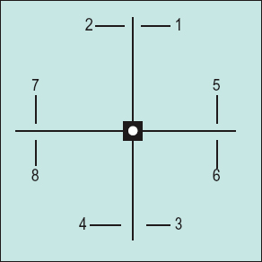
electrooculogram (EOG) Recording of eye movements and eye position provided by the difference in electrical potential between two electrodes placed on the skin on either side of the eye. The EOG consists of two potentials: the standing potential (resting potential, dark phase, dark current) which is evoked by moving the eyes in the dark and originates from the retinal pigment epithelium and the light potential (light rise) which is evoked by moving the eyes in a lighted environment and originates from the photoreceptors. Clinically, the ratio between the light and dark potentials (sometimes also called the Arden index or Arden ratio) is assessed. If that ratio is less than 1.8 it indicates a malfunction of the structures from which the potential originates. The EOG is also used to monitor eye movements (Fig. E3).
See disease, Best’s; fundus flavimaculatus; potential of the eye, resting.
electroretinogram (ERG) Recording of mass electrical response of the retina when it is stimulated by light. It is recorded by placing an electrode in contact with the cornea (often with the aid of a contact lens) or around the eye under the eyelid. A second electrode is placed either on the forehead or the face. The response is complex as many cells of various types contribute to it and varies according to whether the eye is dark or light adapted, the colour of the stimulus, the health of the retina, etc. The curve consists of two major components: a negative a-wave and a positive b-wave. The a-wave originates in the photoreceptors while the b-wave originates in the bipolar and Mueller cells. Both waves also have a photopic and a scotopic component.
pERG indicates that this potential is pattern-elicited. The ERG and pERG are useful indicators of the health of all the layers of the retina and can differentiate between the functioning of the rods and cones.
See alternating checkerboard stimulus; Leber’s congenital amaurosis; potential, early receptor; potentials, oscillatory; retinitis pigmentosa.
electroretinography, multifocal (mfERG) Simultaneous recording of the electroretinogram from small retinal areas (e.g. 103 hexagonal areas in the central 50 degrees of the retina) which are independently light stimulated according to a binary m-sequence (e.g. a random reversal at 75 Hz). This method enables evaluation of specific regions of the retina (e.g. macular degeneration) and mainly of the cone pathway.
elephantiasis oculi 1. Enlargement of the eyelids due to lymphatic obstruction. Syn. elephantiasis palpebral. 2. Extreme exophthalmos.
elephantiasis palpebral See elephantiasis oculi.
elevation of the eye Upward rotation of an eye. It is accomplished by the superior rectus, inferior oblique, lateral rectus (very slightly) and medial rectus (very slightly) muscles. It can be produced voluntarily or by using base-down prisms. Syn. supraduction; sursumduction.
elevator An extraocular muscle involved in rotating the eye upward such as the superior rectus and inferior oblique muscles.
ellipse, Tscherning A graphical representation of the front surface power as a function of total lens power in best-form lenses. There are two possible solutions: (1) Those lenses which are the least curved and represented by the lower portion of the Tscherning ellipse. This portion is called the Oswalt branch of the ellipse. (2) Those lenses which are most curved and represented by the upper portion of the ellipse. This latter portion is called the Wollaston branch of the ellipse.
ellipses, MacAdam Areas in the CIE chromaticity diagram, each in the form of an ellipse within which the colour appears the same. The size of each ellipse varies in the diagram, being larger for greens than for blues or reds. The ellipses are distorted for colour deficient individuals.
ellipsoid 1. The refractile outer portion of the inner member of a rod or cone cell. It is located between the myoid and the outer member of the cell, and contains mitochondria. The myoid is in contact with the external limiting membrane of the retina while the outer member is next to the pigment epithelium. 2. Surface of revolution generated by rotating an ellipse about a major or minor axis.
Elschnig’s membrane; pearls; spots See under the nouns.
embolism, retinal See retinal embolism.
embryotoxon, anterior See arcus, corneal.
embryotoxon, posterior A condition in which a thickened and anteriorly displaced Schwalbe’s line is visible on external examination. The condition is inherited as an autosomal dominant trait.
See gonioscopy; ring of Schwalbe, anterior limiting; syndrome, Alagille; syndrome, Rieger’s.
emedastine See antihistamine.
Emmert’s law See law, Emmert’s.
emmetrope One who has emmetropia.
emmetropia The refractive state of the eye in which, with accommodation relaxed, the conjugate focus of the retina is at infinity. Thus, the retina lies in the plane of the posterior principal focus of the eye and distant objects are sharply focused on the retina. This is the ideal refractive state of the eye. Note: the concept of emmetropia is not simple because accommodation is not inactive when fixating at distance (tonic accommodation). In fact, some authors consider hypermetropia of up to 1.00 D, in a pre-presbyope, as emmetropia.
See accommodation, resting state of; ametropia; distances, conjugate.
emmetropization A process that is presumed to operate to produce a greater frequency of emmetropic eyes than would otherwise occur on the basis of chance. This mechanism would coordinate the development of the various components of the optical system of the eye (e.g. axial length, refracting power of the cornea, depth of the anterior chamber, etc.) to prevent ametropia.
emmetropization theory See theory, emmetropization.
empirical horopter See horopter, empirical.
empiricism The belief that knowledge or behaviour stems from experience, learning or data acquired by observation or experimentation. See nativism; theory, empiricist.
empiricist theory See theory, empiricist.
Emsley’s reduced eye See eye, reduced.
endophthalmitis Inflammation of the intraocular structures. It can occur after a penetrating wound of the eye (either surgical or accidental), bacterial infection, or intraocular foreign bodies.
See panophthalmitis; vitrectomy.
endoscope Instrument designed to examine cavities which are not accessible for direct examination with the eye. It usually incorporates fibre optics to increase the flexibility of the instrument. Examples: a laryngoscope which is introduced through the mouth to examine the larynx; an ophthalmic endoscope to examine the intraocular structures by inserting a fibre optics system through the sclera, as may be used in ocular surgery.
endothelial bedewing; blebs See under the nouns.
endothelial corneal dystrophy See cornea guttata; dystrophy, Fuchs’endothelial; dystrophy, posterior polymorphous.
endothelial polymegethism; polymorphism See under the nouns.
endothelium, corneal See corneal endothelium.
endpiece That part at either end of a spectacle frame front which contains the pivot for the sides. Syn. lug.
enhancement, brightness An increase in brightness resulting either from making a stimulus intermittent, or when a surface is surrounded by a dark area, as compared to when it is surrounded by a light area.
See contrast; effect, Brücke–Bartley.
enlargement Term commonly used in low vision practice to refer to an increase in the size of the retinal image seen by the patient. It can be expressed as the enlargement ratio (ER).
See magnification, lateral; power, equivalent viewing.
enophthalmos Recession of the eyeball into the orbit. It is caused by a degeneration and shrinking of the orbital fat, a tumour, an injury to the orbit or to shortening of the extraocular muscles following excessive resections.
See entropion; exophthalmometer.
entoptic image See image, entoptic.
entoptoscope, blue field An instrument enabling the visualization, especially by patients with a dense cataract, of the shadows of leucocytes flowing in the retinal capillaries and therefore providing a test of macular function. It consists of a very bright light source, an interference filter with a maximum transmission in the blue end of the spectrum, and a diffuser. The instrument is held close to the eye of the patient who is asked to describe his or her observations. The leucocytes appear as flying corpuscles and if many corpuscles (at least 15) are seen moving in the entire field the test is considered positive whereas if none or only a few corpuscles are seen the test is considered negative. Positive responses usually indicate that the patient has good macular function and negative responses usually indicate that the patient has poor macular function. This test is very useful in predicting central vision before cataract extraction.
See angioscotoma; maxwellian view system, clinical.
entrance pupil See pupil, entrance.
entropion Inward turning of the eyelid. It results in the eyelashes rubbing the cornea (as in trichiasis) and this usually causes discomfort. The most common cause of entropion that occurs in old people (called involutional entropion) and only affects the lower eyelid is due to a combination of atrophy and weakening of the tarsus, loss of tone of the subcutaneous tissues and loss of elasticity of the skin. Other causes are scarring (e.g. trachoma, Stevens–Johnson syndrome), burns of the palpebral conjunctiva (called cicatricial entropion) which may affect either the upper or the lower eyelid, or spasm of the orbicularis muscle often resulting from an ocular inflammation or lid infection (called acute spastic entropion) which may subside spontaneously once the original cause has been removed. Temporary relief of entropion may be provided by the taping of the lower eyelid to the cheek but the treatment is usually surgical.
See ectropion; lens, therapeutic soft contact; spectacles, orthopaedic; tarsus; trichiasis.
cicatricial e.; involutional e. See entropion.
congenital e. A rare congenital inversion of the eyelid usually associated with tarsal hypoplasia or microphthalmia. It may be confused with epiblepharon. If treatment is needed it is surgical, although many cases resolve spontaneously with time.
enucleation Removal of an eye from its socket. It is usually performed to reduce pain in a blind eye, when there is a risk of sympathetic ophthalmia following trauma, or when there is a malignant tumour in the eye. Immediately following the operation a spherical implant (made up of hydroxyapatite, polyethylene, or silicone rubber) is placed into the eye socket to be replaced several weeks later by an artificial eye.
See evisceration; eye, artificial.
enzyme A protein substance which catalyses (i.e. enhances a chemical reaction in other bodies without undergoing a change in itself) and is formed by living cells but can act independently of their presence. Example: Enzyme preparations (containing pancreatin, papain or subtilisin A) used to break down tear proteins that become attached to the surface of contact lenses.
See deposits, contact lens; phagocytosis; surfactant; wetting solution.
ephedrine hydrochloride See mydriatic.
epiblepharon A congenital anomaly in which a fold of skin lies across the upper or lower lid margin. In the lower eyelid it causes a turning inward of the eyelashes without causing entropion. The condition commonly resolves itself with facial growth.
See blepharochalasis; entropion, congenital.
epibulbar Situated on the eyeball. Syn. epiocular.
epicanthus A condition in which a fold of skin that stretches from the upper to the lower eyelid partially covers the inner canthus. It is normal in the fetus, in Down’s syndrome and in many infants, especially of oriental origin, where it may give the impression of a convergent strabismus (pseudoesotropia). The condition is normally bilateral. As the bridge of the nose develops, the folds eventually disappear. Syn. epicanthal fold.
See strabismus, apparent.
epicanthus inversus A condition in which a fold of skin that stretches from the lower eyelid upward and toward the nose partially covers the inner canthus. It is often associated with ptosis.
epichoroid Synonym of suprachoroid. See sclera.
epidemiology A branch of health science that deals with the incidence, prevalence, distribution and aetiology of disease in a population.
epidiascope A projector used to project by reflection opaque pictures (such as the page of a book) onto a screen.
epikeratophakia See epikeratoplasty.
epikeratoplasty A surgical procedure on the cornea aimed at correcting ametropia. The patient’s corneal epithelium is removed and a donor’s corneal disc (or lenticule) that was previously frozen and reshaped to produce a new anterior curvature is rehydrated and sutured to Bowman’s membrane. The lenticule can be removed and exchanged to provide a different power. There are many problems associated with this procedure, in particular the surface re-epithelialization. Syn. epikeratophakia; refractive keratoplasty and keratorefractive surgery (both terms also include keratomileusis, keratophakia and radial keratotomy).
See Intacs; keratomileusis; keratophakia; keratectomy, photorefractive; LASIK; LASEK; lenticule.
epilation The removal of hair by the roots as in the case of ingrowing eyelashes. The eyelashes are removed with forceps but, unfortunately, they tend to regrow.
See distichiasis; trichiasis.
epinastine hydrochloride See antihistamine.
epinephrine See adrenaline (epinephrine).
epiocular See epibulbar.
epiphora Overflow of tears due to faulty apposition of the lacrimal puncta in the lacrimal lake, scarring of the puncta, paresis of the orbicularis muscle, obstruction of the lacrimal passage or ectropion. This impairment of the outflow of tears is often unilateral. The main symptoms are discomfort and blurring of vision and sometimes embarrassment. Management depends on the cause. Syn. watery eye (colloquial).
See dacryops; hyperlacrimation.
epiretinal membrane See fibrosis, preretinal macular.
episclera A loose connective and elastic tissue which covers the sclera and anteriorly connects the conjunctiva to it. It is a vascularized tissue whose deeper layers merge with the scleral stroma. It sends connective tissue bundles into Tenon’s capsule. The episclera becomes progressively thinner towards the back of the eye.
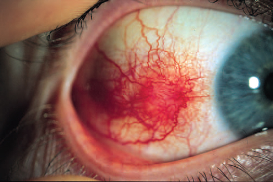
episcleritis Inflammation of the episclera. It is a benign, self-limiting, frequently recurring condition that typically affects adults. The disease is characterized by redness (usually in one quadrant of the globe) and varying degrees of discomfort. There are two types of episcleritis: simple which is the most common and nodular which is localized to one area of the globe forming a nodule. Simple episcleritis usually subsides spontaneously within 1–2 weeks while the nodular type usually takes longer. If the discomfort is intense topical corticosteroids may be used (Fig. E4).
epithelial arcuate lesion; plug; splitting See staining, fluorescein.
epithelial downgrowth Abnormal growth of corneal or conjunctival epithelium into the interior of the eye, as a complication of a penetrating corneal injury and much more rarely following cataract extraction. It may cause secondary glaucoma as a result of cell proliferation through the wound onto the endothelial surface and into the iridocorneal angle.
epithelial keratitis See keratitis, punctate epithelial; keratitis, Thygeson’s superficial punctate.
epithelial microcysts See microcysts, epithelial.
epithelioma A tumour of epithelial cells, ranging from benign to malignant (e.g. carcinoma). Example: epithelioma of the conjunctival epithelium that begins near the limbus and spreads to the fornices and cornea. Treatment usually consists of excision, cryotherapy or both, or even enucleation if the tumour is very advanced.
epithelium, corneal See corneal epithelium.
equation, paraxial See paraxial equation, fundamental.
equator of the crystalline lens The circle formed by the outer margin of the lens. The equator is not smooth, but shows a number of indentations corresponding to the zonular fibres. The indentations tend to disappear during accommodation, when the zonular fibres are loose.
See lens, crystalline; Zinn, zonule of.
equatorial plane of the eye See plane, equatorial.
equivalent oxygen pressure See oxygen pressure, equivalent.
equivalent points See points, nodal.
equivalent power See power, equivalent.
equivalent, spherical A spherical power whose focal point coincides with the circle of least confusion of a spherocylindrical lens. Hence, the spherical equivalent of a prescription is equal to the algebraic sum of the value of the sphere and half the cylindrical value, i.e. sphere + cylinder/2. Example: the spherical equivalent of the prescription –3 D sphere –2 D cylinder axis 180° is equal to –4 D.
erector A lens (for example an erecting eyepiece) or prism system (erecting prism such as a Dove or a Porro prism) placed in an optical system for the purpose of forming an erect image.
error, cylindrical; spherical See prescription.
error of refraction of the eye See ametropia; refractive error.
erythema multiforme A mucocutaneous disease that occurs as a hypersensitivity to drugs (e.g. sulfonamides) or as a consequence of infection. The condition, which principally affects young people, is characterized by the sudden appearance of various erosions of the mucous membranes and epidermis. A common complication is conjunctivitis, which may become severe with cicatrization, abnormal lid margin function, symblepharon, corneal ulceration and vascularization and keratoconjunctivitis sicca. The patient complains of pain, discharge, photophobia and reduced vision if the cornea is involved. Treatment includes cleansing of the eyelids with antibiotic ointment and, if severe, topical steroids. Syn. Stevens–Johnson syndrome (although this term applies to the severe form of the disease).
See syndrome, Stevens–Johnson.
erythrolabe See pigment, visual.
erythromycin See antibiotic.
erythrophobia See chromatophobia.
erythropsia See chromatopsia.
erythropsin See rhodopsin.
eserine See physostigmine.
eso deviation Term referring to either esophoria or esotropia.
See strabismus, convergent.
esodisparity Fixation disparity characterized by a slight overconvergence of the eyes, while still retaining single binocular vision.
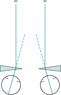
esophoria (E, ESOP, SOP, eso) Turning of the eye inward from the active position when fusion is suspended. If symptomatic, treatment may be by means of base-out prisms, plus spherical lenses or visual training (Fig. E5).
See convergence excess; divergence insufficiency.
esotropia A synonym for convergent strabismus.
See strabismus, accommodative; strabismus, convergent.
blind spot e. See syndrome, Swann’s.
consecutive e. See strabismus, consecutive.
infantile e. See strabismus, infantile.
non-accommodative acquired e. See strabismus, non-accommodative acquired.
Esterman grid; test See test, Esterman.
esthesiometer See aesthesiometer.
ethmoid bone See orbit; sinus, ethomoidal.
ethylenediamine tetraacetic acid (EDTA) A chelating agent used in ophthalmic preparations to remove metals which are essential for the metabolism of bacteria and viruses, and therefore inactivate them. It enhances the bactericidal effect of preservatives and is commonly combined with benzalkonium chloride, chlorhexidine and thiomersalate.
See antiseptic.
etiology See aetiology.
euchromatopsia Normal perception of colours. Syn. euchromatopsy
See chromatopsia.
euryopia Abnormally wide palpebral aperture.
euthyroid See disease, Graves’.
Euthyscope See Visuscope.
eversion , lid Turning of the eyelid inside out so as to expose the palpebral conjunctiva. For the upper lid this is accomplished by grasping the lid by the central eyelashes, pulling it downward and forward and then folding it back over a cotton applicator (or thin plastic rod) placed at the upper margin of the tarsus, while the patient continually maintains downward fixation. Return to the normal lid position is obtained by asking the patient to look up and gently pushing the eyelashes in an outward and downward direction. Foreign bodies and even contact lenses are often lodged under the upper eyelid or in the conjunctival fornix of the upper eyelid. To inspect the superior conjunctival fornix double lid eversion is necessary. Following lid eversion (and usually with local anaesthesia of the conjunctiva), a retractor is placed between the two skin surfaces of the lid with the retractor engaging the tarsus and, after gently pulling outward and upward, the fornix will become visible. Eversion of the lower lid is performed easily by drawing the margin downward while the patient looks upward.
See irrigation; sulcus, subtarsal.
evidence-based healthcare See trial, randomized controlled.
evisceration Removal of the inner contents of the eye with the exception of the sclera. It is usually performed when there is intraocular suppuration or pain in a blind eye. The intraocular contents are immediately replaced by a spherical implant (made of hydroxyapatite, polyethylene, or silicone rubber) and several weeks later by an artificial eye.
See enucleation.
evoked potential See potential, visual evoked cortical.
excavation, physiological See cup, physiological.
excimer laser See laser, excimer.
excyclophoria See cyclophoria.
excyclovergence Rotary movements about their respective anteroposterior axes of one eye relative to the other. If the upper pole of the cornea of one eye moves away from that of the other eye, it is called excyclovergence. If, however, the upper pole of the cornea of one eye moves towards that of the other eye, it is called incyclovergence.
exenteration Removal of the entire contents of the orbit, including the eyeball, the extraocular muscles, the optic nerve, nerves and blood vessels, the orbital fat and connective tissues. It is performed in cases of malignant tumours.
See enucleation; evisceration.
exfoliation of the lens Shedding of the layers of the lens capsule. It often occurs as a result of exposure to prolonged and intense heat.
See cataract, heat-ray; pseudoexfoliation.
exo deviation Term referring to either exophoria or exotropia.
See strabismus, divergent.
exodisparity Fixation disparity characterized by a slight underconvergence of the eyes, while still retaining single binocular vision.
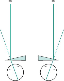
exophoria (X, XOP, exo) Turning of the eye outward from the active position when fusion is suspended. If symptomatic, treatment may be by means of base-in prisms, minus spherical lenses or visual training (Fig. E6).
See convergence insufficiency; divergence excess.
exophoria, physiological The relative exophoria at near, when the heterophoria in near vision is compared to the heterophoria in distance vision. It is, on average, of the order of 3–4 D at a fixation distance of 40 cm. Example: if distance heterophoria is 6 D eso and near heterophoria is 2 D eso, the physiological exophoria is 4 D exo.
exophthalmometer Instrument for measuring the amount of exophthalmos (or enophthalmos). There are several types, the most common being the Hertel exophthalmometer: it measures the distance between the corneal apex and the apex of the deepest angle of the lateral orbital margin of both eyes simultaneously, using either mirrors or prisms and a superimposed millimetre scale. The normal range varies between 12 mm and 21 mm and a difference between the eyes greater than 2 mm is suspicious.
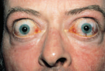
exophthalmos Abnormal protrusion of the eyeball(s) from the orbit, caused by Graves’ ophthalmopathy, endocrine malfunction, paralysis of the extraocular muscles, injury of the orbit, cavernous sinus thrombosis or a tumour behind the eye. The palpebral fissure is usually wider and a rim of sclera may be visible above and below the cornea (Fig. E7). Unilateral displacement is usually referred to as proptosis.
See diplopia, pathological; disease, Graves’; elephantiasis oculi; enophthalmos; syndrome, Apert’s; syndrome, Crouzon’s.
exotropia See strabismus, divergent.
exotropia, consecutive See strabismus, consecutive.
exotropia, paralytic pontine See syndrome, ‘one and one half’.
experiment, Bidwell’s Experiment aimed at producing the complement of a colour stimulus by viewing it through a rotating disc which presents the sequence: black, colour stimulus and white.
See colour, complementary.
experiment, Scheiner’s A demonstration of the refractive changes occurring in the eye when accommodating. The subject observes a target monocularly (such as a simple point of light) through a Scheiner’s disc (an opaque disc with two pinholes separated by a distance less than the pupil diameter). It will be seen singly at only one distance where the eye is focused, because target and retina are then conjugate. If the eye accommodates, two points of light are seen. The principle of this experiment is incorporated in several refractometers.
See Fig. D6; optometer, infrared; optometer, Young’s.
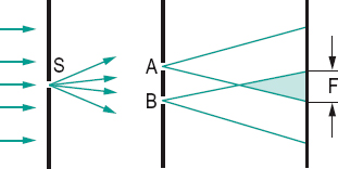
experiment, Young’s Method of producing interference of light which was shown by Young in 1801. He used two coherent beams of light that were produced by passing light through a very small circular aperture in one screen, then through two small circular apertures very close together in a second screen. On a third screen, behind the second screen, there will be two overlapping sets of waves and, if the original source is emitting monochromatic light, interference fringes will appear on the third screen (Fig. E8).
See coherent sources; interference fringes.
exposure keratitis See keratitis, exposure.
exposure meter Light measuring instrument for ascertaining the setting (lens aperture, shutter speed, etc.) of a camera for correct light exposure of the photographic material (CIE).
expressivity The extent to which an inherited trait or disease is manifested in the phenotype. It is a qualitative evaluation unlike penetrance. Syn. expression.
extended object; source; wear lens See under the nouns.
external hordeolum; limiting membrane; ophthalmoplegia See under the nouns.
extinction phenomenon See phenomenon, extinction.
extorsion See torsion.
extraocular muscles See muscles, extraocular.
extrinsic muscles See muscles, extraocular.
exudate A liquid or semisolid which has been discharged through the tissues to the surface or into a cavity. Exudates in the retina are opacities that result from the escape of plasma and white blood cells from defective blood vessels. They usually look grayish-white or yellowish and are circular or ovoid in shape. They are sometimes classified into three groups according to size: (1) punctate hard exudates, which often tend to coalesce. They are found in diabetic retinopathy, Coats’ disease, etc.; (2) exudates of moderate size, such as ‘cotton-wool or soft exudates’ as, for example, in branch/central retinal vein occlusion, hypertensive retinopathy, etc. These ‘exudates’ have ill-defined margins and are actually areas of ischaemia containing cytoid bodies, unlike hard exudates which are generally lipid deposits; (3) larger exudates, as found in the severe forms of retinopathy.
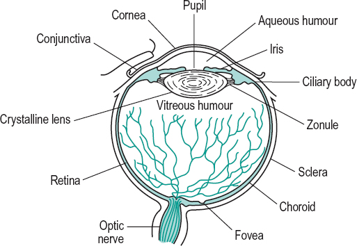
eye The peripheral organ of vision, in which an optical image of the external world is produced and transformed into nerve impulses. It is a spheroidal body approximately 24 mm in diameter with the segment of a smaller sphere (of about 8 mm radius), the cornea, in front. It consists of an external coat of fibrous tissue, the sclera and transparent cornea; a middle vascular coat, comprising the iris, the ciliary body and the choroid; and an internal coat, the retina, which includes the cones and rods photoreceptors. Within the eye, there are the aqueous humour located between the cornea and the crystalline lens, the crystalline lens held by the zonule of Zinn and the vitreous body located between the crystalline lens and the retina. The movements of the eye are directed by six extraocular muscles (Fig. E9). Syn. organ of sight; visual organ.
amaurotic e. See amaurosis.
amblyopic e. An eye which has amblyopia. Syn. lazy eye (colloquial).
aphakic e. An eye without the crystalline lens.
artificial e. A prosthesis made of glass or plastic which resembles the eye and which is placed in the socket after enucleation.
See ocularist; prosthesis, ocular.
axial length of the e. See length of the eye, axial.
e. bank An organization that collects, evaluates, stores and distributes eyes from donors. The eyes are used for corneal transplants and research.
See keratoplasty.
black e. A colloquial term for a swollen or blue-black spot on the skin of the eyelid caused by effusion of blood as a result of a superficial injury in which the skin is not broken. The correct term is ecchymosis of the eyelid. The condition recovers by itself within 2–3 weeks while changing in colour to yellow. Immediately after the injury, application of ice helps minimize the haemorrhage and swelling.
See haematoma.
bleary e. A red and watery eye, with a lacklustre appearance. Lack of sleep is a common cause. Syn. blear eye.
e. blink See blink.
compound e. The eye of arthropods composed of a variable number of ommatidia.
See corneal facet; ommatidium.
crossed e’s. See strabismus, convergent.
cyclopean e. Imaginary eye located at a point midway between the two eyes. When the two visual fields overlap and the impressions from the two eyes are combined into a single impression, the apparent direction of a fixated object appears in a direction that emanates from the cyclopean eye.
dark-adapted e. An eye that has been in darkness and is sensitive to low illumination. Syn. scotopic eye.
deviating e. The non-fixating eye in strabismus or under heterophoria testing. Syn. squinting eye.
dominant e. The eye that is dominant when ocular dominance exists.
See manoptoscope; test, hole in the card.
dry e. This term encompasses various tear film disorders ranging from a mild form causing discomfort, which is usually relieved with artificial tears, to the most common form keratoconjunctivitis sicca.
See keratoconjunctivitis sicca.
equatorial plane of the e. See plane, equatorial.
exciting e. See ophthalmia, sympathetic.
fixating e. The eye that is directed towards the object of regard in strabismus.
glass e. An artificial eye made of glass.
See ocularist; prosthesis, ocular.
e. impression See impression, eye.
e. lens See eyepiece.
light-adapted e. An eye that has been exposed to light and is insensitive to low illumination. Syn. photopic eye.
See adaptation, light; theory, duplicity.
e. movements See movements, eye.
e. patch A piece of material or plastic that is worn over the eye when it has been injured or over the socket when it is missing.
phakic e. An eye that contains the crystalline lens.
See phakic.
photopic e. See eye, light-adapted.
pink e. See conjunctivitis, contagious.
e. position Position of the eye in the orbit, maintained by the extraocular muscles.
See position, primary; position, secondary; position, tertiary.
pseudophakic e. An eye fitted with an intraocular lens implant.
See lens, intraocular.
red e. A colloquial term often used for any condition in which the blood vessels of the conjunctiva or ciliary body are congested. Many conditions result in a red eye (e.g. subconjunctival haemorrhage, pterygium, conjunctivitis, episcleritis, corneal abrasion, corneal erosion, ulcerative keratitis, corneal dendritic ulcer, acute iritis, angle-closure glaucoma, orbital cellulitis, and possibly contact lens wear).
See injection, ciliary; injection, conjunctival.
reduced e. A mathematical model of the optical system of the eye. It consists of a single refracting surface with one nodal point, one principal point and one index of refraction. In the first such model, proposed by Listing in 1853, the refracting surface had a power of 68.3 D and was situated 2.34 mm behind the schematic eye’s cornea. It had an index of refraction of 1.35, a radius of curvature of 5.124 mm and a length of 20 mm. Donders’ reduced eye was even more simplified. It has a power of 66.7 D, a radius of curvature of 5 mm, an index of refraction of 4/3 and anterior and posterior focal lengths of –15 and +20 mm, respectively, with a refracting surface situated 2 mm behind the schematic eye’s cornea. Gullstrand’s reduced eye has a radius of curvature of 5.7 mm, an index of refraction of 1.33, a power of 61 D with the refracting surface situated 1.35 mm behind the schematic eye’s cornea. Emsley’s reduced eye has a power of 60 D, an index of refraction of 4/3 and is situated 1.66 mm behind the schematic eye’s cornea, with anterior and posterior focal lengths of –16.67 and +22.22 mm, respectively.
schematic e. A model consisting of various spherical surfaces representing the optical system of a normal eye based on the average dimensions (called the constants of the eye) of the human eye. There are many schematic eyes, although the most commonly used is that of Gullstrand. A great deal of variation among authors stemmed from the difficulty in giving an index of refraction that would represent the heterogeneous character of the crystalline lens. Gullstrand in fact proposed two schematic eyes, one which he called the exact schematic eye and the other which he called the simplified schematic eye in which the divergent effect of the posterior corneal surface is ignored and the cornea replaced by an equivalent surface; the crystalline lens is homogeneous and the optical system is free from aberrations.
scotopic e. See eye, dark-adapted.
e. shield 1. See occluder. 2. A protective device to cover the eye against injury, glare or in radiotherapy of the face.
sighting-dominant e. The eye that is preferred in monocular tasks, such as looking through a telescope or aiming a firearm.
e. socket The bony orbit which contains the eyeball, the muscles, the nerves, the vessels, the orbital fat and the orbital portion of the lacrimal gland.
e. speculum An instrument designed to hold the eyelids apart during surgery. Syn. blepharostat.
squinting e. See eye, deviating.
e. stone A small, smooth shell or other object that can be inserted beneath the eyelid to facilitate the removal of a foreign body from the eye.
sympathetic e. The uninjured eye in sympathetic ophthalmia that becomes secondarily affected. Syn. sympathizing eye.
wall e. A colloquial term referring to (1) a white opaque cornea or (2) a divergent strabismus.
watery e. See epiphora.
eyeball The globe of the eye without its appendages.
eyebrow A transverse elevation covered with hairs and situated at the junction of the forehead and upper lid. Syn. supercilium.
See ophryosis.
eyecup A small vessel made of glass, plastic or porcelain, used to bathe the eye.
eyeglass 1. Synonym for monocle. In the plural (eyeglasses) it refers to pince-nez or a similar type of eyewear without sides, or to spectacles. 2. The eyepiece of an optical instrument.
eyelashes Rows of stiff hairs (cilia) growing on the margin of the upper and lower eyelids. The upper eyelashes are longer and more numerous and curl upward, while the lower ones turn downward. Syn. cilia; lashes.
See distichiasis; epilation; madarosis; polystichia; trichiasis.
eyelid eversion See eversion, lid.
eyelid twitch See ciliosis; myasthenia gravis; myokymia.
eyelids A pair of movable folds of skin which act as protective coverings of the eye. The upper eyelid extends downward from the eyebrow and is the more moveable of the two. When the eye is open and looking straight ahead, it just covers the upper part of the cornea; when it is closed, it covers the whole cornea. The lower eyelid reaches just below the cornea when the eye is open and rises only slightly when it shuts. Each eyelid consists of the following layers, starting anteriorly: (1) skin, (2) a layer of subcutaneous connective tissue, (3) a layer of striated muscle fibres of the orbicularis muscle, (4) a layer of submuscular connective tissue, (5) a fibrous layer, including the tarsal plates, (6) a layer of smooth muscle, (7) the palpebral conjunctiva. Syn. blephara; lids; palpebrae.
See ablephary; blepharitis; ciliosis; ectropion; entropion; epicanthus; eversion, lid; eyelid lamella; lagophthalmos; ligament, palpebral; myokymia; orbital septum; phthiriasis; sign, Cogan’s lid twitch; sign, Collier’s; sign, Dalrymple’s; sulcus, inferior palpebral; sulcus, superior palpebral; tarsorrhaphy; tarsus; xanthelasma.
e. lamella The eyelid is sometimes conceptualized as consisting of an anterior and a posterior lamella. The anterior lamella consists of the skin, the layer of subcutaneous connective tissue and the layer of striated muscle fibres of the orbicularis muscle. The posterior lamella consists of the tarsal plates, a layer of smooth muscle (Müller’s palpebral muscle), and the palpebral conjunctiva.
e. retractor muscles The eyelid muscles that open the palpebral aperture. The upper eyelid is elevated by the levator palpebrae superioris muscle and the superior tarsal muscle (of Müller) and the lower eyelid is depressed by the inferior tarsal muscle (of Müller).
eyepiece The lens or combination of lenses in an optical instrument (microscope, telescope, etc.) through which the observer views the image formed by the objective. The most common eyepieces are composed of two single lenses or two doublets: the lens or doublet nearer the eye is called the eye lens and the one nearer the objective is called the field lens. The role of the eyepiece is to magnify the image and to reduce the aberrations of the image formed by the objective. Syn. eye lens; eyeglass; ocular.
Huygens’ e. Negative eyepiece used commonly in microscopes. It consists of two planoconvex lenses mounted with their plane surfaces facing the eye. In the most common type the eye lens has a focal length half that of the field lens and the separation is equal to half the sum of the two focal lengths.
negative e. Eyepiece made up of two lenses, in which the first principal focus of the eyepiece lies between the two lenses, such as in a Huygens’ eyepiece.
orthoscopic e. An eyepiece corrected for distortion, and which provides a wide field of view and high magnification. It consists of a triplet field lens and a single eye lens. It is used in high-power telescopes and range finders.
See triplet.
positive e. Eyepiece made up of two lenses in which the first principal focus of the eyepiece lies in front of the field lens, such as in a Ramsden eyepiece.
Ramsden e. Positive eyepiece consisting of two planoconvex lenses mounted with their convex side facing each other and having equal focal lengths. The lenses are usually separated by two-thirds the focal length of either.
eyesight Vision.
eyesize The horizontal dimension of the lens opening of a frame which is bounded by two vertical lines at a tangent to the left and right sides of the opening.
eyestrain See asthenopia.
eyewash Any liquid which is used for bathing the eye. Example: physiological saline.
eyewear See spectacles.
eyewire The rim that surrounds the lens of a spectacle frame.