Dyspnea

Published on 23/05/2015 by admin

Filed under Pulmolory and Respiratory

Last modified 22/04/2025

Print this page

rate 1 star rate 2 star rate 3 star rate 4 star rate 5 star
Your rating: none, Average: 0 (0 votes)

This article have been viewed 3547 times

Chapter 19 Dyspnea

What Is Dyspnea?

In healthy people, breathing is an unconscious activity that is regulated by automatic command by groups of neurons in the brain stem to control cyclic contraction and relaxation of the respiratory muscles. With a perturbation of this process, the affected person may experience breathing difficulty or discomfort. This sensation is considered a symptom and typically is referred to as dyspnea, which literally means “disordered breathing” (dys– + –pnea). In 1999, the American Thoracic Society defined dyspnea as “a subjective experience of breathing discomfort that consists of qualitatively distinct sensations that vary in intensity.” Patients typically cite such discomfort when describing their symptoms: “I am short of breath.” “I can’t get enough air.” “It’s hard to breathe.”

A majority of studies investigating dyspnea have focused on patients with chronic obstructive pulmonary disease (COPD), for two reasons: (1) COPD is the most prevalent respiratory disease, and (2) exertional breathlessness is the major symptom of this condition. Thus, the accumulated knowledge of clinical features of dyspnea and current understanding of the relevant mechanisms and qualities derive in large part from studies involving patients with COPD.

An interesting point is that dyspnea shares many features with pain. Both symptoms are complex neurophysiologic processes that are influenced by physiologic, psychologic, social, and environmental factors. These sensations function as warning signals of potential harm that usually lead the affected individual to reduce activities in order to minimize the complaint and/or to seek medical attention. Like pain, dyspnea can be perceived only by the affected person and has both sensory (how bad is it?) and affective (how does it feel?) components. Because both dyspnea and pain are under behavioral control, any emotional state may worsen these experiences to a degree that may be out of proportion to the magnitude of physiologic impairment. For example, high levels of anxiety and panic attacks are associated with increased breathlessness and more intense pain.

Dyspnea is an important problem in the elderly population, often with a major impact on quality of life. It is estimated that more than 30% of those 65 years of age or older without known cardiorespiratory disease report breathlessness with various activities of daily living, including walking on a level surface or up an incline. An analysis of 124 patients over 70 years of age who were randomly selected from a large family medicine practice revealed that up to 37% had moderate to severe dyspnea, and that dyspnea was associated with poor perceived health, more anxiety and depression, and impaired daily functioning. With any physical activity, including exercise, older people exhibit higher levels of ventilation than those typical for younger persons performing the same amount of physical work. Not clear, however, is whether this increased ventilatory demand in older people is a direct result of the aging process in the respiratory system (secondary to a decrease in lung elasticity, increase in chest wall stiffness, and decrease in respiratory muscle strength) or a consequence of sedentary life style, deconditioning, and possible weight gain, which frequently occur with advancing age in populations of developed countries.

Gender differences in the prevalence and severity of dyspnea among patients with COPD have been documented. For example, women are more likely to report severe dyspnea compared with men despite significantly fewer pack-years of smoking cigarettes and similar frequencies of coughing. In the National Emphysema Treatment Trial involving 1053 patients with severe COPD (emphysema phenotype), women reported greater dyspnea than that described by men when findings were controlled for lung function, age, pack-years of smoking, and proportion of the lung affected by emphysema. Whether this gender difference in dyspnea relates to physiologic differences (compared with men, women have smaller airway lumina, with disproportionately thicker airway walls) is uncertain.

Mechanisms of Dyspnea

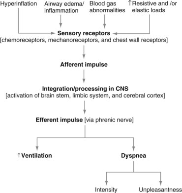
The neurophysiologic pathways that mediate the control of breathing (to supply oxygen, to eliminate carbon dioxide, and to maintain acid-base balance) also are relevant to the mechanisms of dyspnea (Figure 19-1). In simple terms, nerve fibers (sensory receptors) send electrical signals (afferent impulses) to the spinal cord, which in turn transmits these signals to the brain.

The brain interprets these signals as a sensation (dyspnea). Outgoing commands from the brain may then elicit an appropriate response—the affected person may stop the offending activity, for example, or may use a rescue inhaler in an attempt to relieve the breathing difficulty.

Presented next is an overview of the neurophysiology of dyspnea, with details of the important components of the relevant pathways.

Afferent Impulses

Afferent information from sensory receptors are transmitted to brain stem respiratory centers that automatically adjust breathing and also may project to higher brain areas for direct assessment of the status of various stimuli. Well-characterized examples include the following: The glossopharyngeal nerve transmits impulses for peripheral chemoreceptors; the vagus nerve transmits information for rapidly adapting, slowly adapting, and C-fiber lung mechanoreceptors; and cervical spinal nerves 3 to 5 (C3 to C5) transmit sensory information accrued by mechanoreceptors from the diaphragm.

Two different pathways have been proposed to process respiratory sensations to the cerebral cortex. One pathway reflects discriminative processing—that is, awareness of the spatial, temporal, and intensity components (e.g., how bad is it?). With activation of respiratory muscle receptors, afferent information is relayed into the brain stem medulla and is then projected to the ventroposterior thalamus area; from here, projections ascend to the primary and secondary somatosensory cortex. These structures are thought to process the intensity component of dyspnea.

A second pathway reflects affective processing (e.g., how does it feel?). With activation of airway and lung receptors, afferent information is relayed by the vagal nerve to the brain stem medulla and is then projected to the amygdala and medial dorsal areas of the thalamus; from here, projections ascend to the insular and cingulate cortex. These structures are part of the limbic system, which forms the inner border of the cortex and contains rich interconnections among the cerebral cortex, thalamus, and brain stem, and are thought to process the affective component of dyspnea.

The Language of Dyspnea

To help dyspneic patients describe their experience more accurately, questionnaires have been developed that allow for the selection of specific descriptors of breathlessness. It appears that the descriptors selected by patients relate in part to the underlying mechanisms contributing to dyspnea. For example, chest tightness is relatively specific to bronchoconstriction in patients with asthma but is not typically reported by patients with COPD. This perception of tightness presumably is due to activation of sensory receptors located in large airways and can be relieved with use of bronchodilator therapy.

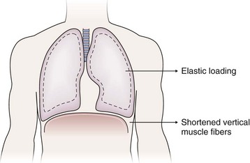
The sense of respiratory work or effort commonly is reported by patients with various conditions including asthma, COPD, interstitial lung disease, and neuromuscular disease. The descriptor “work/effort” of breathing difficulty probably is related to activation of respiratory muscle afferents imposed by mechanical loads (airway narrowing → added resistance; parenchymal edema/infiltrates → added elastance) imposed by certain diseases as well as respiratory muscle weakness. For example, in patients with COPD, the lungs typically hyperinflate during the performance of physical tasks. This dynamic hyperinflation results in two major consequences that contribute to dyspnea: (1) an added elastic load and (2) functional weakening of the diaphragm by shortening of the vertical muscle fibers (Figure 19-2). Although patients with acute asthma initially may experience chest tightness, they typically report that the increased work or effort of breathing develops as airway narrowing progresses as a result of subsequent lung hyperinflation.

A perception of not being able to take in enough air has been described as air hunger, or “unsatisfied inspiration.” This experience is not specific to any particular disease or stimulus. A consistent finding is that patients with various cardiorespiratory conditions generally report greater breathing difficulty during inspiration than with expiration. A clustering of reported sensations such as smothering, suffocating, and air hunger has been documented in patients with panic disorder and idiopathic hyperventilation syndrome who do not have any cardiopulmonary or neuromuscular disease. These findings are consistent with an increase in ventilatory drive.

The three qualities of dyspneic sensations, as described, do not explain all clinical features of breathlessness. Moreover, multiple pathways may combine to contribute to dyspnea in an individual patient. For example, in a patient with cardiogenic pulmonary edema, bilateral pleural effusions, cardiac cachexia, and arterial hypoxemia, dyspnea may be due to activation of carotid body neurons (hypoxemia), stretch receptors (interstitial edema), and Golgi tendon organs and spindle fibers (muscle weakness).

Acute Dyspnea

Initial Evaluation

The rapidity with which dyspnea develops is clinically important. Although a standard definition of acute dyspnea does not exist, potentially life-threatening cardiopulmonary processes often are heralded by unprecedented, severe dyspnea minutes to hours in duration. Whenever possible, details should be sought about the circumstances under which dyspnea began, whether it is associated with other symptoms, and how it has progressed. Knowledge of medications and comorbid conditions also is helpful. Dyspnea in the prehospital environment independently predicts a nearly seven-fold likelihood of hospital admission from the emergency department. Studies have shown that delays in seeking clinical attention for acute exacerbations of asthma, COPD, and congestive heart failure (CHF) are associated with a higher frequency of hospital admissions and worse outcomes.

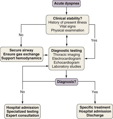
With any clinical encounter, the focus of the initial assessment is on whether or not the patient is stable (Figure 19-3). If this evaluation reveals evidence of hemodynamic insult or lability, hypotension may need to be treated promptly with intravenous fluids, vasopressors, and/or vasodilators. Airway patency and adequacy may be threatened by a depressed level of consciousness, aspiration, or trauma. Endotracheal intubation may become necessary in such instances and when gas exchange derangements cannot be rectified by supplemental oxygen or noninvasive positive-pressure ventilation. Once these basic support elements are addressed, diagnostic testing can safely proceed.

Differential Diagnosis

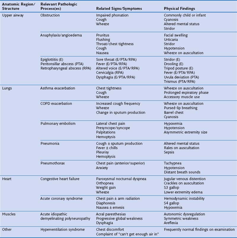
One approach to the differential diagnosis for acute dyspnea is to consider how processes in certain anatomic regions contribute to this symptom (Table 19-1). Obstruction is the most common mechanism for dyspnea arising from upper airway problems. Stridor, a variably high-pitched, harsh inspiratory noise caused by turbulent airflow, often can be heard in the context of an aspirated foreign body and edema of the epiglottis and laryngeal soft tissues. Prompt evaluation and management of upper airway blockage are critical, because the airway is endangered. Exacerbations of asthma and COPD typically are manifested as bronchospasm, wheeze, and cough. Sputum production is common to exacerbations of COPD associated with acute bronchitis and pneumonia, but its physical characteristics alone are not useful in predicting a causative pathogen. Pleuritic chest pain is sharp, incisive, breath-taking discomfort caused by irritation of the parietal pleural nerve supply along the thoracic cavity. This type of pain can accompany pulmonary embolism, pneumonia with or without pleural effusion, and pneumothorax.

In an acute coronary syndrome (ACS) or CHF, dyspnea is caused by pulmonary venous hypertension and interstitial fluid accumulation. Sudden dyspnea without chest discomfort is the presenting feature of myocardial infarction in 4% to 14% of events. Papillary muscle rupture with mitral valve incompetence, florid pulmonary edema, and shock complicates myocardial infarction in approximately 7% of affected patients. Neuromuscular diseases more commonly cause chronic, progressive dyspnea, but ventilatory failure can develop over just a few hours in acute idiopathic demyelinating polyneuropathy (AIDP). Dyspnea is a frequent consequence of severe abdominal distention, such as that seen with bowel obstruction. The increased pressure of the abdominal cavity restricts diaphragm excursion, thereby decreasing functional residual capacity. Abdominal pain restricts ventilation as a consequence of muscle splinting, which also can cause atelectasis.

Physical Examination

Inspection of the patient in respiratory distress may be quite revealing. Stigmata of adrenergic excess commonly are present, including hypervigilance, diaphoresis, and tachycardia. The breathing pattern frequently is rapid and shallow, which foreshortens responses to clinical questioning. An exception is the deep and sometimes bradypneic pattern of Kussmaul respirations seen with severe metabolic acidosis, typical of diabetic ketoacidosis. Engagement of accessory inspiratory muscles (e.g., scalenes, intercostals, and sternocleidomastoids) often signals respiratory failure. This finding alone is associated with a nearly three-fold risk of death and a roughly doubled requirement for posthospital care in patients admitted through the emergency department for COPD exacerbation. Among adult asthmatics who were noted to use accessory breathing muscles at the time of hospitalization, percent-predicted FEV1 values were lower than in patients who were not activating these muscles. Pursed-lip breathing (exhaling against partially occluded lips) can be seen in persons with airflow obstruction. In patients with moderate to severe COPD, pursed-lip breathing facilitates the recruitment of accessory muscles, decreases the electromyographic propensity for diaphragm fatigue, and improves tidal volume and arterial oxyhemoglobin saturation. Dyspnea observed in the context of altered mental status and cyanosis is worrisome, because taken together, these findings attest to profound gas exchange problems.

Biomarkers

Brain Natriuretic Peptides

Peripheral blood concentrations of brain-type natriuretic peptides (BNPs) commonly are used in emergency medicine to distinguish CHF from other causes of acute dyspnea. BNP is one of the natriuretic peptides released from cardiac myocytes with ventricular stretching and pressure overload. A prospective, multicenter study published in 2002 called the Breathing Not Properly trial evaluated the test characteristics and diagnostic accuracy of BNP for CHF in 1586 patients who presented for urgent evaluation of acute dyspnea. The clinical diagnosis of CHF was adjudicated by two cardiologists who were blinded to BNP results. At a cutoff value of 100 pg/mL, the diagnostic accuracy for CHF was 83%. Patients with BNP levels of 100 pg/mL or higher were nearly 30 times more likely than those with lower values to have CHF. Other clinical correlates of CHF, such as cephalization of pulmonary vessels on chest radiograph (odds ratio, 10.7), crackles on chest auscultation (odds ratio, 2.2), and jugular venous distention (odds ratio, 1.8), also were helpful.

A related natriuretic peptide called amino-terminal pro-BNP (NT-proBNP) also has been shown in the emergency department setting to strongly suggest CHF as the correct diagnosis for acute dyspnea. Nonetheless, elevated natriuretic peptide measurements do not independently secure a CHF diagnosis. Atrial arrhythmias, cardiomyopathies, regurgitant valvular disease, pulmonary arterial hypertension, obstructive sleep apnea, pulmonary embolism, and even sepsis can cause abnormal results on BNP assays. An echocardiogram can be beneficial when BNP levels return to within an indeterminate range.

Thoracic Imaging

Chest Radiograph

Plain chest radiography is perhaps the most widely utilized imaging modality for the evaluation of acute dyspnea. Depending on patient stability and mobility, adequate views in anteroposterior or standing posteroanterior projections can be obtained using portable techniques. The chest radiograph complements the workup for an airway foreign body but has some limitations. Retrospective studies involving children have shown that the chest radiograph has a sensitivity of 73% to 85% and a specificity of 9% to 45% for foreign body, suggesting that direct visualization is warranted in most instances.

Certain technical caveats apply to the use of the chest radiograph to diagnose pneumothorax. The air collection appears as a lucent region between the chest wall and the line of the visceral pleura. This interface can be difficult to visualize in the supine patient, because air accumulates in subpulmonic regions. This pattern of localization causes the so-called deep sulcus sign. Chest radiography cannot be used to estimate the size of a pneumothorax, and there is no difference between expiratory and inspiratory views in terms of diagnostic yield.

The chest radiograph is insensitive for the diagnosis of pulmonary embolism. For example, a wedge-shaped peripheral opacity (Hampton’s hump) and abrupt pulmonary arterial cut-off (Westermark’s sign) were observed on the chest radiograph in only 22% and 14%, respectively, of pulmonary embolism cases confirmed by angiography.

Treatment

Further details on the treatment of acute dyspnea caused by the aforementioned processes are presented elsewhere in this book. Two interventions, oxygen therapy and noninvasive positive-pressure ventilation (NIPPV), merit a brief discussion here because of their application to several of these processes. Table 19-2 presents a list of processes that cause acute dyspnea and their specific therapies.

Table 19-2 Specific Therapies for Acute Dyspnea by Etiologic Condition

Etiologic Condition Therapeutic Intervention(s)/Agent(s)
Aspirated foreign body Endotracheal intubation
Fiberoptic or rigid bronchoscopy with removal
Anaphylaxis/angioedema Antihistamines
Subcutaneous epinephrine
Systemic corticosteroids
Epiglottitis (E) Endotracheal intubation (PTA/RPA)
Peritonsillar abscess (PTA) Broad-spectrum antibiotics (PTA/RPA)
Retropharyngeal abscess (RPA) Incision and drainage (PTA/RPA)
Asthma exacerbation Supplemental oxygen
Inhaled β2-adrenergic agonists by nebulizer or MDI
Systemic corticosteroids
COPD exacerbation Supplemental oxygen
Inhaled β2-adrenergic agonists by nebulizer or MDI
Inhaled anticholinergic agents by nebulizer or MDI
Systemic corticosteroids
Antibiotics for purulent sputum
Noninvasive positive-pressure ventilation
Pulmonary embolism Systemic anticoagulation
Catheter-based thromboembolectomy
Pneumonia Supplemental oxygen
Antibiotics
Airway clearance techniques
Immunization
Pneumothorax Thoracostomy tube placement
Supplemental oxygen
Congestive heart failure Supplemental oxygen
Diuretics
Systemic vasodilators
Inotropic agents
Acute coronary syndrome Percutaneous transluminal coronary angioplasty ± stent
Antiplatelet agents
Lipid-lowering therapies
Diuretics
Acute idiopathic demyelinating polyneuropathy Mechanical ventilatory support
Intravenous immunoglobulin infusion
Systemic corticosteroids
Hyperventilation syndrome Anxiolytic medications
Psychiatric evaluation

COPD, chronic obstructive pulmonary disease; MDI, metered dose inhaler.

Chronic Dyspnea

Chronic dyspnea has been defined as lasting longer than 1 month. Prevalence statistics for chronic dyspnea vary by setting and population. In a cross-sectional survey from Australia, 8.9% of the nearly 5500 respondents acknowledged exertional breathlessness. Environmental tobacco smoke exposure independently increased the likelihood of dyspnea by a factor of 1.45 among 4197 Swiss never-smokers. In 2009, dyspnea was the most common symptom or reason for referral encountered by European hospital-based internists, constituting 19% of admissions. Chronic dyspnea accounts for 3% to 25% of general ambulatory practice visits and has a significant impact among the elderly.

Patients with chronic dyspnea can be disproportionately burdened with comorbid conditions. A 2006 telephone survey of 1003 patients with COPD, 61% of whom reported moderate or severe dyspnea, identified coexistent hypertension (55%), hypercholesterolemia (52%), depression (37%), cataracts (31%), and osteoporosis (28%). Retrospective and prospective data suggest that patients with COPD have a tendency to sustain falls. Idiopathic pulmonary fibrosis, a disease in which the vast majority of patients experience breathlessness, is associated with an excessive risk of cardiac ischemia, venous thromboembolism, and obstructive sleep apnea. From the U.S. Third National Health and Nutrition Examination Survey (NHANES III), researchers interested in asthma and obesity found that the proportion of subjects citing dyspnea during hill climbing rose as obesity worsened. The prevalence of airflow obstruction fell as body mass index increased, yet the most severely obese subjects were using bronchodilators more frequently. These observations led investigators to conclude that asthma may be overdiagnosed in severely obese patients.

Differential Diagnosis

Causes of chronic dyspnea are listed in Table 19-3. Conditions such as COPD and CHF (see Table 19-1) may have both acute and chronic phases. Relatively few studies have tried to identify those diseases that most commonly underlie chronic dyspnea in the outpatient setting. A prospective evaluation of 85 patients at a university-based pulmonary clinic showed that asthma (29%), COPD (14%), interstitial lung disease (14%), and cardiomyopathy (10%) explained two thirds of cases. Although the study investigators emphasized a rational diagnostic approach that incorporated the responses to disease-specific therapies, an average of 6.2 tests were conducted per patient, and no participants had pulmonary vascular disease or muscle weakness. Another investigation of 72 consecutive patients with chronic dyspnea and unrevealing history, physical examination, chest radiograph, and spirometry data found that 36% had pulmonary disease, 14% had cardiac disease, and 19% had primary hyperventilation. Only 3% had an extrathoracic reason for their breathlessness.

Table 19-3 Differential Diagnosis of Chronic Dyspnea

Anatomy Processes Risk Factors
Upper airway Vocal cord dysfunction Head, neck, and lung cancer
Neck surgery
Head trauma
Endotracheal intubation
Viral infection
Psychiatric disorder
Subglottic stenosis Endotracheal intubation
ANCA-positive vasculitis
Partially obstructing lesion(s) Cancer
Granulomatous inflammation
Thyroid Thyrotoxicosis Autoimmunity
Hypothyroidism Viral infection
Blood Anemia Chemotherapy
Chronic kidney disease
Chronic gastrointestinal blood loss
Heart Systolic heart failure Myocardial infarction
Diastolic heart failure Hypertension, obesity
Pericardial disease Pericarditis
Valvular disease Mitral and aortic insufficiency
Intracardiac shunt Patent foramen ovale, ventricular septal defect
Lungs    
Airways COPD Tobacco abuse, α1-antitrypsin deficiency
Asthma Atopy, genetic predisposition
Cystic fibrosis Heritable genetic defect
Parenchyma Interstitial lung disease Idiopathic interstitial pneumonia
Pneumotoxic drug reaction
Connective tissue disease
Vasculature Pulmonary arterial hypertension Idiopathic pulmonary arterial hypertension
Chronic venous thromboembolism
Vasculitis
Obesity-hypoventilation syndrome
Arteriovenous malformation  
Pleura Pleural effusion Systolic heart failure
Cancer
Infection
Hepatic hydrothorax
Thorax Kyphoscoliosis Congenital
Osteoporosis
Respiratory muscles Mechanical loading Morbid obesity
Pregnancy
Dyskinesia/dystonia  
Neurodegenerative disease  
Integrated Deconditioning Sedentary lifestyle
Nutritional deficiency
Obesity
Other Hyperventilation syndrome Anxiety

ANCA, antineutrophil cytoplasmic antibodies; COPD, chronic obstructive pulmonary disease.

Dyspnea often affects patients without obvious heart or lung disease; therefore, the clinician must consider various etiologic conditions. Some of these processes, listed in Table 19-3, warrant further discussion, because dyspnea could be overlooked as a presenting symptom. Moderate to severe anemia can limit tissue oxygen delivery but does not lead to oxyhemoglobin desaturation. Thus, the mechanism by which a low hemoglobin concentration provokes dyspnea probably is related to impaired muscle energetics and a compensatory increase in ventilation and cardiac output. Patients with many forms of advanced cancer indeed experience significant, albeit temporary, dyspnea relief from blood transfusions and erythropoietic growth factor support. Respiratory muscle weakness has been implicated as the cause of chronic dyspnea in patients with hypothyroidism and thyrotoxicosis, on the basis of responses to specific medical therapy. Limited data are available regarding the prevalence of dyspnea among patients with selected neuromuscular diseases. Roughly 40% of patients with amyotrophic lateral sclerosis, 23% of patients with post-poliomyelitis, and 12% of patients with multiple sclerosis complain of dyspnea. Both weight gain and a sedentary lifestyle are common causes of exertional dyspnea in those residing in developed countries.

Medical History

Processes that cause chronic breathlessness are likely to have evolved to some extent before a patient presents for evaluation. Accordingly, it is incumbent on the clinician to ask about the ways in which the patient’s dyspnea has changed. Questions might focus on how frequently dyspnea occurs, how long each episode lasts, how intense each episode is, and defining factors that both trigger and relieve it. Eliciting which activities of daily living provoke dyspnea can facilitate longitudinal assessment. This determination also sheds light on whether the patient is modifying behaviors as dyspnea worsens. Unless the clinician specifically asks about activities that the patient has stopped because of breathing difficulty, it can appear that breathlessness only modestly affects that patient’s functional status. Perspectives on the patient’s dyspnea from spouses, relatives, and friends frequently are useful and should be sought.

As discussed earlier, the correct diagnosis may be suggested by patient-selected descriptors of dyspnea, but it also can be substantiated by the presence or absence of associated symptoms. For example, asthmatic persons generally experience episodes of wheezing, chest tightness, and dyspnea with or without antecedent exposure to a discrete trigger such as an aeroallergen or cold air. A report of wheezing is nonspecific for asthma, however, because it can signify COPD, upper airway obstruction, or CHF. Many asthmatics also cough during bronchospasm periods, but asthma explains a chronic cough only about 25% of the time. The elderly asthmatic patient may describe exertional dyspnea and no other respiratory symptoms—a possible correlate of nonreversible airflow obstruction.

Paroxysmal nocturnal dyspnea and orthopnea may suggest CHF. A systematic review of studies investigating emergency department diagnosis of CHF found that a history of paroxysmal nocturnal dyspnea increased the pretest probability of CHF in the dyspneic patient by a factor of 2.6. Data from the Cardiovascular Health Study suggest that orthopnea and paroxysmal nocturnal dyspnea are relatively specific (87.5% and 89.8%, respectively) but somewhat insensitive (42.8% and 37.6%, respectively) indicators of CHF. Orthopnea also may be due to respiratory muscle weakness and abdominal “loading,” as with ascites and obesity.

Physical Examination

A focused physical examination frequently yields sufficient clues about the origin of chronic breathlessness. The investigation should begin with an appraisal of the patient’s voice. Someone with “breathy” dysphonia whose voice tends to wane during prolonged speech could have vocal cord dysfunction. In the setting of chronic dyspnea, stridor most commonly is caused by the development of benign or malignant lesions or focal stenoses. The diagnostic utility of jugular venous pressure elevation for detection of decompensated CHF has been extensively studied. The sensitivity and specificity of this finding for high pulmonary capillary occlusion pressure (i.e., pulmonary venous hypertension) are 70% and 79%, respectively. The jugular venous pressure can be difficult to measure in persons with an obese habitus and can be elevated as a consequence of pulmonary arterial hypertension, tricuspid regurgitation, pericardial constriction, or occlusion of the superior vena cava. Examination of the neck also should involve palpation for thyroidomegaly and lymphadenopathy, because dyspnea can result from tracheal compression.

Inspection of the spine and thorax can provide a clear reason for why a patient experiences persistent dyspnea. Severe kyphosis reduces total lung capacity, limits alveolar ventilation, and eventually impairs gas exchange. The anteroposterior dimension of the thorax can be exaggerated in patients with COPD, but this adaptation to lung hyperinflation lacks sensitivity for this diagnosis. Central adiposity in the morbidly obese patient reduces functional residual capacity (FRC) and reserve volume, thereby increasing resistance to airflow and decreasing lung compliance. Collapse of small peripheral airways in the lung bases of obese patients can lead to ventilation-perfusion mismatch and breathlessness.

Crackles on lung auscultation can reflect several pathologic processes, all of which should be considered along with other aspects of the examination. The negative predictive value of crackles for interstitial lung disease and CHF has been reported as 98% and 89%, respectively, indicating that a majority of patients without crackles will not have these conditions. The sensitivity of crackles for CHF can be as low as 29%, even when echocardiography is used to affirm or refute presence of left ventricular dysfunction. In one study of 57 patients with pleural effusions, 49 of whom were dyspneic, dullness to percussion and decreased breath sounds were individually more suggestive of pleural fluid than crackles. Wheezes similarly are useful in diagnosing asthma and COPD, but their absence cannot exclude airflow obstruction. Emphysema is suggested by diminished breath sounds in the upper lung fields, sometimes associated with indistinct heart tones. The S3 gallop on heart auscultation is highly specific for elevated left ventricular end-diastolic pressure.

Diagnostic Testing

Details from the history and physical examination should inform the selection of diagnostic tests that further refine decision-making. One approach involves categorizing these modalities according to their ability to investigate relevant anatomy and physiology (Table 19-4). Various guidelines on management of COPD recommend that the diagnosis be determined by a value of 70% or less for the ratio of postbronchodilator forced expiratory volume in 1 second (FEV1) to forced vital capacity (FVC). However, the FEV1/FVC ratio declines with aging, and the use of a “fixed” ratio may lead to overdiagnosis of COPD among patients older than 60 years of age. We concur with the American Thoracic Society–European Respiratory Society recommendation that airflow obstruction be diagnosed by a FEV1/FVC value below the lower limit of normal for the specific patient.

Table 19-4 Diagnostic Testing for Chronic Dyspnea

Test Diagnostic Utility
Spirometry Diagnose and quantify airflow obstruction and restriction
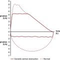
Flow-volume loop Diagnose upper airway obstruction
Single-breath diffusion capacity for carbon monoxide (DLCO) Reduced in emphysema, interstitial lung disease, and pulmonary hypertension
Can be reduced in anemia
Can be increased in alveolar hemorrhage, asthma
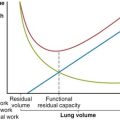
Lung volume determination Confirms restrictive lung diseases
Bronchoprovocation testing Diagnose airway hyperreactivity
Maximal inspiratory and expiratory mouth pressures Evaluate neuromuscular weakness
Chest computed tomography Interstitial lung disease
Airway caliber well delineated; can identify endobronchial lesions
Excellent characterization of pleural space and mediastinum
Echocardiography Diagnose and quantify ventricular function
Evaluate valvular incompetence, pericardium
Cardiopulmonary exercise testing Diagnose cardiac dysfunction, ventilatory limitation, oxygen desaturation, deconditioning, psychogenic dyspnea
Complete blood count Diagnose anemia

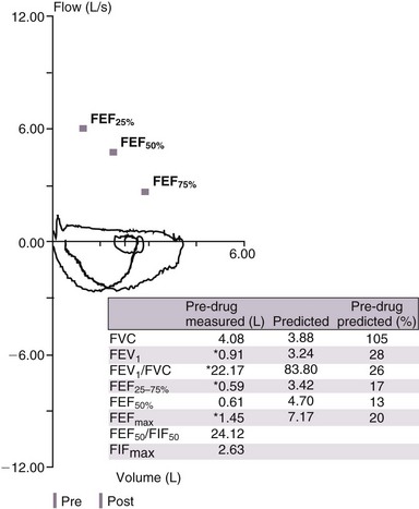
A low FVC may be due to air trapping in a patient with airflow obstruction or may point to a restrictive lung process that can be confirmed by measurement of lung volumes. Analysis of the flow-volume loop can demonstrate evidence of upper airway obstruction (Figure 19-4). In cases of suspected asthma, a negative result on direct bronchoprovocation testing essentially rules out this diagnosis. However, false-positive results can be seen in normal persons as well as those with sarcoidosis or vocal cord dysfunction. Measurement of maximal inspiratory mouth pressure is sensitive for detection of respiratory muscle weakness.

Many cardiac causes of chronic breathlessness can be identified by echocardiography. In addition to quantifying left ventricular systolic function, this procedure provides information about peak systolic pulmonary artery pressure, pericardial structure, and valvular function. Intravenous administration of agitated saline solution coupled with echocardiography (i.e., contrast echocardiography) is more than 98% sensitive for detecting pulmonary arteriovenous malformations. In a study of nearly 18,000 patients without established coronary artery disease who were referred for radionuclide myocardial perfusion testing, evidence of inducible ischemia was similar for patients with self-reported dyspnea and for those with typical angina pectoris, prompting the investigators to conclude that a more systematic appraisal of dyspnea should occur at the time of stress testing referral.

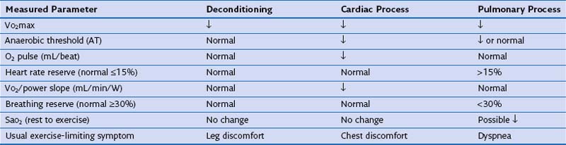
The etiology of chronic dyspnea will sometimes remain elusive despite a careful history and physical examination and multiple diagnostic endeavors. Then, cardiopulmonary exercise testing should be considered to provoke the patient’s dyspnea with comprehensive assessment of the oxygen transport system on an integrated level. Pulmonary gas exchange, cardiac function, and metabolic activity can be measured noninvasively (Table 19-5). Patients also are asked to rate dyspnea and leg discomfort throughout the exercise test. The results of cardiopulmonary exercise testing usually are able to distinguish cardiac dysfunction and ventilatory limitation but cannot always discriminate between cardiac disease and deconditioning.

Treatment

Disease-specific therapies generally are successful in mitigating chronic dyspnea, although this may not be the case with multiple concurrent processes or disease progression. Dyspnea is a prominent feature of CHF, yet most studies of various interventions for CHF consider death or utilization of health care resources as outcome metrics. Good-quality evidence supports exercise training to improve functional status in patients with CHF. Diuretics, inotropes, and vasodilators are mainstays of a management strategy of regulating volume status and controlling symptoms in CHF.

For patients with COPD, the combination of an inhaled corticosteroid and a long-acting β-agonist has been shown to improve lung function, relieve respiratory symptoms, and modestly reduce risk of death. Large trials of inhaled long-acting anticholinergic drugs also show a positive impact on dyspnea associated with COPD. Supplemental oxygen is indicated for patients with COPD who have resting hypoxemia, although its effect on mortality and symptoms in patients who display only exertional oxyhemoglobin desaturation is currently being investigated. Specific criteria to select patients with emphysema who could benefit from lung volume reduction surgery are well established. Pulmonary rehabilitation has been shown to have multiple benefits, including decreasing the severity of dyspnea, enhancing exercise tolerance, reducing the frequency of exacerbations, and improving quality of life. Interstitial lung disease is a broad category of infiltrative pulmonary disorders for which disease-modifying and symptom-relieving treatments are needed.