Dysphagia and weight loss in a middle-aged man

Published on 10/04/2015 by admin

Filed under Surgery

Last modified 22/04/2025

Print this page

rate 1 star rate 2 star rate 3 star rate 4 star rate 5 star
Your rating: none, Average: 0 (0 votes)

This article have been viewed 930 times

Problem 12 Dysphagia and weight loss in a middle-aged man

You realize that his symptoms warrant investigation, even in the absence of any physical signs, and you go on to arrange some investigations.

Blood results are as follows:

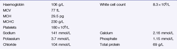
A further investigation is performed and a representative film is shown (Figure 12.1).

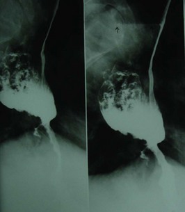
Based on the radiological findings, the patient is referred for another investigation (Figure 12.2).

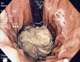
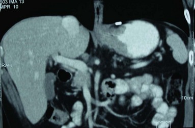
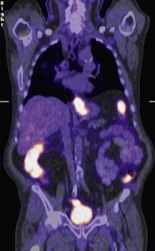
The lesion is biopsied and confirmed to be a moderately differentiated adenocarcinoma. The patient is referred for further staging investigations (Figure 12.3 and Figure 12.4.).

Various imaging investigations are performed and do not show any evidence of tumour dissemination. The tumour appears to be confined to the stomach wall, although there may be some thickening in the immediately adjacent tissues.

The patient and his diagnostic tests are discussed at a multidisciplinary meeting. The treatment recommendation is for neoadjuvant chemotherapy before and after surgical resection.

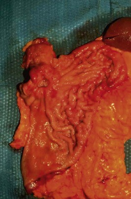
Following three cycles of chemotherapy (over 3 months) and 4 weeks of stabilization, the patient underwent surgery. Figure 12.5 shows the operative view of the upper abdomen.

The tumour showed extensive transmural infiltration by a moderately differentiated adenocarcinoma with involvement of the lymph nodes along the left gastric artery. The patient underwent three more cycles of chemotherapy 2 months following his gastrectomy. He initially did very well but died of disseminated disease 18 months later.

Answers

A.1 From the history there are several features which point to a diagnosis of a gastrointestinal malignancy. Dysphagia and weight loss appearing for the first time in someone over 50 should cause concern and prompt investigation. This patient also has risk factors (alcohol and smoking) for both oesophageal and gastric cancer.

Other diagnoses to consider include:

A.2 Investigations should include:

A.3 The blood results show a mild microcytic anaemia with a low MCV and MCHC. This is typical of iron deficiency anaemia and consistent with bleeding from occult gastrointestinal pathology. The other blood tests are normal.

A.4 The contrast study is a barium meal and this shows narrowing at the junction of the oesophagus and stomach. There is hold-up of contrast in the distal oesophagus, and the oesophagus appears dilated above the gastro-oesophageal junction. These appearances are typical of either achalasia or pseudoachalasia (i.e. a distal oesophageal cancer, or a lesion in the proximal stomach).

This man needs an endoscopic examination of his upper gastrointestinal tract. In most circumstances the imaging investigation of choice for a patient with these symptoms would be an endoscopy. However, barium meal examinations are still frequently performed by general practitioners as the initial investigation of upper digestive tract symptoms.

A.5 The patient has undergone an endoscopic examination of the upper digestive tract. This image shows a polypoid tumour extending from the squamo-columnar junction into the proximal stomach (the endoscope has been retroflexed to obtain this view). These tumours are classified according to their location in relation to the squamo-columnar junction:

The patient therefore has a Type III tumour.

A.6 The first image shows a CT scan of the abdomen (with intravenous and oral contrast). There is thickening of the wall of the proximal stomach and no evidence of metastatic deposits. CT scans of the chest, abdomen and pelvis (particularly spiral scans) are capable of providing highly accurate information on:

The CT is less helpful in the detection of peritoneal deposits. If there is evidence of metastatic disease then no further investigations are required. CT has limitations in detecting nodal disease (N stage) and is not very accurate at assessing the depth of penetration of tumours through the stomach wall (T stage). CT will generally under-stage both the T and N stage in gastric cancer.

The second image is a positron emission tomography (PET) scan which demonstrates a hot spot in the proximal stomach, corresponding to the proximal gastric cancer.

Positron emission tomography (PET) scans have become increasingly useful in gastric cancers. They are more sensitive than CT scans and EUS in detecting distant metastases, such as bony metastases. This nuclear medicine imaging technique detects uptake from a positron-emitting radionuclide tracer (commonly fluorine-18) that is introduced into the body on fluorodeoxyglucose (FDG) molecules prior to the scan. Fluorine-18 is a glucose analogue that is preferentially taken up by rapidly growing malignant tumours. PET scans will not detect primary gastric cancers in up to 30% of cases (usually those with signet ring cell features), and in these select cases may not be useful in detecting the presence of any metastatic disease.

A.7 Further investigations are needed to accurately stage the disease and then determine the best treatment options. These include: Staging laparoscopy to rule out the presence of small peritoneal deposits. These are common in gastric cancer and cannot be easily detected by CT or EUS. Endoscopic ultrasound scanning (EUS) can give accurate information about depth of penetration of the tumour through the stomach wall and also on the extent of any lymph node involvement. Where available, EUS is a useful adjunct to CT in preoperative staging of gastric cancer.

A transthoracic echocardiogram and pulmonary function tests (arterial blood gas and spirometry) will help determine the patient’s general state of health.

A.8 The treatment of gastric cancer depends on:

Curative treatment may be considered for:

Treatment options include:

Very early and localized cancers that have not invaded the gastric submucosa (early gastric cancer) can be removed endoscopically. These sorts of tumours are rare outside of Japan.

In most parts of the world the majority of patients with gastric cancer have incurable disease at the time of presentation. In contrast, gastric cancer is relatively common in Japan where screening programmes are used to detect the disease at a much earlier stage and a high percentage of patients are cured by surgery.

While some individuals can be cured of more advanced cancers, the role of surgery is usually to relieve symptoms and palliate obstruction or bleeding. Improved methods of palliation have decreased the need for surgical intervention. These procedures include endoscopic ablation of the tumour by laser or argon beam coagulation, palliative chemotherapy and palliative radiotherapy. Self-expanding metal stents can be used to relieve obstruction.

A.9 The location of cancers of the oesophagus and stomach may directly interfere with the patient’s nutritional status. While treatment is ongoing, it is important to ensure the patient is capable of meeting his/her nutritional requirements. The advice of a dietician should be sought prior to the commencement of treatment. In many cases, supplementation with high energy and protein drinks will be sufficient (e.g. Sustagen). If the tumour has partially occluded the lumen of the oesophagus or stomach, placement of a naso-gastric feeding tube (or naso-enteric feeding tube for a distal gastric tumour) will prevent further weight loss and malnutrition while the patient is undergoing treatment. For more advanced tumours (i.e. those with complete occlusion of the lumen), a surgically placed feeding tube into the proximal jejunum may be necessary.

A.10 This is a total gastrectomy specimen in which all of the stomach has been removed along with the omentum and spleen. There is thickening of cardia and fundus from tumour infiltration. The mucosa in the body/distal stomach looks relatively normal while in the proximal stomach, ulceration is evident (in the centre of the tumour) and this probably represents transmural infiltration (T3).

The prognosis for gastric cancer remains poor with overall 5-year survival of 10–15% in most Western countries. This reflects the advanced stage of the disease at presentation and the increasing age of the population; many patients with gastric cancer are elderly and frail and not suitable for curative surgical treatment. In Japan the overall 5-year survival is 50% or above. This reflects diagnosis at an earlier stage and more effective surgical treatment.

The incidence of gastric cancer is decreasing dramatically in the West. In developing countries the incidence remains high and is particularly so in Japan, China, East Asia and Latin America. The reason for the fall in incidence in the West is related to environmental factors. Helicobacter pylori is associated with gastric cancer. The prevalence of Helicobacter in Western communities has steadily declined as public health and hygiene have improved. This has coincided with the reduction in gastric cancer. Dietary changes with an increase in protein relative to carbohydrate, increasing food hygiene and refrigeration and increasing consumption of fresh fruit and vegetables have probably also decreased the incidence of gastric cancer.

The distribution of gastric cancer in the West is changing. Cancers used to be prevalent in the distal stomach. It is these cancers that are becoming less common whereas, for reasons unknown, the incidence of cancers of the cardia (together with adenocarcinoma of the distal oesophagus) has increased dramatically over the last two decades.

In surgical series in the West survival for potentially curative surgery is around 50%. Early gastric cancer (confined to the mucosal layer of the stomach) can be cured by surgery but as the tumour spreads through the gastric wall and involves lymph nodes the likelihood of a cure lessens.

Further Information

, www.cancer.gov/cancertopics/wyntk/stomach. A web page from the National Cancer Institute with links providing current information on many different aspects of gastric cancer (in the form of a web-based information booklet)

, www.surgical-tutor.org.uk. A surgical resource with extensive up-to-date information on gastric cancer

, www.helico.com. The website of the Helicobacter Foundation, founded by Dr Barry Marshall