Dysphagia
Dysphagia means difficulty in swallowing and should be distinguished from pain on swallowing. Dysphagia may be associated with ingestion of solids or liquids, or both. Pain on swallowing is odynophagia, which in itself does not interfere with the act of swallowing.
Causes
Acquired
In the Wall
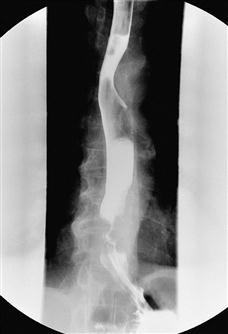
Figure 14 Barium swallow showing oesophageal carcinoma.
The barium swallow reveals an irregular narrow stricture at the distal oesophagus with ‘shouldering’ where the contrast tapers into the stricture.
Outside the Wall
Neuromuscular Disorders
History
Congenital
Acquired
In the lumen
There may be a history of ingestion of a foreign body such as a coin (children) or false teeth (elderly). Occasionally the history may not be forthcoming. In the case of a food bolus, it is unusual for this to cause dysphagia without there being some form of underlying stricture.
In the wall
With a caustic stricture, there is usually a history of caustic ingestion, except in the psychiatrically disturbed, where the history may not be apparent. There will be sudden onset of pain and dysphagia, which may improve with appropriate treatment only to recur after several months due to a stricture. Patients with inflammatory stricture due to gastro-oesophageal reflux associated with a hiatus hernia will have a history of retrosternal burning pain and acid reflux, which is worse on recumbency or bending down. The dysphagia is usually of gradual onset and the patient may localise the site of dysphagia to the level of the lower end of the sternum. Oesophageal candidiasis may cause dysphagia and this usually occurs in the immunocompromised patient. Achalasia is a disorder where there is degeneration of the oesophageal myenteric plexus resulting in loss of peristaltic contraction in the oesophagus and failure of the lower oesophageal sphincter to relax in response to swallowing. It usually presents between 30 and 50 years of age. The dysphagia may be intermittent and then gets progressively worse. It may be worse for liquids than for solids. Fluid regurgitation at night may result in aspiration pneumonitis. With carcinoma, the dysphagia is usually of rapid onset. Initially, it is for solids, then for fluids. There may be associated weight loss, anorexia and symptoms of anaemia. There may be a history of achalasia or Barrett’s oesophagus. Dysphagia with food sticking at the upper end of the oesophagus in a middle-aged woman may suggest Plummer–Vinson syndrome. This is due to a web in the upper oesophagus (post-cricoid web). The condition is pre-malignant. A history of radiotherapy to chest or mediastinum may suggest an irradiation stricture. With scleroderma, the patient may have noticed changes in the skin, around the lips, in the fingers (sclerodactyly) or may have a past history of Raynaud’s phenomenon. Chagas’ disease is extremely rare and is associated with degeneration of the myenteric plexus associated with trypanosomal infection. The symptoms are identical to those of achalasia.
Outside the wall
The presence of a large goitre will be obvious. With the pharyngeal pouch, patients are usually of middle age or elderly. They may have noticed a swelling, usually in the left posterior triangle of the neck. They may also have dysphagia localised behind the manubrium associated with the pouch pressing on the oesophagus. On lying down, there is regurgitation of food with coughing. The patient may also have halitosis. With bronchial carcinoma, there may be direct pressure on the oesophagus from the tumour or via secondary spread to the mediastinal lymph nodes. There may be a history of bronchial carcinoma or the patient may present with haemoptysis. With dysphagia from mediastinal lymphadenopathy, the patient may have noticed enlarged swellings at other sites, e.g. axilla or groin. Dysphagia from pressure of an enlarged left atrium may be associated with mitral stenosis and there may be a past history of this. With a paraoesophageal (rolling) hernia, the dysphagia may be intermittent, due to a full stomach pressing on the adjacent oesophagus. Hiccups may occur due to irritation of the diaphragm.
Neuromuscular
There will usually be a history of Guillain–Barré syndrome, poliomyelitis, motor neurone disease, myasthenia gravis or a CVA.
Others
Globus is a subjective feeling of a lump or foreign body in the throat. The term ‘globus hystericus’ was previously used because it was felt to be psychogenic but it is now considered that a globus sensation may still indicate organic pathology. Investigation is required to exclude the true causes of dysphagia.
Examination
With oesophageal atresia, an orogastric tube is passed and it will arrest at the site of the obstruction.
In many cases of dysphagia, there will be nothing to find on examination. A goitre is usually an obvious swelling that moves on swallowing. With a pharyngeal pouch, there may be a palpable swelling low down in the posterior triangle of the neck (usually left), which gurgles on palpation. With carcinoma, there may be signs of weight loss, a palpable liver due to metastases, or cervical lymphadenopathy due to metastases. Koilonychia, angular cheilitis and glossitis are clinical features associated with Plummer–Vinson syndrome. With irradiation stricture, there may be changes in the skin consistent with previous radiotherapy. With scleroderma, there may be calcinosis of the subcutaneous tissue, Raynaud’s phenomenon, sclerodactyly and telangiectasia. With dysphagia, due to an enlarged left atrium in mitral stenosis, there may be signs of mitral stenosis, e.g. peripheral cyanosis, malar flush, left parasternal heave, tapping apex beat, opening snap and mid-diastolic murmur best heard at the apex. A variety of neurological abnormalities will be associated with dysphagia of neuromuscular origin.
General Investigations
■ FBC, ESR
Hb ↓ associated with carcinoma but may also occur with oesophagitis associated with peptic strictures. Anaemia may also be associated with Plummer–Vinson syndrome. ESR ↑ in malignancy and scleroderma.
■ LFTs
Alkaline phosphatase↑in liver secondaries.
■ CXR
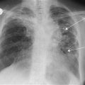
Foreign body if radio-opaque. Air-fluid level in achalasia. Gastric air bubble in chest in paraoesophageal hernia. Hilar lymphadenopathy. Bronchial carcinoma. Widened mediastinum with aortic aneurysm. Large left atrium (double shadow behind the heart) – mitral stenosis.
Specific Investigations
■ Barium swallow
Pharyngeal pouch (never OGD as it may perforate the pouch), stricture, achalasia, external compression.
■ OGD
Foreign body. Food bolus. Candidiasis. Benign versus malignant stricture. Plummer–Vinson syndrome – post-cricoid web. Biopsy – benign lesion versus malignant lesion, absence of myenteric plexus in achalasia.
■ CT
Goitre. Mediastinal nodes. Spread of malignancy. Tumour staging. Aortic aneurysm. Dysphagia lusoria (abnormally placed arteries causing external compression).