[level-membership-for-opthalmology-category]7.1
Dry Age-Related Macular Degeneration
Clinical Features:
The hallmark feature of dry AMD is the presence of drusen, which are yellow-colored subretinal deposits that range in size and appearance (Fig. 7.1.1). Small, fine drusen without other manifestations such as RPE changes or atrophy should not be considered AMD. Additionally, there can be varied pigmentary changes within the RPE. Advanced forms of AMD feature atrophy of the RPE with eventual GA, which can occur in the presence or absence of drusen (Fig. 7.1.2 and Fig. 7.1.3).
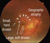
Figure 7.1.1 Color fundus photograph of dry AMD with many drusen of varying size intermixed with RPE hyperpigmentation and hypopigmentation. There is an area of geographic atrophy (arrowheads), which is difficult to appreciate clinically.
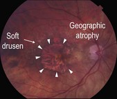
Figure 7.1.2 Color fundus photograph of a large area of central geographic atrophy (arrowheads) and surrounding soft drusen.
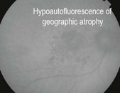
Figure 7.1.3 Fundus autofluorescence image (corresponding to Figure 7.1.1) highlights areas of geographic atrophy as distinct areas of hypoautofluorescence.
OCT Features:
Drusen are identified on OCT by their characteristic appearance as discrete elevations of the RPE layer at the level of Bruch’s membrane (Figs. 7.1.4 and 7.1.5). Drusen may be of varying size and contour. Drusen can be described histopathologically as basal linear, when the deposits occur between the basement membrane of the RPE and Bruch’s membrane (more typical), or basal laminar, when the deposits occur between the plasma membrane of the RPE and the basement membrane of the RPE. Basal laminar drusen are also called cuticular drusen and are characteristically small and regular shaped in a diffuse arrangement within the macula. It is difficult to distinguish between basal linear and basal laminar drusen on OCT. GA is identified by absence of the outer retinal layers and RPE, which leads to a reverse shadowing effect (Fig. 7.1.6).
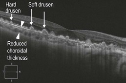
Figure 7.1.4 OCT (corresponding to Figure 7.1.1) showing features of dry AMD. There are many discrete, round, hill-shaped elevations below the RPE, which are basal linear drusen (arrows). Drusen exhibit a medium-intensity reflectance pattern on OCT. The choroidal thickness is typically less than normal in AMD (space between arrowheads).
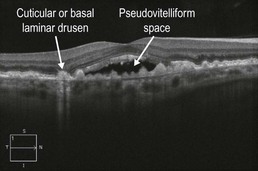
Figure 7.1.5 OCT showing basal laminar or cuticular drusen. An associated pseudovitelliform detachment is present in the macula, which can be seen in the setting of cuticular drusen, even in the absence of choroidal neovascularization.
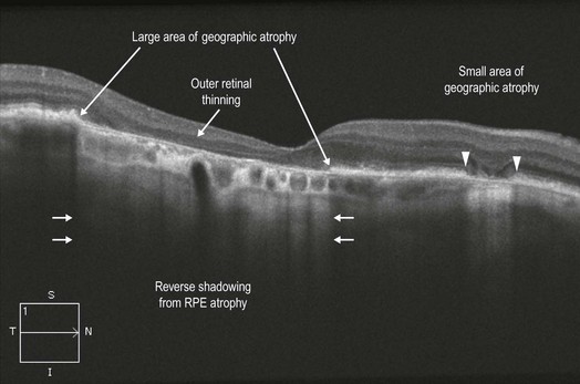
Figure 7.1.6 OCT (corresponding to Figure 7.1.2) showing larger (between arrows) and smaller (between arrowheads) areas of GA. In areas of GA, there is loss of the outer retinal layers and RPE. Due to absence of the RPE in these areas, the OCT signal is able to penetrate more deeply, which exaggerates the typical signal pattern so that there is a ‘reverse’ shadowing effect.
Ancillary Testing:
Color fundus photographs and fundus autoflourescence can be helpful to highlight and track areas of GA (Fig 7.1.3).
[/level-membership-for-opthalmology-category][not-level-membership-for-opthalmology-category]7.1
Dry Age-Related Macular Degeneration
Clinical Features:
The hallmark feature of dry AMD is the presence of drusen, which are yellow-colored subretinal deposits that range in size and appearance (Fig. 7.1.1). Small, fine drusen without other manifestations such as RPE changes or atrophy should not be considered AMD. Additionally, there can be varied pigmentary changes within the RPE. Advanced forms of AMD feature atrophy of the RPE with eventual GA, which can occur in the presence or absence of drusen (Fig. 7.1.2 and Fig. 7.1.3).
Figure 7.1.1 Color fundus photograph of dry AMD with many drusen of varying size intermixed with RPE hyperpigmentation and hypopigmentation. There is an area of geographic atrophy (arrowheads), which is difficult to appreciate clinically.
[/not-level-membership-for-opthalmology-category]
