Drowning

Published on 14/03/2015 by admin

Filed under Emergency Medicine

Last modified 22/04/2025

Print this page

rate 1 star rate 2 star rate 3 star rate 4 star rate 5 star
Your rating: none, Average: 0 (0 votes)

This article have been viewed 1041 times

Chapter 29 Drowning

PATHOPHYSIOLOGY

The concepts of ‘wet’ and ‘dry’ drowning, based on the presumption that a variable degree of laryngospasm occurs which, in some patients, prevents any aspiration of water or other substances into the airway, bronchial tree and lungs, were discarded at the World Congress on Drowning.

The International Liaison Committee on Resuscitation (ILCOR) advisory statement on drowning describes an involuntary period of laryngospasm secondary to the presence of liquid in the oropharynx or larynx. It has been described that most patients aspirate less than 4 mL kg–1 of fluid and that approximately 10–20% of individuals maintain tight laryngospasm until cardiac arrest occurs and inspiratory efforts have ceased. This has been confirmed by autopsy studies, with 10% of patients showing no aspiration and approximately 20% of patients presenting with normal chest radiographs. Other sources suggest that drowning probably never occurs without aspiration to some degree.

What appears clear is that initial voluntary breath holding precedes a variable degree of aspiration of water, followed eventually by apnoea, with the common pathway in all drowning being profound hypoxia associated with acidosis and hypercapnoeia. This hypoxia not only leads to unconsciousness, loss of airway reflexes and aspiration, but also leads to the cardiovascular effects of drowning. These include extreme bradycardia, ventricular fibrillation and asystole, which are often exacerbated by hypothermia in cold waters. Hypothermia is common in child victims of drowning due to their large surface area:body mass ratio and, although hypothermia has been associated with a poor prognosis after drowning as it is related to the duration of submersion, it has also been associated with a better prognosis, particularly in children, presumably due to a protective effect on cerebral organ function with the rapid onset of low temperatures. There have been several cases reported of survival of both children and adults following submersion in cold water for up to 66 minutes.

Following early survival, lethal hypoxia may still develop. However, in these later instances it is due to the effects of surfactant disruption and abnormal function, atelectasis and intrapulmonary shunting. This profound secondary hypoxia may itself lead to respiratory failure and cardiac arrest.

In some groups of patients, aspiration of sand, silt, stagnant water, sewage and vomitus may result in bronchial occlusion, bronchospasm, inflammatory damage to alveolar capillary membranes, abscess formation and pneumonia. Late, atypical pneumonias may be caused by less common pathogens such as Aeromonas, Pseudallescheria and Burkholderia.

OUTCOME

The commonest cause of death in hospitalised drowning patients is post-hypoxic encephalopathy. Other common causes of death are acute respiratory distress syndrome (ARDS), multi-organ dysfunction syndrome (MODS) or sepsis syndrome.

Generally speaking, judging prognosis after a drowning incident is difficult, but there are recognised associations with particular outcomes. Pointers towards poor prognosis include:

Individuals with any of these features have been reported to survive without disability, although the chances of successful resuscitation to a favourable neurological outcome are usually slim. Importantly, age has no independent association with outcome.

Examination

Examination should follow a systematic course as described by ABCDE. Treatment should follow the same schema and should be delivered in systems when any abnormality is identified.

Drowning victims may be classified into one of four groups based on presenting physical examination:

MANAGEMENT

Key to the management of drowning patients, as with other critically ill or injured patients, is assessment and intervention in systems defined by ABCDE. This system of management fundamentally relies at its core on the maximisation of perfusion, ensuring the delivery of oxygenated blood to the brain and other vital organs.

Prehospital management differs very little from emergency department management in that the priorities are identical. However, as with most critically ill or injured patients, minimising any delay and striving for rapid transport with essential life-saving interventions being performed are the keys to optimum survival.

If no resuscitative efforts have been initiated, and there is apnoea or no cardiac output, commence cardiopulmonary resuscitation (CPR) compression ratio of 30:2; early defibrillation where appropriate; correction of any likely causative factors, which in this case would clearly include hypoxia and hypothermia; and regular intravenous adrenaline (epinephrine).

Airway

The airway should be checked for foreign material and suction performed under direct vision. A look, listen and feel process will allow assessment of airway patency and respiratory effort. Basic airway manoeuvres such as a jaw thrust will be useful, whereas chin-lift and head-tilt should be reserved for patients with a clear history of non-trauma.

Clearance and maintenance of a patent airway, preferably by the most definitive means available, are of primary importance. Ideally this would be by either immediate intubation with a cuffed endotracheal tube or a rapid sequence induction (RSI). If this skill is not immediately available, time should not be wasted making several attempts at this while the patient becomes more hypoxic. A suitable alternative may be a laryngeal mask airway (LMA), although there is an increased risk of aspiration of gastric contents, particularly in drowning patients who may have swallowed copious amounts of water. Newer versions of the LMA with an embedded channel for a gastric tube may facilitate safer airway management utilising suction aspiration of stomach contents.

Manual in-line immobilisation should be employed during airway management if there is a history or a suspicion of trauma, particularly in diving accidents. Adjuncts such as rigid cervical collars, sandbags and tape assist in immobilising an at-risk cervical spine, although their limitations should be known, particularly in children where an immobilised and

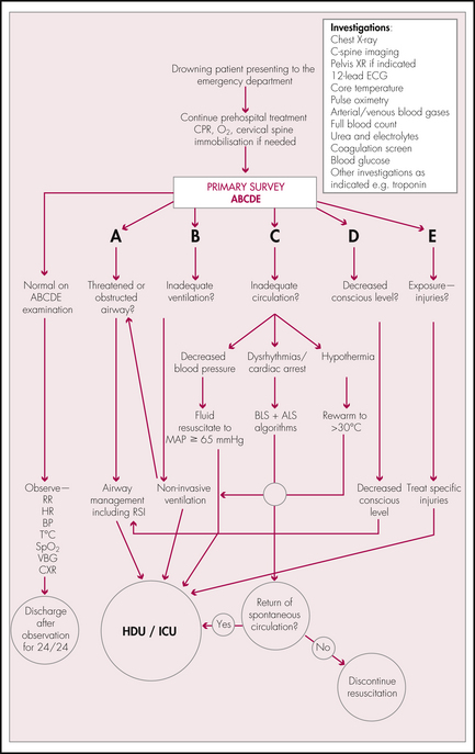
restrained head and neck may lead to a mobile body. Care must be taken not to manage a potential cervical spine injury to the detriment of efficient airway management.

Breathing

Adequate oxygenation and ventilation are required to reverse hypoxia and acidosis, which may have a mixed respiratory and metabolic aetiology. If the patient is conscious and the airway is protected, apply high flow oxygen at 15 L min–1 via a non-rebreather mask. Unconscious patients with definitive airway management in situ should have an initial FiO2 of 1.0 maintained. If patients are obtunded, management of the airway is essential as described above. The potential for aspiration is high in drowning patients due to swallowed water, and a low threshold must be maintained for RSI and intubation.

Awake patients with adventitious signs in their chest suggesting aspiration or acute pulmonary oedema may benefit from non-invasive ventilation (NIV), delivered as either continuous positive airway pressure (CPAP) or bi-level positive airway pressure (BiPAP), which has been shown to be effective at reducing mortality and preventing intubation in selected other patient groups. Although ‘fluid overload’ may be possible in drowning victims, which would mandate loop diuretics such as frusemide for treatment of symptoms and signs of pulmonary oedema, the probable cause is more likely to be hypoxia- and acidosis-induced acute myocardial dysfunction with fluid maldistribution, and therefore NIV and potentially high-dose nitrate infusion may be the most appropriate treatment.

Conscious patients should be monitored by means of continuous pulse oximetry, and ideally side-stream end-tidal CO2 (EtCO2) monitoring may give evidence of hypoventilation. This equipment is not common; however, it has been shown to be efficacious in monitoring patients at risk for compromised ventilation. Ventilated patients should routinely have EtCO2 monitored and ventilation titrated to allow maximum cerebral perfusion; in the presence of head injury, maintenance of EtCO2 at 35 mmHg is appropriate.

Indications for RSI and intubation may be:

Intubated patients should have an orogastric tube inserted to treat intragastric swallowed fluid and air. Have a high index of suspicion for tension pneumothorax if there is hypotension resistant to fluid resuscitation, particularly in the setting of potentially serious trauma associated with drowning. Small pneumothoraces may be managed by observation; however, if patients are intubated and ventilated, positive pressures may turn a simple pneumothorax into a tension pneumothorax, and placement of intercostal catheters may be an early necessity.

Circulation

Patients in cardiac arrest following drowning who require basic or advanced life support and are not hypothermic have a poor prognosis. Routine BLS/ALS should be implemented.

Cardiac arrhythmias are secondary to hypoxia, acid–base disturbance and hypothermia, rather than electrolyte disturbances secondary to massive fluid aspiration, and any type of cardiac arrhythmia may be observed, particularly bradycardia and atrial fibrillation which are statistically the most common abnormalities.

Venous access should be urgently obtained with a large-bore cannula. Cardiac arrhythmias should be treated according to ALS algorithms. Boluses of crystalloid or colloid fluid are useful in maintaining perfusion of vital organs, although importantly attempts should not be made to attain ‘normal’ blood pressures, particularly in the presence of potential serious trauma or covert blood loss. A mean blood pressure of 65 mmHg will be sufficient to maintain cerebral and renal perfusion.

Rewarming is essential (see the ‘Hypothermia’ section in Chapter 32): wet clothing should be removed, the skin should be dried and the patient should be wrapped in warm blankets with aluminium foil outside this. Intravenous fluids should be warmed to 40°C; ventilator circuits should contain a humidifier/warmer to ensure ventilatory gases are also close to body temperature. Children lose heat quickly due to the large surface area:body mass ratio and therefore careful management of temperature is mandatory.

A low threshold for intraosseous cannulation should be maintained, especially in cardiac arrest or critical condition post drowning; this may be achieved in adults as well as children. Care should be taken with central venous cannulation as the hypothermic myocardium may be sensitive and prone to dysrhythmias. Rescuscitation should not be ceased until the core temperature is above 30°C, and there are still no signs of life or organised cardiac function.