Dome-Shaped Macula
OCT Features:
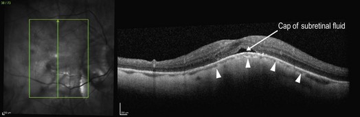
A vertical line scan is more helpful than a horizontal scan in diagnosing dome-shaped macula. Within the convexity of a posterior staphyloma, there is an inward bowing of the sclera within the central macula (Fig. 9.4.1). The overlying macula follows the same contour as the sclera and there is often a cap of subretinal fluid (hyporeflective space), in the absence of any CNV or CSCR. The choroid is typically thin and the underlying sclera can usually be imaged well with standard spectral domain OCT protocols. However, both enhanced depth and swept source imaging techniques offer the ability to visualize deeper structures and are a better choice for assisting in this diagnosis, if available.
Ancillary Testing:
Fluorescein can be helpful to rule out the presence of a concomitant CNV or CSCR.


