Chapter 61 Disorders of the female genitalia
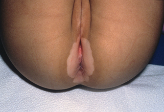
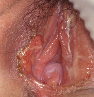
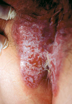
Nonneoplastic epithelial disorders of the vulva
1. What is lichen sclerosus (also known as lichen sclerosus et atrophicus)?
Lichen sclerosus (LS) (Fig. 61-1) is an inflammatory condition that primarily affects the superficial dermis. The disease process results in thinned or atrophic white papules and plaques of the skin. LS primarily affects the anogenital region, but it can also present on the trunk or extremities. Lichen sclerosus of the vulva most commonly affects postmenopausal women, but it can also develop in 7% to 15% of prepubertal females.
2. Describe the clinical signs of lichen sclerosus of the vulva.
The characteristic findings associated with lichen sclerosus include white, thinned, crinkled skin (it is frequently described as “cigarette paper” atrophy). Often there are areas of ecchymoses in the involved skin. The majority of the vagina is usually unaffected; however, a patient may have involvement of the vaginal introitus, leading to stenosis. One also may find fusion of the labia minora, phimosis of the clitoral hood, and fissures. LS can also simultaneously involve the perianal area, which then forms a “figure-of-eight” pattern.
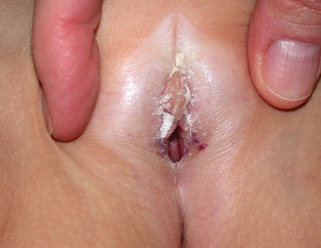
Val I, Almeida G: An overview of lichen sclerosus, Clin Obstet Gynecol 48(4):808–817, 2005.
4. Is there a need to biopsy lichen sclerosus of the vulva?
Yes. Biopsy of the affected site can aid in differentiating lichen sclerosus from other disorders, such as vitiligo (Fig. 61-2) and lichen planus. Hyperkeratotic, ulcerated, or nodular lesions should be sampled to rule out vulvar intraepithelial neoplasia (VIN) or squamous cell carcinoma of the vulva. The risk of LS becoming malignant is 4% to 6%.
5. How does one treat lichen sclerosus?
The most widely accepted first-line treatment is use of a high-potency topical corticosteroid, most commonly clobetasol proprionate. Other less common therapies used for recalcitrant LS include intralesional corticosteroids, retinoids, oral potassium para-aminobenzoate, chloroquine, tacrolimus/pimecrolimus, cyclosporine, and photodynamic therapy with topical 5-aminolevulinic acid. LS in children often will resolve at the time of puberty.
8. Are there different variants of lichen planus that affect the vulva?
Yes. Variants that affect the vulva include erosive lichen planus, papulosquamous lichen planus, and hypertrophic lichen planus. The most common variant found in the vulva is erosive lichen planus. This variant is characterized by erosions on a background of violaceous papules and plaques. The skin of the vulva may also appear shiny, reticulated and white. Rarely, oral-vulval-vaginal lichen planus may also be seen. Of the approximate 1% of the population with oral lichen planus, 25% have vulvovaginal disease.
9. What are the clinical symptoms of vulval lichen planus?
Patients most commonly present with complaints of vulvovaginal burning, pruritus, postcoital bleeding, and dyspareunia. Up to 70% of patients also have vaginal lesions. There may be an associated purulent vaginal discharge and chronic erosions of the vaginal epithelium can lead to the development of extensive adhesions leading to vaginal narrowing and obliteration.
Lewis FM: Vulval lichen planus, Br J Dermatol 138(4):569–575, 1998.
11. How do you treat lichen planus of the vulva?
12. Describe lichen simplex chronicus of the vulva.
Lichen simplex chronicus is an end-stage response that results from chronic scratching or rubbing of the skin. The skin becomes thickened, lichenified (exaggeration of the normal skin lines), and will often show excoriations. Any condition that causes pruritus of the skin, in combination with the patient scratching the involved area, will ultimately result in the development of lichen simplex chronicus. The primary skin condition is most commonly eczema.
13. Psoriasis can be present on the vulva. How does it present?
Psoriasis of the vulva presents as beefy red symmetrical plaques. The silvery scale associated with psoriatic lesions on other areas of the body is typically absent. A patient may have inverse psoriasis of the inguinal creases, referred to as inverse psoriasis. Skin lesion of psoriasis in this area may be more inflamed and have undergone maceration secondary to friction and moisture. Vulvar pruritus and burning may be prominent symptoms.
14. How is psoriasis of the vulva treated?
Table 61-2. Condyloma Accuminatum versus Molluscum Contagiosum
CONDYLOMA ACCUMINATA | MOLLUSCUM CONTAGIOSUM | |
---|---|---|
Clinical appearance | Soft fleshy cauliflower-like papules. Can be very small with a dome shape and thus difficult to differentiate from molluscum contagiosum | Small, dome-shaped, typically flesh-colored papules with a central umbilication when squeezed on the lateral edges |
Etiology | Human papilloma virus (HPV) HPV 6 and 11 are responsible for 90% of these lesions |
DNA poxvirus—molluscum contagiosum virus (MCV). MCV-1 is most prevalent, MCV-2 is most commonly associated with sexually transmitted molluscum contagiosum |
Transmission | Sexually transmitted Highly contagious |
Sexually transmitted in adults (this disease is common in children on nongenital skin, and is not thought to be sexually transmitted) Highly contagious; known to be spread through fomites (e.g., wet towels, etc.) |
Autoinoculation | Yes | Yes |
Treatment | Imiquimod Podophyllin Trichloroacetic acid Laser ablation Cryosurgery |
Imiquimod Cantharidin Trichloroacetic acid Curettage Laser ablation Cryosurgery Expectant management (many lesions will spontaneously resolve within two years) |
Vaccine? | Quadrivalent vaccine for immunity against HPV 6, 11, 16, and 18 | None |
Vasculitic disease of the vulva
18. What is Behçet’s disease?
Behçet’s disease (Fig. 61-3) is a chronic relapsing vasculitis of unknown etiology that results in ocular, mucocutaneous, genital, pulmonary, neurologic, gastrointestinal, and articular involvement. Oral and genital lesions are manifested as painful aphthous-like ulcers, and may be the earliest signs of this disease. Behçet’s disease is most prevalent along the “silk road,” which spans from Japan and China to the Mediterranean Sea (countries such as Turkey), but it is also seen in the Unites States, though uncommonly. The disease usually presents in the third decade of life.
Neoplastic disorders of the vulva
20. What is the most common cancer of the vulva?
Ninety percent of vulval cancer is squamous cell carcinoma (SCC). The remaining 10% of malignancies involving the vulva include melanoma, basal cell carcinoma, Paget’s disease, and other rare cancers.
Key Points: Disorders of the Female Genitalia
1. Lichen sclerosus commonly involves the anogenital area, forming a “figure-of-eight” pattern on the skin.
2. There is a low but present risk (4% to 6%), of squamous cell carcinoma arising in lichen sclerosus.
3. Patients with vulvar lichen planus also commonly have vaginal lesions that may lead to significant morbidity secondary to vaginal adhesions and obliteration.
21. Is squamous cell carcinoma of the vulva associated with a precancerous state?
Yes. Vulvar intraepithelial neoplasia (VIN) is the term used to describe dysplasia of vulvar epithelial cells. VIN is further divided into VIN usual type, which is associated with high-risk human papilloma virus (HPV) strains, such as HPV 16 and HPV 18, and differentiated VIN, which is associated with chronic vulvar dermatoses, particularly lichen sclerosus. VIN is associated with vulvar pruritus and burning. The lesion may be described as a raised papule or nodule that is erythematous or white.
24. What is the second most common vulvar malignancy?
Malignant melanoma. It is most common in elderly women in the seventh or eighth decade of life. Unlike other cutaneous melanomas that develop in sun-exposed areas, and are thus thought to be related to ultraviolet light exposure, melanomas of the vulva are thought, possibly, to be associated with chronic inflammatory disease, chemical irritants, viral infections (including HPV), and genetic susceptibility.
De Simone P, Silipo V, Buccini P, et al: Vulvar melanoma: a report of 10 cases and review of the literature, Melanoma Res 18:127–133, 2008.
26. Are melanomas of the vulva more aggressive than other cutaneous melanomas?
No. Melanomas found in the vulvar area do not behave differently than melanomas on other areas of the body. Vulvar melanomas tend to be diagnosed at a later stage than other cutaneous melanomas that are on skin that is more visible and examined more frequently than the vulva. The 5-year survival rate is between 36% and 60%.
27. What is the treatment of melanoma of the vulva?
Traditionally, treatment of vulvar melanoma involves surgical management in the form of a radical vulvectomy with bilateral inguinal–femoral lymph node dissection. However, this disfiguring therapy, which leads to a high rate of morbidity, has not been shown to increase the survival rates or lower the recurrence rate over more conservative management. More conservative surgical treatment includes wide local excision for early stage disease. The use of adjuvant chemotherapy varies according to the institution.
28. What is Paget’s disease of the vulva?
Paget’s disease (Fig. 61-4) is a neoplasia of the vulva that consists of adenocarcinoma-type cells that invade the epidermis, the appendages of the skin, and occasionally the dermis. It represents 1% of vulvar neoplasms. However, 25% of the time that extramammary Paget’s disease is present, it is associated with an underlying adenocarcinoma (most commonly of the skin adnexa or Bartholin gland) or at a distant site (e.g., the breast, genitourinary system, or the intestinal tract). Thus, it is prudent to search for other possible malignancies if Paget’s disease of the vulva is diagnosed.
31. How does one treat Paget’s disease of the vulva?