[level-membership-for-dermatology-category]
Chapter 18 Disorders of pigmentation
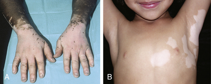
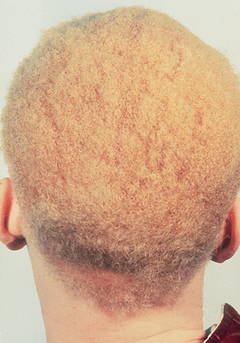
Leukoderma: partial or complete loss of skin pigmentation
Halder RM, Chappell JL: Vitiligo update, Semin Cutan Med Surg 28:86–92, 2009.
Key Points: Racial Differences in Pigmentation
Key Points: Pigmentation Disorders
Key Points: Sun-tanning
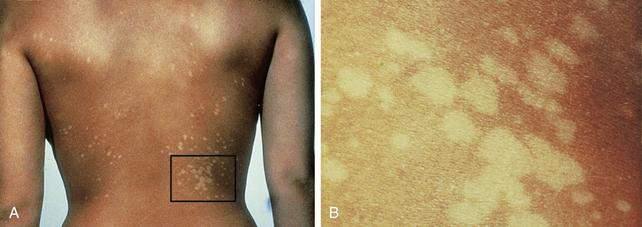
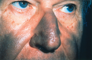
Melanoderma: abnormal darkening of the skin
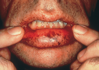
A benign condition characterized by the rapid development of hundreds of lentigines widespread over the skin surface in adolescents or young adults has been described. However, the presence of multiple lentigines at a young age is suggestive of autosomal dominant disorders, especially Moynahan’s syndrome or Peutz-Jeghers syndrome (Fig. 18-4).
Litt J, editor: Litt’s drug eruption reference manual, ed 15, New York, 2009, Informa Healthcare.
Table 18-2. Classification of Dermal Hyperpigmentation
| INCREASED MELANOCYTE NUMBER | INCREASED MELANIN | NONMELANIN PIGMENTS |
|---|---|---|
| Genetic | ||
| Mongolian spot Nevus of Ota Nevus of Ito |
− − − |
− − − |
| Chemical or Drug | ||
| − − − − − − − − − |
Fixed drug eruption − − − − − − − − |
Silver Mercury Bismuth Gold Antimalarials Phenothiazines Minocycline Amiodarone |
| Endocrine or Metabolic | ||
| − − |
Chronic nutritional deficiency Melasma |
− Ochronosis |
| Physical | ||
| − | Erythema ab igne | − |
| Inflammation and Infection | ||
| − | Macular amyloidosis | − |
| − | Erythema dyschromicum perstans | − |
| − | Postinflammatory hyperpigmentation | − |
[/level-membership-for-dermatology-category][not-level-membership-for-dermatology-category]
Chapter 18 Disorders of pigmentation
Leukoderma: partial or complete loss of skin pigmentation
Halder RM, Chappell JL: Vitiligo update, Semin Cutan Med Surg 28:86–92, 2009.
Key Points: Racial Differences in Pigmentation
Key Points: Pigmentation Disorders
Key Points: Sun-tanning
[/not-level-membership-for-dermatology-category]
