Disorders of nails
Congenital disease
A number of usually rare congenital conditions can affect the nails. In the nail–patella syndrome, the nails (and patellae) are absent or rudimentary. The nails in pachyonychia congenita are thickened and discoloured from birth. Nail dystrophy is a feature of dystrophic epidermolysis bullosa (p. 90). Racket nails, characterized by a broad short thumbnail, is the commonest congenital nail defect. It is dominantly inherited and more common in women.
Dermatoses
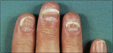
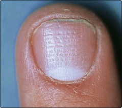
The nails are commonly involved in skin disease (Figs 1 and 2), and are routinely assessed in a dermatological examination. Details are given in Table 1, and a differential diagnosis of the changes is shown in Table 2. Treatment is aimed at the associated dermatosis; care of the hands (p. 38) is especially important.
| Dermatosis | Nail changes |
|---|---|
| Alopecia areata | Fine pitting, roughness of nail surface |
| Darier’s disease | Longitudinal ridges, triangular nicks at distal nail edge |
| Eczema | Coarse pitting, transverse ridging, dystrophy, shiny nails due to rubbing |
| Lichen planus | Thinned nail plate, longitudinal grooves, adhesion between distal nail fold and nail bed (pterygium), complete nail loss |
| Psoriasis | Pitting, nail thickening, onycholysis (separation of nail from nail bed), brown discoloration, subungual hyperkeratosis |
Table 2 Differential diagnosis of nail changes in dermatoses and systemic disease
| Change | Description of nail | Differential diagnosis |
|---|---|---|
| Beau’s lines | Transverse grooves | Any severe systemic illness that affects growth of the nail matrix |
| Brittle nails | Nails break easily, usually at distal margin | Effect of water and detergent, iron deficiency, hypothyroidism, digital ischaemia |
| Colour change | Black transverse bandsBlueBlue–greenBrownBrown ‘oil stain’ patchesBrown longitudinal streakRed (’splinter haemorrhages’)White spotsWhite transverse bandsWhite/brown ‘half and half’ nailsWhite (leuconychia)YellowYellow nail syndrome (Fig. 5) | Cytotoxic drugsCyanosis, antimalarials, haematomaPseudomonas infectionFungal infection, stain from cigarette smoke, chlorpromazine, gold, Addison’s diseasePsoriasisMelanocytic naevus, malignant melanoma, Addison’s disease, racial variantInfective endocarditis, traumaTrauma to nail matrix (not calcium deficiency)Heavy metal poisoningChronic renal failureHypoalbuminaemia (e.g. associated with cirrhosis)Psoriasis, fungal infection, jaundice, tetracyclineDefective lymphatic drainage – pleural effusions may occur |
| Clubbing | Loss of angle between nail fold and nail plate, bulbous fingertip, nail matrix feels spongy | Respiratory: bronchial carcinoma, chronic infection, fibrosing alveolitis, asbestosis Cardiac: infective endocarditis, congenital cyanotic defects Other: inflammatory bowel disease, thyrotoxicosis, biliary cirrhosis, congenital |
| Koilonychia | Spoon-shaped depression of nail plate | Iron deficiency anaemia; also lichen planus and repeated exposure to detergents |
| Nail fold telangiectasia | Dilated capillaries and erythema at nail fold | Connective tissue disorders including systemic sclerosis, systemic lupus erythematosus, dermatomyositis |
| Onycholysis | Separation of nail from nail bed | Psoriasis, fungal infection, trauma, thyrotoxicosis, tetracyclines (photo-onycholysis) |
| Pitting | Fine or coarse pits may be seen in nail bed | Psoriasis, eczema, alopecia areata, lichen planus |
| Ridging | Transverse (across nail)Longitudinal (up/down) | Beau’s lines (see above), eczema, psoriasis, tic-dystrophy, chronic paronychiaLichen planus, Darier’s disease |
Infections
Bacterial or fungal infection may involve the nail fold (paronychia) or the nail itself.
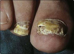
Onychomycosis (tinea unguium)
Fungal infection of the nails (onychomycosis) increases with age – children are seldom affected. Toenails, especially the big toenails (Fig. 3), are involved more than fingernails. The process usually begins at the distal nail edge and extends proximally to involve the whole nail. The nail separates from the nail bed (onycholysis), the nail plate becomes thickened, crumbly and yellow, and subungual hyperkeratosis occurs. Several – but almost never all – of the toenails may be involved. Tinea pedis often coexists and, if the fingernails are diseased, Trichophyton rubrum infection of the hand is usually seen. Treatment is with oral terbinafine (Lamisil) or itraconazole (Sporanox).
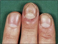
Chronic paronychia
Chronic paronychia of the fingernails due to Candida albicans is often seen in wet workers. The cuticle is lost, the proximal nail fold becomes boggy and swollen (Fig. 4) and light pressure may extrude pus. The nail plate becomes irregular and discoloured. Gram-negative bacteria may be co-pathogens and turn the nail a blue–green colour. Management is directed towards keeping the hands dry, applying an imidazole lotion or cream to the nail fold twice daily, or oral itraconazole for 14 days.
Systemic disease
Nail changes not infrequently indicate an underlying internal medical disorder. Table 2 shows some systemic associations.
Tumours
 Viral warts. Periungual warts are common. Treatment is similar to that for warts elsewhere (p. 53).
 Periungual fibromas. These are seen in patients with tuberous sclerosis (p. 92) and appear at or after puberty.


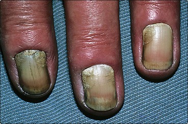
Fig. 5 Yellow nail syndrome.
The nails grow very slowly, lymphatic drainage is abnormal, and pleural effusions may occur.
Disorders of nails
 Congenital nail problems are uncommon except for racket nails.
 Sports trauma often results in subungual haematoma, onychogryphosis or onycholysis.
 Common dermatoses, e.g. psoriasis, lichen planus and eczema, have distinctive nail changes.


 Acute paronychia is usually bacterial: antibiotics are given.
 Systemic diseases may cause nail changes that help in diagnosis.

