Ear Disorders
Ear disorders are common in general practice. Earache is a frequent complaint. It may represent intrinsic ear disease or it may be due to referred pain from the oral cavity or pharynx. Causes of deafness are dealt with on p. 94.
Causes
Local
Middle Ear
Referred Pain
History
Local
External ear
Trauma may cause subperichondrial haematoma. This is usually caused by a shearing blow. The patient will present with bruising and a swelling. If the swelling is not drained, repeated trauma occurs, and this will lead to the condition of ‘cauliflower ear’, often seen in rugby players. Avulsion of the ear is rare but may occur in major trauma. Occasionally, cellulitis and swelling of the ear occur due to bites, either animal or human. The patient may complain of a swelling on the lobe of the ear. Enquire whether there has been any recent ear-piercing, as inclusion dermoids may result. Ulcers may arise on the pinna as a result of malignant disease, the most common being squamous cell carcinomas and rodent ulcers.
External auditory meatus
The patient may complain of irritation, discharge, pain and occasionally deafness. Inflammation may spread out onto the pinna. There may be a history of poking the ear with a matchstick or hairgrip to remove wax. It may occur in those who swim frequently at public baths. With a furuncle (boil) in the external auditory meatus, the patient will present with extreme pain and throbbing. The pain is made worse by inserting an auriscope into the meatus. There may be a history of a foreign body inserted into the external auditory meatus. The patient is most commonly a child. Seeds or pips may have been inserted into the ear. In adults, it is often matchsticks, cotton-wool buds or paperclips that have been inserted to remove wax from the ear. Occasionally an insect gets into the ear. There is usually pain and discharge. Malignant disease of the external auditory meatus is rare. When advanced, it may cause intractable pain, bloodstained discharge and may invade the middle ear, facial nerve or the temporomandibular joint. The patient may present with a facial palsy.
Middle ear
Acute otitis media usually occurs in children. There may be a preceding history of common cold, tonsillitis, adenoiditis or infectious disease of childhood. In adults, there may be a history of sinusitis, trauma, air-flight or temporal bone fracture. The patient presents with severe earache, often with deafness, and appears flushed and ill. Chronic otitis media presents with a history of deafness, discomfort in the ear, tinnitus and occasionally problems with the balance. Acute mastoiditis is a complication of otitis media. It is rare nowadays because of antibiotics. Occasionally, children still present with pain, discharge (creamy and profuse) and deafness, with swelling behind the ear.
Referred pain
Check for a history of dental problems. In the elderly, there may be carcinoma of the posterior third of the tongue. A classical picture of this used to be the elderly man spitting blood with a piece of cotton wool in his ear. Check for a history of tonsillitis or pharyngitis or difficulty in swallowing due to quinsy. Occasionally a foreign body lodged in the pharynx may be responsible. Other malignant disease of the pharynx may cause referred pain. Check for a history of dysphagia.
Neurological
Herpes zoster of the geniculate ganglion may give rise to lesions of the external auditory meatus, pinna and sometimes the palate. Glossopharyngeal neuralgia presents with very severe pain radiating from throat to tongue and into the ear.
Examination
Local
External ear
With trauma, there may be obvious subperichondrial haematomas. Bites will result in cellulitis and swelling of the ear. Inclusion dermoids usually follow ear-piercing and present as lumps in the lobe of the ear adjacent to the area of piercing. Malignant disease will usually present as an ulcer, either with a typical appearance of a rodent ulcer or with the everted edges of a squamous cell carcinoma.
External auditory meatus
There is often inflammation at the entry to the meatus. The meatus is tender and there is moist debris, oedema and redness of the meatal wall on examination with an auriscope. A furuncle (boil) is extremely painful. Examination with the auriscope will reveal the tense, tender boil or even pus in the external auditory meatus. A foreign body is usually obvious on examination with the auriscope, as is malignant disease.
Middle ear
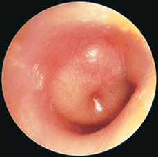
With acute otitis media, the patient is usually flushed and ill, often with a temperature of 39–40°C. Examination of the tympanic membrane shows loss of lustre and disruption of the light reflex. There is redness, fullness and bulging of the drum. There may be signs of perforation with otorrhoea, with a mucoid, purulent or bloodstained discharge. Chronic otitis media shows fluid in the middle ear (glue ear), with discoloration and often retraction of the tympanic membrane. Tuning-fork test will reveal conductive deafness. In acute mastoiditis the child is usually ill with a high pyrexia. There is tenderness over the mastoid process and post-auricular swelling. The tympanic membrane may be red and bulging or perforated with discharge.
Referred pain
Examine for dental problems. Examine the posterior third of the tongue for carcinoma. Inspect the throat with a mirror, looking for tonsillitis, pharyngitis, quinsy, foreign body or malignancy.
Neurological
With herpes zoster, there will be vesicles in the meatus, on the pinna and sometimes on the palate and fauces. This will have been preceded by severe otalgia. With glossopharyngeal neuralgia, there may be a trigger area in the throat in the same way as there is a trigger area that may initiate trigeminal neuralgia.
General Investigations
The majority of ear disorders can be diagnosed on clinical examination alone.
■ Hb, FBC, ESR
Hb ↓ malignant disease. WCC ↑ acute otitis media. Acute mastoiditis. ESR ↑ malignancy and infection.
■ Swab
C&S. Otitis externa. Otitis media with discharge. Furuncle. Tonsillitis.
■ Biopsy
Malignant disease of the pinna and external auditory meatus. Carcinoma of the posterior third of the tongue.
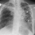
■ Skull X-ray
Opacity and coalescence of the air cells in mastoiditis.