Chapter 18 Diseases of the placenta and membranes
ABNORMAL PLACENTATION
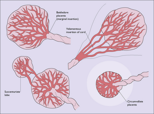
The shape of the definitive placenta is determined at the time the placenta forms, the variations found (Fig. 18.1) mostly having no clinical significance.
GESTATIONAL TROPHOBLASTIC DISEASE
Gestational trophoblastic disease is uncommon affecting 1/750 pregnancies of women of non-Asian ethnicity but 1/380 of those from Asia. It is potentially lethal, but with treatment a 98% cure rate is attainable. The disease occurs in two forms (Box 18.1).
Hydatidiform mole
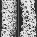
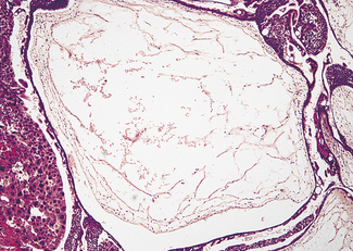
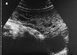
The tumour may have completely or partially replaced the placenta (Fig. 18.2). In the complete form, hydropic swelling and vesicle formation is associated with trophoblastic proliferation and a paucity or absence of blood vessels within the villi (Fig. 18.3). No fetus can be found. Five per cent of complete moles undergo malignant change.
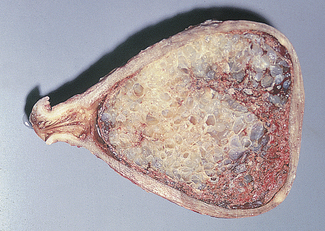
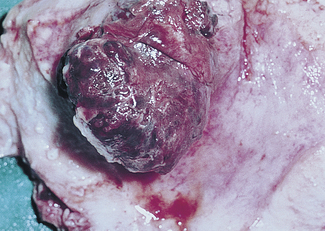
Fig. 18.2 Benign trophoblastic tumour (hydatidiform mole): gross specimen. Note the grape-like masses.
Malignant trophoblastic disease
In choriocarcinoma (Fig. 18.4) the tumour is characterized by sheets of trophoblastic cells, both syncytio- and cytotrophoblasts, with few or no villi formed.
Diagnosis of benign gestational trophoblastic disease
Follow-up
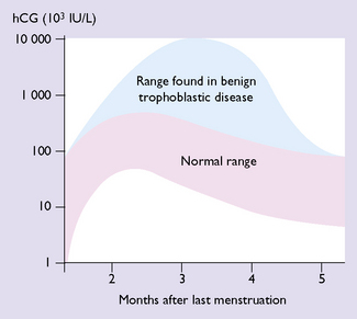
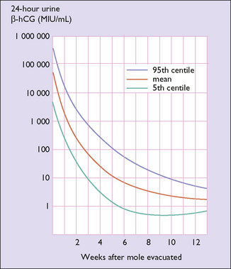
Follow-up is important as the disease persists in between 5 and 10% of cases, often developing a malignant form. Follow-up is primarily by serial β-hCG assays as outlined in Box 18.2 and Figure 18.7. Many countries have a molar register so compliance with follow-up can be monitored.
Malignant gestational trophoblastic disease
Malignant gestational trophoblastic disease is best managed at special centres, where meticulous follow-up is conducted. Treatment is by chemotherapy (Table 18.1) and follow-up is shown in Box 18.3.
| Non-Metastatic mg/kg IM | Metastatic mg/kg IM | |
|---|---|---|
| Initial course | ||
| Methotrexate at 48-hour intervals for 4 doses | 1 | 1.5 |
| Folic acid (citrovorum factor) on alternate days, 24 hours after the injection of methotrexate | 0.1 | 0.15 |
| Subsequent courses | ||
Box 18.3 Principles of the management of malignant trophoblastic disease