[level-membership-for-dermatology-category]
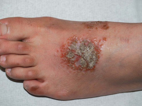
Discoid eczema
Nummular eczema
Management strategy
 Other disorders may need to be excluded, especially mycoses, psoriasis, Bowen’s disease, mycosis fungoides, sarcoidosis.
 A medication and alcohol history should be taken.


 Tar-based treatments and impregnated bandages to minimize the effects of scratching may help.
 Sedating antihistamines before retiring will help nocturnal scratching and minimize excoriation.
 Systemic immunosuppressive therapies are usually not required.
Specific investigations


First-line therapies
 Topical corticosteroids ± antibacterial agents
 Emollients
 Tar-based preparations
 Oral antibiotics
 Oral antihistamines
 Topical doxepin
Second-line therapies
 Phototherapy (broadband or narrowband UVB, 311 nm)
 Photochemotherapy (psoralen plus UVA, PUVA)
 Topical immune modulators
 Cyclosporine
 Intralesional corticosteroid injection
 Oral corticosteroids
Formal trials of these treatments in discoid eczema are lacking and therefore the evidence gradings are weak; however, all are useful in AD or other dermatitis (see gradings in Atopic dermatitis, Chapter 18), and are therefore likely to be effective in discoid eczema. Personal experience is that narrowband UVB or cyclosporine are useful if required.
Half-side comparison study on the efficacy of 8-methoxypsoralen bath–PUVA versus narrow-band ultraviolet B phototherapy in patients with severe chronic atopic dermatitis.
Der-Petrossian M, Seeber A, Honigsmann H, Tanew A. Br J Dermatol 2000; 142: 39–43.
Third-line therapies
 Azathioprine
 Methotrexate
 Mycophenolate mofetil
 Hypnosis
 Infliximab
As for second-line therapies, formal trials of these treatments in discoid eczema are lacking and therefore the evidence gradings are weak; however, all are useful in AD or other dermatitis (see gradings in atopic dermatitis, Chapter 18), and are therefore likely to be effective in discoid eczema.
[/level-membership-for-dermatology-category][not-level-membership-for-dermatology-category]
Discoid eczema
Nummular eczema
Management strategy
Other disorders may need to be excluded, especially mycoses, psoriasis, Bowen’s disease, mycosis fungoides, sarcoidosis.
A medication and alcohol history should be taken.
Tar-based treatments and impregnated bandages to minimize the effects of scratching may help.
Sedating antihistamines before retiring will help nocturnal scratching and minimize excoriation.
Systemic immunosuppressive therapies are usually not required.
