Difficulties with postoperative fluid balance in a 58-year-old man

Published on 10/04/2015 by admin

Filed under Surgery

Last modified 22/04/2025

Print this page

rate 1 star rate 2 star rate 3 star rate 4 star rate 5 star
Your rating: none, Average: 0 (0 votes)

This article have been viewed 883 times

Problem 1 Difficulties with postoperative fluid balance in a 58-year-old man

The provisional diagnosis is this patient probably has small bowel obstruction secondary to adhesions.

Blood is collected for haematological and biochemical analysis and an IV cannula placed for fluid replacement. Abdominal and chest radiographs are ordered. A urinary catheter is inserted and 350 mL concentrated urine drains. A NG tube is placed and 800 mL faeculent fluid is drained within the first 2 hours. His blood results are as follows:

Investigation 1.1 Blood results

Haemoglobin 165 g/L
White cell count 9.6 × 109/L
Platelets 350 × 109/L
Sodium 149 mmol/L
Potassium 3.4 mmol/L
Urea 10.0 mmol/L
Creatinine 0.12 mmol/L
Chloride 112 mmol/L
Bicarbonate 29 mmol/L
Glucose 4.4 mmol/L
Bilirubin 19 µmol/L
Total protein 65 g/L
Globulins 27 g/L
Albumin 38 g/L
ALT 25 U/L
AST 39 U/L
ALP 74 U/L
GGT 17 U/L
LDH 110 U/L
Amylase 65 U/L
Calcium 2.16 mmol/L
Phosphate 1.15 mmol/L
Uric acid 0.21 mmol/L
Cholesterol 3.6 mmol/L

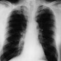
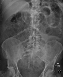
His abdominal X-ray is shown in Figure 1.1.

Over the next 12 hours the patient’s symptoms fail to improve. His pain becomes more severe, initially more frequent and then constant. He develops increasing abdominal distension and tenderness. He becomes pyrexial.

Conservative management has not been successful and an emergency operation is arranged.

At operation there are multiple adhesions involving the small bowel. These are released. A small segment of small bowel is strangulated, necrotic, but not perforated. This is resected and a primary anastomosis formed. A drain is placed.

The morning after his operation the patient looks reasonably well and is afebrile. He continues on prophylactic intravenous antibiotics and is on patient-controlled narcotic analgesia. His blood pressure is 130/90 mmHg and his pulse rate is 90 bpm. His fluid balance chart for the 24 hours since admission is as follows:

Investigation 1.2 Fluid balance chart on admission

Fluid Input  
IV fluids 4200 mL
Fluid Output  
Urine total 400 mL
Urine last 4 hours 22/16/12/0 mL
Nasogastric tube 700 mL
Wound drain 200 mL

With a bolus of 500 mL isotonic saline the urine output is increased to 50 mL over the next hour. A maintenance regimen of 1 L of isotonic saline is ordered, to be followed by 1 L dextrose 5% at 100 mL/hour and a replacement regimen of isotonic saline at 50 mL/hour using an infusion pump.

On review the following morning the patient had been given an additional 500 mL of dextrose 5% overnight when his urine output fell over a 4-hour period to less than 20 mL/hour. He now feels thirsty and there is loss of skin turgor. His abdomen is mildly distended, with absent bowel sounds, but soft and non-tender. His blood pressure is 110/65 mmHg on lying and 90/60 mmHg on sitting. His pulse rate is 100 bpm and he has a dry tongue. His fluid balance chart for the previous 24 hours shows the following:

Investigation 1.3 Fluid balance 24 hours later

Fluid Input  
IV fluids 3700 mL
Fluid Output  
Urine total 800 mL
Urine last 4 hours 15/13/9/8 mL
Nasogastric tube 2800 mL
Wound drain 400 mL

The early morning electrolytes are as follows:

Investigation 1.4 Electrolytes

Sodium 138 mmol/L
Potassium 2.7 mmol/L
Chloride 102 mmol/L
Bicarbonate 29 mmol/L
Urea 7.0 mmol/L
Creatinine 0.08 mmol/L

Answers

A.1 Colicky abdominal pain, vomiting, abdominal distension and absolute constipation are symptoms and signs suggesting a diagnosis of intestinal obstruction. At initial presentation there is hyperactivity of the bowel leading to colicky pain and tinkling bowel sounds on auscultation. A prolonged period of obstruction can ultimately lead to a paralysis of the bowel with a disappearance of colicky pain and visible peristalsis.

The commonest cause of small bowel obstruction in developed countries is postoperative adhesions (accounting for up to 60% of cases). Abdominal wall hernias account for the majority of the remaining cases of obstruction, with malignancy and inflammatory bowel disease being other causal factors to consider.

Operative procedures most closely associated with subsequent adhesive bowel obstruction are appendicectomy, colorectal surgery and gynaecological operations.

Initially, obstruction of the small bowel leads to proximal dilatation due to accumulation of gastrointestinal secretions and swallowed air. This leads to increased peristaltic and secretory activity in the intestine (both above and below the level of obstruction), which can lead to initially frequent loose stools and flatus. As the degree of obstruction becomes more severe then constipation ensues and dilatation of the proximal small bowel leads to vomiting. The development of fever can occur late and may be associated with bowel ischaemia.

This bowel dilatation is associated with compression of the mucosal lymphatics leading to significant bowel wall oedema. This, combined with increased secretory activity, leads to large third space fluid shifts with loss of electrolytes and proteins into the intestinal lumen.

The pain is often described as ‘crampy’ and ‘intermittent’. This is more prevalent in simple obstruction. Development of more constant pain can be an indication of a more serious complication – suggesting strangulation or ischaemia of the bowel.

Small bowel obstruction can be either partial or complete, simple or strangulated. Uncomplicated obstruction due to adhesions most commonly involves the small bowel. Initially this is best treated conservatively as many episodes will subside spontaneously. Strangulated obstructions are surgical emergencies and if not appropriately treated can lead to vascular compromise – ultimately creating bowel ischaemia and perforation.

A.2 This man is clinically dehydrated and has signs suggesting small bowel obstruction. Initial treatment consists of the following measures:

A.3 The supine radiograph shows features of small bowel obstruction. There are distended small bowel loops in the centre of the abdomen with prominent valvulae conniventes. The bowel wall between the loops is thickened and oedematous. No air is seen in the colon or rectum.

However, in up to 30% of cases these radiological features may not be present on a plain abdominal X-ray. In such cases CT scanning can be useful in detecting small bowel obstruction as well as defining the lead point of obstruction and detecting possible aetiological factors or other causes of acute abdominal pain.

The blood results show the following:

A.4 The key requirement is to correct any pre-existing fluid deficit prior to surgery.

Calculating the exact fluid deficit in patients can be difficult. However, an estimation of the degree of fluid deficit can be made by analysis of clinical signs. A patient who is thirsty, has dry mucous membranes, loss of skin turgor, tachycardia and postural hypotension is likely to have a loss of 10–15% of total body water. Fluid losses less than 5–10% of body water can be difficult to detect clinically. Once fluid losses exceed 15–20% there is marked circulatory collapse.

The volume of fluid required can be extrapolated from lean body weight. This patient weighs 90 kg but is overweight. His lean (metabolically active) body mass is nearer 70 kg. Fat is a relatively metabolically inert tissue and the percentage of fat relative to lean mass tends to increase with age. Calculations based on body weight alone in obese patients are likely to overestimate fluid needs.

In a 70 kg lean man who has thirst, dry mucous membranes, tachycardia and a slight postural fall in pressure, it is likely that he has a 10% loss of total body water. Body water itself is 60% of body weight and volume deficit is 10% × 60% of 70 kg, or 10% of 42 L = 4.2 L.

Vomiting or nasogastric aspiration results in loss of electrolyte rich fluid. Similarly, large occult losses occur in intestinal obstruction. Several litres of fluid may be sequestered into the gut contributing to hypovolaemia. These losses are largely isotonic and infusion of isotonic saline would be appropriate with potassium supplementation if serum levels indicate low levels.

Generally, half of this estimated loss is replaced quickly and then the patient re-assessed before replacing the rest, in this case 2 L of isotonic saline over 1 hour.

Resuscitation should be guided by improvement of clinical signs. The most important guide is a resumption of normal urine output (greater than 0.5 mL/kg/h), stabilization of blood pressure, and reduction of tachycardia.

Careful monitoring of the patient’s vital signs during this time is critical, particularly in the elderly or in patients who have associated cardiac disease. In these cases the use of a central venous catheter to measure central venous pressure may be helpful. It may be necessary to nurse the patient in a high dependency unit.

A.5 Fluid requirements in the postoperative period are intended to supply basal maintenance requirements and replace any ongoing losses (e.g. from pre-existing surgical problems, surgical drains, pyrexia).

Normal fluid losses (replaced with maintenance fluids) can be predicted with reasonable accuracy. Under normal circumstances fluid is lost through urinary losses, the gastrointestinal tract and insensible losses.

Fluid losses from the urinary tract are regulated by aldosterone and antidiuretic hormone (ADH). A fall in glomerular perfusion will trigger an increase in aldosterone leading to sodium retention within the kidney. ADH leads to retention of water in the renal tubules. Normal urinary losses are around 1.5–2.0 L/day.

The gastrointestinal tract secretes a large amount of electrolyte rich fluid into the gut. After digestion and absorption the remaining material enters the colon where much of the remaining water is reabsorbed. Approximately 300–400 mL are lost into the faeces each day.

In insensible losses air is humidified as it is inspired into the respiratory system and much of this water is lost with expiration. Fluid is also lost from the skin and these insensible losses can reach levels up to 700 mL per day. However, approximately 300 mL of fluid are produced by endogenous metabolism – leading to a total ‘insensible’ loss of 400 mL/day. Insensible losses may also be increased by pyrexia or tachypnoea.

Under normal circumstances a 70 kg man needs 2.5–3.0 L of water, 100–150 millimoles (mmol) of sodium and 60–90 mmol of potassium to replace normal losses of water and electrolytes. These maintenance requirements can be met by appropriate intravenous volumes of isotonic (0.9%) saline and 5% dextrose. Isotonic saline contains 154 mmol of sodium ions in 1 L, thus 0.6–1.0 L will provide enough sodium replacement for 24 hours. A further 2.0–2.4 L of 5% dextrose will make up the additional water. An addition of 20 mmol of potassium to each L of fluid will provide sufficient potassium.

When patients undergo surgery this leads to an endocrine response with an increased production of ADH and aldosterone. This in turn leads to renal conservation of sodium and water and by way of exchange, loss of potassium. Potassium is released by damaged tissues during surgery and any reduction in urine output may also lead to impairment of potassium excretion. Usually more potassium is released by tissue damage than is lost in the urine and therefore any potassium supplementation is best guided by serum electrolyte levels in the postoperative period.

Replacement fluid is intended to replace any ongoing losses recorded in a defined period of time. In a previously well patient undergoing emergency surgery this period is usually the previous 24 hours but in seriously ill patients where there have been major fluid shifts or where there are elements of cardiac or renal failure, intravenous fluid adjustment may be required on a more frequent basis. In the acutely ill general surgical patient fluid replacement is usually on a volume for volume basis with fluid of a similar electrolyte composition to that being lost. Nasogastric losses tend to be lower in sodium but not potassium and are typically replaced with 0.45–0.9% saline with 20 mmol/L of potassium. Gastrointestinal losses (either through a stoma or diarrhoea) have a slightly higher sodium level and similar potassium level – and can be replaced with normal (0.9%) saline with 20 mmol/L of potassium.

Where large volumes of fluid may be required then regular measurement of electrolyte levels is needed to monitor and tailor electrolyte replacement. More importantly, regular clinical examination remains critical in assessing fluid status (blood pressure, pulse rate and urine output).

A.6 The assessment for fluid requirements should begin with a careful clinical examination and a calculation of fluid losses and gains over the previous 24 hours. These show that the patient has lost more fluid than he has received; he is in negative fluid balance and hypokalaemic. He has clinical features of recurrent hypovolaemia, with oliguria, tachycardia, postural hypotension and loss of skin turgor. His mildly distended abdomen suggests likely ileus with ongoing sequestration of fluid into the small bowel.

His fluid requirements include maintenance and replacement fluids, including fluid to restore his existing hypovolaemia and correction of the hypokalaemia. A fluid bolus of up to 2 L of normal saline per hour should be given to restore urine output to at least 50 mL/hour. During this time the patient must be frequently assessed to ensure urine output returns and avoid heart failure by over-expansion of the intravascular compartment.

Potassium is predominantly an intracellular ion, and provided there is no acid–base disturbance, the serum potassium concentration reflects the total body pool. A serum potassium between 2.5 and 3.0 mmol/L represents a potassium deficit of 200–300 mmoL. The aim should be to replace 25% of this within 6 hours and 50–75% over 24 hours. Using a peripheral line, rates of replacement should not exceed 10 mmol/hour because of the risk of cardiac arrhythmia. With central access, 20 mmol/hour can be replaced without cardiac risk. Rates should never exceed 30 mmol/hour without cardiac monitoring. In this case with a potassium level of 2.7 mmol/L at least 150 mmol of replacement will be required over 24 hours along with ongoing maintenance to compensate for ongoing loss. After the initial replacement, losses may still persist. Further replacement should be guided by repeated serum potassium estimation.

Maintenance fluids are prescribed as already described: 2 L 5% dextrose (with 20 mmol potassium per L). Replacement should be the same in volume and electrolyte content as the losses they replace; particular attention should be paid to increased nasogastric losses, which have been 3.5 L in the previous 24 hours. There is likely to be additional loss from small bowel fluid sequestration secondary to ileus. These losses have been inadequately replaced. In general terms the gastrointestinal losses need to be replaced, litre for litre, with isotonic saline. Sodium (50–100 mmol/L) and chloride (100–140 mmol/L) losses into the stomach and small intestine can be matched reasonably accurately with isotonic saline. Potassium (5–15 mmol/L) must be added to the replacement fluids. In reality, losses of this magnitude in such a complex clinical setting will be replaced on an hour-by-hour basis and serum electrolyte concentrations monitored frequently.

Revision Points

Fluid Balance

Approximately 60% of body weight is made up of water.

In a 70 kg man, this is 42 litres (L):

Volume changes may be due to:

Fluid management requires understanding and calculation of three principal factors:

Some examples of abnormalities include:

The aim of perioperative fluid administration is to maintain adequate intravascular volume and thereby optimize oxygen delivery to the tissues. Patients’ fluid requirements can rarely be predicted accurately and can change dramatically from hour to hour. Prescribing using a formulaic approach without regular clinical review can lead to excessive fluid administration. Successful fluid management relies on careful anticipation of expected fluid losses and appropriate replacement. Repeated clinical assessment with measurement of pulse, blood pressure and urine output, to determine whether patients are hypovolaemic or overloaded, must be combined with regular measurement of electrolyte concentrations.